



Abstract
Computed tomography (CT) image reconstruction is faced with challenge of balancing image quality and radiation dose. Recent unrolled optimization methods address low-dose CT image quality issues using convolutional neural networks or self-attention mechanisms as regularization operators. However, these approaches have limitations in adaptability, computational efficiency, or preservation of beneficial inductive biases. They also depend on initial reconstructions, potentially leading to information loss and error propagation. To overcome these limitations, Kernel Basis Attention Primal-Dual Network (KBA-PDNet) is proposed. The method unrolls multiple iterations of the proximal primal-dual optimization process, replacing traditional proximal operators with Kernel Basis Attention (KBA) modules. This design enables direct training from raw measurement data without relying on preliminary reconstructions. The KBA module achieves adaptability by learning and dynamically fusing kernel bases, generating customized convolution kernels for each spatial location. This approach maintains computational efficiency while preserving beneficial inductive biases of convolutions. By training end-to-end from raw projection data, KBA-PDNet fully utilizes all original information, potentially capturing details lost in preliminary reconstructions. Experiments on simulated and clinical datasets demonstrate that KBA-PDNet outperforms existing approaches in both image quality and computational efficiency.
Keywords
Introduction
Computed Tomography (CT) has revolutionized medical imaging, providing invaluable insights for diagnosis, treatment planning, and disease monitoring. 1 Its ability to produce high-resolution, three-dimensional images of internal structures has made it an indispensable tool across various medical specialties. However, the increasing use of CT scans has raised concerns about cumulative radiation exposure to patients. 2 To address these concerns, low-dose CT protocols have been developed, aiming to reduce radiation exposure while maintaining diagnostic image quality. This approach, while beneficial for patient safety, presents significant challenges in image reconstruction. Low-dose scans inherently produce noisier and more artifact-prone data, potentially compromising diagnostic accuracy. Consequently, there is a pressing need for advanced reconstruction methods that can effectively balance the trade-off between radiation dose reduction and image quality preservation, ensuring that the benefits of CT imaging can be maximized while minimizing potential risks to patient health.
Traditional approaches to CT image reconstruction have relied on analytical methods such as Filtered Back Projection (FBP) and iterative techniques incorporating Total Variation (TV) regularization. 3 FBP, while computationally efficient, often produces suboptimal results in low-dose settings due to its sensitivity to noise and limited ability to incorporate prior knowledge. 4 TV regularization, on the other hand, has shown promise in preserving edge information and reducing noise, but tends to over-smooth images, potentially obscuring fine details critical for accurate diagnosis. 5 Moreover, these methods struggle to adapt to the complex, non-linear noise characteristics inherent in low-dose CT data, leading to a compromise between noise suppression and structural preservation. As the demand for lower radiation doses increases, the limitations of these traditional approaches become more pronounced, necessitating the development of more sophisticated reconstruction techniques capable of extracting maximum information from limited and noisy projection data.
Recent advancements in deep learning have sparked a paradigm shift in CT image reconstruction, offering promising solutions to the challenges posed by low-dose imaging. One approach involves using convolutional neural networks (CNN), such as U-Net, for post-processing FBP reconstructions. 6 While effective in noise reduction, these methods are limited by their reliance on initial reconstructions, potentially propagating and amplifying errors. A more sophisticated approach, inspired by unrolled optimization techniques, has led to the development of learned iterative reconstruction methods. These methods incorporate deep neural networks into the reconstruction process itself, often by unrolling a fixed number of iterations of traditional iterative algorithms. 7 Notable examples include ADMM-Net, 8 which unrolls the alternating direction method of multipliers, and Learned Primal-Dual, 9 which adapts the primal-dual hybrid gradient method. These approaches have demonstrated superior performance by learning both the regularization and the optimization process. Further refinements have been proposed, such as the DREAM-Net 10 and ISTA-Net, 11 which respectively unroll the ACID algorithm 12 and the iterative shrinkage-thresholding algorithm. Additionally, Pan et al. 13 introduced an Iterative Residual Optimization Network specifically designed for limited-angle tomographic reconstruction, enhancing reconstruction accuracy under challenging acquisition conditions. Wang et al. 14 developed a Hybrid-Domain Integrative Transformer Iterative Network tailored for Spectral CT Imaging, effectively leveraging both spatial and spectral information to achieve superior image quality. Furthermore, Wu et al. 15 proposed a Multi-channel Optimization Generative Model for stable ultra-sparse-view CT reconstruction, significantly improving reconstruction stability and detail preservation in scenarios with extremely limited projection data. These methods benefit from their ability to directly incorporate the CT forward model and noise characteristics into the learning process, enabling more accurate reconstructions from low-dose data.
Despite the progress made by deep learning-based methods in CT image reconstruction, several limitations persist when addressing low-dose CT scenarios. Many current approaches rely heavily on initial reconstructions, such as FBP, as inputs to their networks. This dependency can propagate errors and artifacts from the initial reconstruction, potentially limiting the quality of the final output. Furthermore, these methods often struggle to fully utilize the rich information contained in raw projection data, as they operate on already processed images. Another significant challenge is the trade-off between computational efficiency and reconstruction quality. Some advanced iterative methods achieve high-quality results but at the cost of increased computational complexity, making them less practical for clinical applications where rapid image reconstruction is crucial. Conversely, more efficient methods may sacrifice image quality or struggle to adapt to the complex noise patterns characteristic of low-dose CT.
To address these challenges, Kernel Basis Attention Primal-Dual Network (KBA-PDNet), a novel approach for low-dose CT image reconstruction is proposed. The core idea of KBA-PDNet is to unroll a proximal primal-dual optimization process, replacing traditional proximal operators with learnable Kernel Basis Attention (KBA) modules. This design enables the network to directly process raw projection data, eliminating the need for initial reconstructions and allowing for end-to-end training. The key innovation of the method lies in the KBA module, 16 which combines the strengths of convolutional operations and attention mechanisms. By learning a set of kernel bases and dynamically fusing them based on local image structures, KBA achieves adaptive spatial processing while maintaining computational efficiency. This approach preserves the beneficial inductive biases of convolutions, such as translation invariance, while offering the flexibility to capture complex, location-specific features. Furthermore, the end-to-end training paradigm of KBA-PDNet allows for joint optimization of the data fidelity term and the learned regularization, enabling the network to fully exploit the information content in raw projection data. This holistic approach facilitates better handling of the noise characteristics and physical models inherent in low-dose CT imaging, potentially leading to more accurate and robust reconstructions.
Related work
Post-processing methods
Deep learning-based post-processing methods have gained significant attention in low-dose CT reconstruction due to their ability to effectively reduce noise and artifacts. These methods typically operate on initially reconstructed images, often obtained through FBP. Chen et al. 17 pioneered this approach by proposing a three-layer CNN to enhance low-dose CT images. Their method demonstrated superior performance in noise reduction compared to traditional iterative reconstruction techniques. Building upon this work, Jin et al. 6 introduced a more advanced CNN architecture, FBPConvNet, which combined the strengths of FBP with the power of U-Net. This method showed remarkable improvements in image quality, particularly in preserving structural details while suppressing noise. A significant advancement in post-processing came with the introduction of residual learning. Chen et al. 18 proposed a residual encoder-decoder CNN (RED-CNN) that learned to predict the noise component, which was then subtracted from the low-dose CT image. This approach proved effective in maintaining image resolution while reducing noise and artifacts. Generative adversarial networks (GANs) have also been explored for CT image enhancement. Wolterink et al. 19 developed a GAN-based method that learned to transform low-dose CT images to match the quality of routine-dose CT images. Their approach demonstrated the potential of adversarial learning in capturing complex noise patterns and producing more realistic high-quality images.
While these post-processing methods have shown impressive results, they are inherently limited by the quality of the initial reconstruction. They may struggle to recover information lost in the initial reconstruction process, particularly in cases of severe noise or undersampling.
Unrolled optimization approaches
Unrolled optimization approaches have emerged as a powerful paradigm in CT reconstruction, combining the interpretability of traditional iterative methods with the learning capacity of deep neural networks. Adler and Öktem 9 introduced the Learned Primal-Dual algorithm, which unrolls a fixed number of iterations of a primal-dual optimization method. This approach demonstrated significant improvements over traditional methods in both image quality and computational efficiency. Building on this concept, Chun et al. 20 proposed Learned Experts’ Assessment-based Reconstruction Network (LEARN), which incorporates a set of learned “experts” into the unrolled framework, allowing for adaptive regularization. The ADMM-Net, introduced by Yang et al., 21 unrolls the alternating direction method of multipliers (ADMM) algorithm, replacing key operations with learnable modules. This method showed impressive performance in compressed sensing MRI reconstruction and has inspired similar approaches in CT. Aggarwal et al. 22 developed MoDL (Model-Based Deep Learning), which combines model-based reconstruction with deep learning. By unrolling a variable splitting algorithm, MoDL achieves a balance between data consistency and learned regularization, demonstrating robust performance across different acceleration factors in MRI reconstruction.
More recent works have further refined unrolled optimization approaches, addressing specific challenges in CT reconstruction. Zhang et al. 23 proposed CLEAR, a comprehensive learning framework that combines adversarial learning with unrolled optimization. This method enhances subtle structures in low-dose CT imaging, demonstrating improved image quality and preservation of fine details. Wu et al. 24 introduced DRONE, a dual-domain residual-based optimization network for sparse-view CT reconstruction. By integrating domain transformations and residual learning into the unrolled framework, DRONE achieves superior performance in preserving structural details while suppressing artifacts. In another work, Wu et al. 25 developed a deep embedding-attention-refinement network for sparse-view CT reconstruction. This approach incorporates attention mechanisms and embedding techniques into the unrolled optimization process, effectively capturing long-range dependencies and improving reconstruction quality. Hu et al. 26 presented DIOR, a deep iterative optimization-based residual learning method specifically designed for limited-angle CT reconstruction. DIOR addresses the challenges of missing angle information through a carefully designed unrolled network architecture, showing promising results in this particularly challenging scenario. Xia et al. 27 proposed MAGIC, a manifold and graph integrative convolutional network for low-dose CT reconstruction. This method uniquely combines manifold learning and graph convolutional networks within an unrolled framework, effectively capturing both local and global image features. Most recently, Pan et al. 28 introduced an iterative residual optimization network for limited-angle tomographic reconstruction. This approach focuses on iteratively refining the reconstruction through residual learning, demonstrating improved performance in limited-angle scenarios.
These recent advancements highlight the ongoing evolution of unrolled optimization approaches in CT reconstruction. They showcase the trend towards integrating sophisticated learning techniques, domain-specific knowledge, and novel network architectures to address the unique challenges of different CT reconstruction scenarios.
Methods
The pursuit of high-quality image reconstruction from low-dose CT scans presents unique challenges that demand innovative solutions. Traditional approaches often rely on initial reconstructions, such as FBP, which can propagate errors and limit the potential for high-fidelity outputs. Moreover, the complex nature of CT image features requires a reconstruction method that can adaptively process spatial information while maintaining computational efficiency.
To address these challenges, the KBA-PDNet, a novel approach that reimagines CT image reconstruction is proposed. The method is designed to operate directly on raw projection data, bypassing the need for initial reconstructions and potentially unlocking finer details that might otherwise be lost. The core of our network architecture, illustrated in Figure 1, leverages an unrolled optimization framework, integrating specially designed modules that combine the strengths of convolutional operations with the flexibility of attention mechanisms. This unique combination allows for adaptive feature extraction and efficient information propagation throughout the reconstruction process.

Architecture of the proposed KBA-PDNet.
The network unrolls the primal-dual optimization process, with each iteration incorporating KBA modules.
Problem formulation
The CT image reconstruction problem can be mathematically formulated as an inverse problem. Let


Typically, for low-dose CT reconstruction, we choose the data fidelity term as the weighted least squares:

The regularization term

Combining equations (2), (3), and (4), we obtain the complete optimization problem:

The primal-dual algorithm
Building upon the CT reconstruction problem formulation presented earlier, a primal-dual optimization
29
framework is employed to solve the constrained minimization problem. This approach is particularly effective for handling the non-smooth regularization term and the large-scale nature of CT reconstruction. The primal-dual algorithm addresses the optimization problem by reformulating it into a saddle-point problem. Considering the primal Eq. (5), this problem can be rewritten in its primal-dual form:


The primal-dual algorithm then proceeds by alternating updates between the primal and dual variables. The general form of these updates can be expressed as:


The proximal operator for the dual variable (Eq. (7)) has a closed-form solution as the projection onto the unit ball in the infinity norm. This projection can be computed element-wise by clipping the values of
This primal-dual algorithm provides a solid foundation for iterative CT reconstruction. However, the choice of step sizes, the number of iterations, and the exact form of the proximal operators can significantly impact the reconstruction quality and convergence speed.
Unrolling process and iteration structure
Building upon the primal-dual algorithm described previously, a unrolling process and iteration structure of the proposed KBA-PDNet is introduced. This approach transforms the iterative optimization algorithm into a learnable neural network, allowing for end-to-end training and improved reconstruction performance.
The unrolling process involves fixing the number of iterations and replacing the proximal operators with learnable neural network modules. In KBA-PDNet, the primal-dual algorithm for K iterations is unrolled, where each iteration corresponds to a stage in the network. The iteration structure is defined as follows:


The unrolled network maintains the overall structure of the primal-dual algorithm, alternating between updates of the dual variable u and the primal variable

Kernel basis attention (KBA) module
The KBA module, as illustrated in Figure 2(a), is a key innovation in our KBA-PDNet, designed to enhance adaptive spatial processing while maintaining computational efficiency. This module combines the strengths of convolutional operations and attention mechanisms, offering a novel approach to feature extraction in CT image reconstruction.

Architecture of the KBA module. (a) Overall structure of the KBA module, showing the parallel branches for attention generation and feature transformation. (b) Detailed illustration of the KBAntion weights to create adaptive kernels for each spatial location.
At the core of the KBA module is a set of learnable kernel bases
As shown in Figure 2(b), the KBA operates on an input feature map 
The feature transformation branch processes the input through a separate convolutional path:


The KBA module offers several advantages over traditional convolutions and self-attention mechanisms. It can adapt to different spatial locations and image features, potentially capturing more complex and varied patterns. Compared to self-attention, KBA maintains the local receptive field and translation equivariance of convolutions, which are beneficial properties for image processing tasks. Furthermore, by using a shared set of kernel bases, KBA achieves adaptive processing with lower computational complexity than methods that generate entirely new kernels for each location.
The final output of the KBA module is combined with the input through a residual connection:
Loss function
After defining the network architecture with the KBA module, we need to specify an appropriate loss function to guide the training process. For our KBA-PDNet, we adopt a compound loss function that combines mean squared error (MSE) and perceptual loss:

The perceptual loss 
Experiments and results
To evaluate the performance of our KBA-PDNet, we conducted experiments using the dataset from “the 2016 NIH-AAPM-Mayo Clinic Low-Dose CT Grand Challenge”. 30 This dataset contains 5936 full-dose CT images from 10 patients. For our experiments, we randomly selected 4000 images from 8 patients for the training set and 1000 images from the remaining 2 patients for the test set. All images were resized to 512 × 512 pixels.
We simulated projection data using the distance-driven method. 28 The geometry of our simulated CT system included a source-to-isocenter distance of 595 mm and a source-to-detector distance of 1085.6 mm. The detector had 888 elements, each with a size of 1.0239 mm. We simulated 984 projection views over a 360-degree range.
To simulate realistic low-dose conditions, we added Poisson noise and electronic noise to the projection data y to obtain the noisy projections ŷ:
In our KBA-PDNet, we set the kernel size
For comprehensive evaluation, we compared our KBA-PDNet with five state-of-the-art methods. These include FBP, a classical analytical reconstruction method widely used in clinical practice; FBPConvNet,
32
a deep learning-based post-processing method that uses a U-Net architecture to enhance FBP reconstructions; LPD,
9
the Learned Primal-Dual Reconstruction method, which unrolls the primal-dual optimization algorithm into a neural network; LEARN,
20
a learned experts’ assessment-based reconstruction network for sparse-data CT; MAGIC,
27
a manifold and graph integrative convolutional network for low-dose CT reconstruction; and DREAM,
10
a deep residual learning framework for sparse-view CT reconstruction. For FBPConvNet training, we utilized the Adam optimizer with an initial learning rate of
To quantitatively evaluate the performance of different methods, we employed several metrics. These include PSNR, which measures the ratio between the maximum possible signal power and the power of distorting noise; SSIM, which assesses the perceived quality of reconstructed images compared to the ground truth; and Normalized Root Mean Square Error (NRMSE), which provides a normalized measure of the difference between reconstructed and ground truth images. Additionally, we utilized the Noise Power Spectrum (NPS) to analyze the noise texture in the reconstructed images. The NPS is calculated as
Validation with the simulated data
Figure 3 presents the reconstruction results of an abdominal CT image at 10% dose level using different methods. The FBP reconstruction exhibits severe noise and prominent streak artifacts, significantly compromising image quality and diagnostic value. FBPConvNet noticeably reduces noise but tends to over-smooth fine details, particularly in low-contrast regions. LPD shows improved performance in noise suppression while better preserving structural details, though some fine textures are still lost. LEARN demonstrates good ability in preserving image structures but may sometimes introduce slight blocky artifacts. MAGIC demonstrates good noise reduction but occasionally introduces artificial patterns in homogeneous areas. DREAM achieves a good balance between noise suppression and detail preservation, but some edge structures appear slightly enhanced.

Comparison of abdominal CT reconstruction results at 10% dose level.
Our proposed KBA-PDNet achieves the best visual quality among all methods at this ultra-low dose level. It effectively suppresses noise while preserving fine anatomical structures. The region indicated by the red arrow in Figure 3 showcases the superior performance of KBA-PDNet in preserving subtle liver vasculature, which is barely visible or distorted in other methods’ results. The blue arrows point to two potential lesions, which are clearly delineated in the KBA-PDNet reconstruction while being obscured by noise or over-smoothing in other methods. Quantitatively, KBA-PDNet outperforms other methods in terms of both PSNR and SSIM metrics, as shown in Table 1. The improvements are particularly significant at this 10% dose level, demonstrating the robustness of our KBA mechanism in capturing and preserving image features under extreme low-dose conditions.
The quantitative results for the 35% dose level reconstructions.

To quantitatively assess the local reconstruction accuracy and noise characteristics, we selected four homogeneous ROIs as indicated by blue dotted boxes in Figure 3. For each ROI, we calculated the mean CT values and their standard deviations (SDs) across different reconstruction methods. The results are presented in Figure 4.

Quantitative comparison of mean CT values and standard deviations for four ROIs across different reconstruction methods.
The blue dots represent mean values, and the red error bars indicate ±1 standard deviation. The dashed blue line in each plot represents the ground truth mean value. ROIs 1–4 correspond to the regions marked in Figure 3. Figure 4 reveals several important trends across the reconstruction methods. FBP reconstructions show significant deviations in both mean values and SDs, reflecting the high noise level and potential biases introduced by this method. FBPConvNet, while reducing the SD considerably, tends to alter the mean values slightly, possibly due to its post-processing nature. LPD and LEARN demonstrate improved accuracy in mean values compared to FBP and FBPConvNet, but still show some discrepancies in SDs. MAGIC and DREAM both achieve good consistency in mean values across all ROIs, with MAGIC showing slightly lower SDs. Notably, our proposed KBA-PDNet consistently produces mean values closest to the ground truth across all four ROIs. Moreover, the SDs of KBA-PDNet reconstructions are the most comparable to those of the ground truth, indicating that our method not only accurately reconstructs the CT values but also preserves the natural texture and noise characteristics of the original image.
Figure 5 showcases the reconstructions of a thoracic slice using different methods at 5% dose level, displayed in the abdomen window. All methods demonstrate effective noise reduction, but key differences emerge in their ability to preserve fine structures. Two regions of particular interest are highlighted by red arrows. In these areas, KBA-PDNet exhibits superior preservation of subtle lung structures, while other methods tend to over-smooth these details to varying degrees.

Comparison of thoracic CT reconstruction results at 5% dose level.
To further illustrate the denoising performance and structural preservation capabilities of different methods, Figure 6 presents the absolute difference images between each reconstruction and the ground truth.

Absolute difference images between reconstructions and ground truth for the thoracic slic.
The difference images in Figure 6 clearly demonstrate that KBA-PDNet achieves the smallest overall deviation from the ground truth. It effectively suppresses noise while maintaining intricate structural details, particularly in the lung parenchyma and around the bronchial walls. Other methods, while reducing noise, show more pronounced differences in these critical areas. Local PSNR and SSIM values were calculated with results presented in Table 1. KBA-PDNet consistently outperforms other methods across all ROIs in both metrics, with particularly notable improvements in regions containing fine lung structures.
Figure 7 presents the reconstruction results of an abdominal CT slice at 2.5% dose level, comparing various methods with the reference image. At this higher dose level, all reconstruction methods show improved image quality compared to lower dose scenarios, with reduced noise and artifacts overall. However, distinct differences in performance are still observable among the methods. FBP reconstruction, while benefiting from the increased dose, still exhibits noticeable noise and streak artifacts, particularly in the soft tissue regions. These artifacts can potentially obscure fine anatomical details. FBPConvNet demonstrates a significant improvement over FBP, effectively reducing noise. However, it tends to over-smooth some regions, potentially losing some fine texture information, as seen in the zoomed-in area. LPD and LEARN both show good noise suppression while preserving more structural details than FBPConvNet. LPD appears to maintain slightly sharper edges, while LEARN seems to achieve a more balanced noise reduction across the image. MAGIC exhibits strong performance in preserving fine details, especially visible in the zoomed-in region. It maintains a good balance between noise suppression and detail preservation, though some low-contrast features may be slightly affected. DREAM achieves impressive noise reduction while maintaining most of the image structure. However, some subtle details in the soft tissue areas might be slightly smoothed. Our proposed KBA-PDNet demonstrates superior performance among all methods. It effectively suppresses noise while preserving fine anatomical structures. The zoomed-in area clearly shows that KBA-PDNet maintains the intricate details of soft tissue structures, which are critical for accurate diagnosis. The edges of organs and blood vessels are well-defined, and the overall image quality closely resembles the reference image.

Comparison of abdominal CT reconstruction results at 2.5% dose level.
Figure 8 presents the NPS maps for femoral CT reconstructions at 35% dose level across different methods, offering valuable insights into their noise characteristics and frequency content. The FBP method exhibits the highest overall noise power, particularly in mid to high-frequency ranges, as evidenced by the large red and orange areas in the center of its spectrum. FBPConvNet shows improvement over FBP with reduced high-frequency noise, but still retains significant power in the mid-frequency range. LPD and LEARN demonstrate further noise reduction, with more compact high-power regions in the center, indicating better preservation of low-frequency structures while suppressing high-frequency noise. MAGIC and DREAM show excellent noise suppression across all frequencies, with DREAM particularly effective in the high-frequency ranges. Our proposed KBA-PDNet method, however, stands out with the most compact and symmetrical NPS map among all methods. It effectively suppresses noise across all frequency ranges while maintaining a well-defined central region, suggesting an optimal balance between noise reduction and preservation of structural information. This progression from FBP to KBA-PDNet clearly illustrates a trend of increasing noise suppression and better preservation of relevant frequency content, with learning-based methods significantly outperforming traditional FBP.

NPS maps of femoral CT reconstructions at 2.5% dose level for different reconstruction methods.
The quantitative results for the 35% dose level reconstructions are presented in Table 1. The quantitative results in Table 1 illustrate the performance of various reconstruction methods across different dose levels. SSIM and PSNR are critical indicators of reconstruction quality, with higher values reflecting superior performance. At the highest dose level of 10%, all deep learning-based methods outperform the traditional FBP method, demonstrating the effectiveness of advanced reconstruction techniques in enhancing image quality. As the dose level decreases to 5% and further to 2.5%, the challenge of maintaining image fidelity and structural integrity intensifies due to increased noise and artifacts. KBA-PDNet consistently achieves the highest scores across all dose levels, with SSIM values of 0.965, 0.885, and 0.748 for 10%, 5%, and 2.5% doses, respectively. Similarly, PSNR values follow the same trend, indicating that KBA-PDNet excels in both aspects of image quality and structural preservation.
Ablation experiment
To gain deeper insights into the contributions of individual components within KBA-PDNet, a series of ablation studies were conducted. These experiments aim to validate the core design choices and provide guidance for future improvements. The investigation focused on several key aspects: the effectiveness of the KBA module, the impact of the number of kernel bases, the role of the attention mechanism, and the influence of iteration count. All ablation studies were performed on the 10% dose level dataset to ensure evaluation under the most challenging low-dose scenarios.
The effectiveness of the KBA module
We first assessed the contribution of the KBA module to the network's performance. For this purpose, we designed a baseline model where the KBA module was replaced with standard convolutional layers, and compared it to the full KBA-PDNet. Figure 9 illustrates the performance comparison between these two models in terms of PSNR and SSIM across different training epochs.

Performance comparison between KBA-PDNet and the baseline model (standard convolution) in terms of PSNR (left) and SSIM (right) over training epochs for 10% dose level CT reconstruction.
As evident from Figure 9, KBA-PDNet consistently outperforms the baseline model throughout the entire training process. After 100 training epochs, KBA-PDNet achieves a PSNR of 37.7 dB, surpassing the baseline model by 2.1 dB. Similarly, in terms of SSIM, KBA-PDNet reaches 0.956, compared to 0.934 for the baseline model. This significant performance gap clearly demonstrates the effectiveness of the KBA module.
Notably, KBA-PDNet not only exhibits superior final performance but also demonstrates faster convergence and more stable performance improvements throughout the training process. For instance, after just 40 training epochs, KBA-PDNet already achieves a PSNR of 34.2 dB, a level of performance that the baseline model only reaches after 80 epochs.
The impact of the number of kernel bases
To investigate the impact of the number of kernel bases in the KBA module, we conducted experiments varying this parameter from 8 to 64. Figure 10 illustrates how PSNR and SSIM metrics change as the number of kernel bases increases.

Impact of the number of kernel bases on reconstruction quality. Left: PSNR vs. number of kernel bases. Right: SSIM vs. number of kernel bases. Results are shown for 10% dose level CT reconstruction.
As shown in Figure 10, both PSNR and SSIM demonstrate a clear upward trend as the number of kernel bases increases. With 8 kernel bases, the network achieves a PSNR of 36.8 dB and an SSIM of 0.942. Doubling the number of bases to 16 results in significant improvements, with PSNR increasing to 37.3 dB and SSIM to 0.950. Further increasing the number of kernel bases to 32 yields additional gains, with PSNR reaching 37.7 dB and SSIM improving to 0.956. The performance continues to improve with 64 kernel bases, albeit with diminishing returns, achieving a PSNR of 37.9 dB and an SSIM of 0.958. These results indicate that a larger number of kernel bases allows the network to capture a wider range of image features and patterns, leading to improved reconstruction quality. However, the rate of improvement slows down as the number of bases increases beyond 32, suggesting a trade-off between performance gains and computational complexity.
The influence of iteration count
To investigate the impact of the number of unrolled iterations on the performance of KBA-PDNet, we conducted experiments varying the iteration count from 1 to 20. Figure 11 illustrates how PSNR and SSIM metrics change as the number of iterations increases for different dose levels (10%, 5%, and 2.5%).
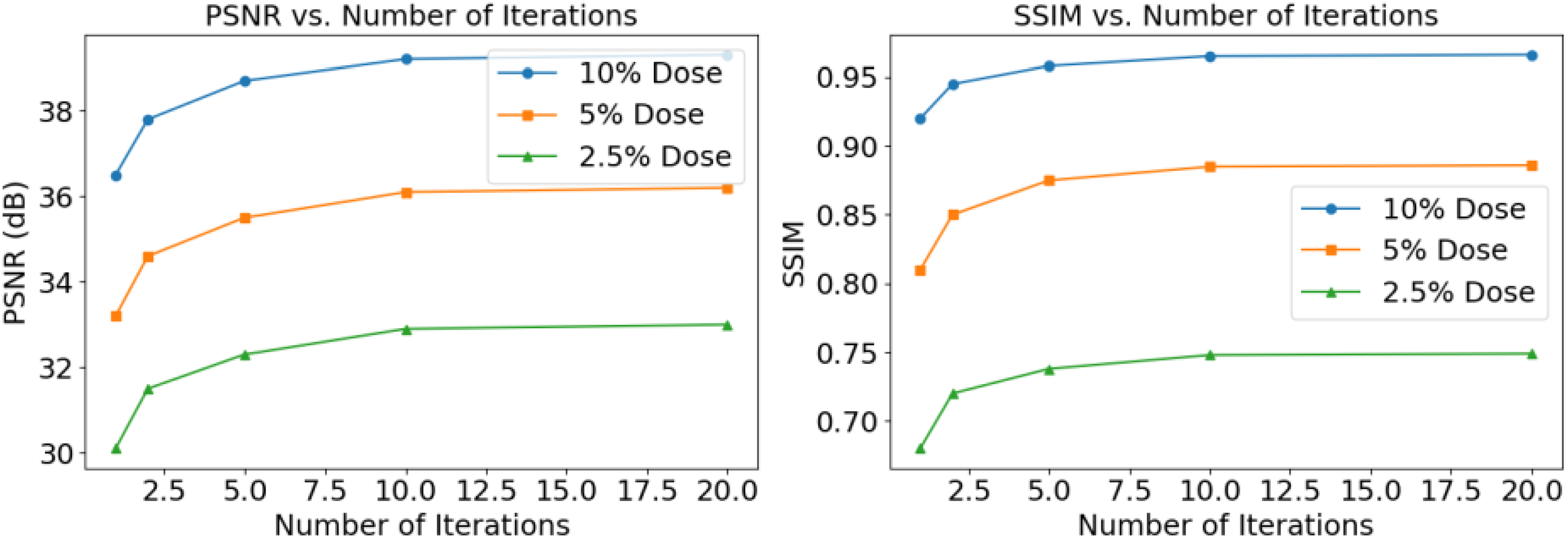
Impact of the number of iterations on reconstruction quality. Left: PSNR vs. number of iterations. Right: SSIM vs. number of iterations. Results are shown for 10%, 5%, and 2.5% dose levels.
As evident from Figure 11, both PSNR and SSIM demonstrate significant improvements as the number of iterations increases, with the most substantial gains observed in the first few iterations. For all dose levels, the performance metrics show a rapid increase from 1 to 5 iterations, followed by a more gradual improvement up to 10 iterations, after which the gains become marginal.
At the 10% dose level, PSNR improves from 36.5 dB with a single iteration to 39.2 dB with 10 iterations, while SSIM increases from 0.920 to 0.965. Similar trends are observed for the 5% and 2.5% dose levels, with the 10-iteration model consistently outperforming those with fewer iterations. Interestingly, the performance improvement from 10 to 20 iterations is minimal across all dose levels. For instance, at the 10% dose level, increasing from 10 to 20 iterations only yields a 0.1 dB gain in PSNR and a 0.001 increase in SSIM. This suggests that 10 iterations provide a good balance between reconstruction quality and computational efficiency. These results indicate that unrolling multiple iterations in KBA-PDNet is crucial for achieving high-quality reconstructions, especially in low-dose scenarios. The iterative refinement process allows the network to progressively improve its estimates, leading to better noise suppression and detail preservation. However, the diminishing returns beyond 10 iterations suggest that this configuration strikes an optimal balance between performance and computational cost for practical applications. Based on these findings, we chose 10 unrolled iterations as the default configuration for our KBA-PDNet.
The role of the attention mechanism
To evaluate the contribution of the attention mechanism in KBA-PDNet, we conducted an ablation study comparing the full KBA-PDNet (with attention) to a variant where the attention component was removed. This experiment aims to quantify the impact of the attention mechanism on reconstruction quality across different dose levels.
As evident from Figure 12, the attention mechanism consistently improves reconstruction quality across all dose levels. The performance gains are particularly pronounced at lower dose levels, highlighting the attention mechanism's effectiveness in challenging low-dose scenarios. At the 10% dose level, the attention mechanism improves PSNR from 38.498 dB to 39.207 dB, a 1.84% increase. The SSIM also improves from 0.948 to 0.965, representing a 1.79% increase. These improvements, while modest, are significant given the already high performance at this dose level.

Comparison of reconstruction quality with and without the attention mechanism. Left: PSNR comparison. Right: SSIM comparison. Results are shown for 10%, 5%, and 2.5% dose levels.
The benefits of the attention mechanism become more apparent at lower dose levels. At the 5% dose level, we observe a 1.30% improvement in PSNR (from 35.610 dB to 36.073 dB) and a 1.96% improvement in SSIM (from 0.868 to 0.885). The most substantial gains are seen at the 2.5% dose level, with a 4.74% increase in PSNR (from 31.371 dB to 32.857 dB) and a remarkable 12.14% improvement in SSIM (from 0.667 to 0.748). These results demonstrate that the attention mechanism plays a crucial role in the performance of KBA-PDNet, particularly in challenging low-dose conditions. The attention mechanism's ability to adaptively focus on relevant features allows the network to better suppress noise while preserving important structural details. This adaptability becomes increasingly valuable as the dose level decreases and the signal-to-noise ratio deteriorates. The more pronounced improvements at lower dose levels suggest that the attention mechanism helps the network to more effectively leverage the limited information available in low-dose scans. It allows the model to prioritize the most relevant features for reconstruction, leading to better noise suppression and detail preservation in these challenging scenarios.
Computational efficiency
In addition to reconstruction quality, computational efficiency is a critical factor for the practical deployment of CT reconstruction algorithms in clinical settings. To evaluate the computational efficiency of KBA-PDNet compared to existing state-of-the-art methods, we measured three key metrics: inference time per image, the number of learnable parameters, and computational complexity quantified by Floating Point Operations per Second (FLOPs). All measurements were conducted on the same hardware configuration comprising an NVIDIA RTX 3090 GPU and using Python 3.8 with the PyTorch library. Table 2 presents the quantitative comparison of computational efficiency across all evaluated methods.
Computational efficiency of CT reconstruction methods.

From Table 2, several observations can be made regarding the computational efficiency of KBA-PDNet relative to other methods. KBA-PDNet achieves an inference time of 0.30 s per image, which is faster than LPD (0.45 s), LEARN (0.40 s), and DREAM (0.50 s), and comparable to MAGIC (0.35 s). This indicates that KBA-PDNet offers a favorable balance between speed and reconstruction quality, making it suitable for clinical environments where rapid image processing is essential. KBA-PDNet has 8.5 million parameters, which is fewer than FBPConvNet (11.7 M), LPD (12.3 M), LEARN (14.5 M), and DREAM (10.2 M), while slightly higher than MAGIC (9.8 M). A reduced number of parameters contributes to lower memory consumption and faster training times, enhancing the model's scalability and deployability. With 2.5 billion FLOPs, KBA-PDNet requires fewer computational resources compared to LPD (3.8 G), LEARN (4.2 G), and DREAM (3.0 G), and is slightly more demanding than MAGIC (2.9 G). The relatively low FLOPs indicate that KBA-PDNet is computationally efficient, enabling quicker processing without significant sacrifices in image quality.
Discussion
In this study, we introduced KBA-PDNet, a novel deep learning-based method for low-dose CT image reconstruction that integrates unrolled optimization techniques with a Kernel Basis Attention (KBA) module. Our experiments demonstrated that KBA-PDNet generally outperforms existing state-of-the-art methods in terms of quantitative metrics such as PSNR and SSIM, as well as visual quality, particularly under challenging low-dose scenarios. The KBA module plays a crucial role in achieving adaptive spatial processing, effectively balancing the strengths of convolutional operations and attention mechanisms. Additionally, our ablation studies highlighted the importance of the number of kernel bases and the role of the attention mechanism in enhancing reconstruction quality.
However, there are limitations to our approach that warrant further investigation. While KBA-PDNet performs well at the tested lowest dose level of 2.5%, its performance under even more extreme low-dose conditions remains to be explored. Extremely low-dose scenarios may introduce higher levels of noise and artifacts, potentially challenging the network's ability to maintain image fidelity. Furthermore, our method relies on high-quality training data, and its generalizability to different types of CT scanners or varying patient populations needs to be validated. The interpretability of the network's decision-making process also remains an open question, as understanding the influence of the KBA module on specific reconstruction features could provide deeper insights into its operational mechanisms. Future work should focus on addressing these limitations by testing KBA-PDNet across a broader range of dose levels, incorporating diverse datasets, and enhancing the interpretability of the model. Additionally, exploring adaptive learning techniques and continuous training paradigms could further improve the network's robustness and applicability in dynamic clinical environments.
Moreover, the underlying principles of KBA-PDNet hold promise for a variety of related fields beyond medical imaging. For instance, in microbiological image analysis, as demonstrated by Zhang et al., 33 and histopathology image classification, highlighted by Chen et al., 34 adaptive attention mechanisms could enhance feature extraction and classification accuracy. In feature extraction and cell image analysis, as explored by Kulwa et al. 35 and Rahaman et al., 36 the ability of KBA-PDNet to dynamically focus on relevant spatial features could improve the precision and reliability of extracted features. Additionally, its applicability extends to video analysis 37 and X-ray image analysis, 38 where efficient and adaptive processing of complex image data is crucial. By leveraging the strengths of KBA-PDNet, these fields can achieve enhanced image processing capabilities, leading to more accurate diagnostics and analyses.
Conclusion
KBA-PDNet represents a meaningful advancement in low-dose CT image reconstruction, offering a solution that effectively balances image quality with reduced radiation exposure. Its innovative KBA module enables adaptive and efficient processing of raw projection data, contributing to improved reconstruction performance compared to existing methods. The demonstrated computational efficiency, characterized by reduced inference time and a manageable number of learnable parameters, underscores its potential for practical clinical applications where rapid and reliable image reconstruction is essential.
Footnotes
Funding
The author(s) disclosed receipt of the following financial support for the research, authorship, and/or publication of this article: This work was supported by the 2023 high-level talent research workstation project of Chongqing Preschool Education College (Preschool teachers full cycle development support system, grand No.: 2023GZZ-009).
Declaration of conflicting interests
The author(s) declared no potential conflicts of interest with respect to the research, authorship, and/or publication of this article.
Data availability statement
Data will be made available on request.*