



Abstract
Background
Recurrent anterior shoulder dislocation is a common shoulder problem, usually caused by a force from the front when the shoulder joint is abducted and externally rotated. In the present study, we investigated the effect of arthroscopic subscapularis augmentation using the long head of the biceps tendon on shoulder motion after restoring anterior stability of the joint in patients with 13.5–20% of scapular glenoid defects.
Methods
Fifty patients admitted to our department with recurrent anterior shoulder dislocation between April 2017 and July 2021 were retrospectively analyzed. The patients were divided into two groups (groups A and B, n = 25 each) with comparable age, sex, hand dominance, and articular glenoid bone loss. Patients in group A were treated with arthroscopic Bankart repair and subscapularis augmentation, whereas those in group B underwent arthroscopic long head of the biceps transposition and subscapularis augmentation. All patients in both groups were followed up for more than 1 year, with a mean follow-up period of 20.1 ± 0.7 months (range, 13–28 months). The primary outcomes were changes in the visual analog scale score, Rowe classification, and Constant–Murley shoulder outcome score.
Results
None of the patients in either group had experienced recurrent dislocation at 1-year follow-up. The visual analog scale scores decreased, and the Rowe and Constant–Murley scores improved significantly compared to the preoperative scores. Significant differences were observed in the forward flexion, abduction, and internal rotation angles of the shoulder joint in both groups at 1-year follow-up compared to baseline. The postoperative forward flexion (P = 0.143), abduction (P = 0.778), and internal rotation angles (P = 0.609) did not differ significantly between the two groups. At 1-year follow-up, the loss of angles of external rotation at the side and external rotation at 90° abduction in group B exhibited significantly less angular loss than group A.
Conclusion
Arthroscopic subscapularis augmentation using the long head of the biceps transposition technique was effective at restoring anterior stability in patients with 13.5–20% scapular glenoid defects. It was more effective at restoring the external rotational function of the shoulder joint than arthroscopic Bankart repair and subscapularis augmentation.
Keywords
Introduction
Recurrent anterior shoulder dislocation is a common shoulder injury, usually caused by a force from the front when the shoulder joint is abducted and externally rotated. 1 Various factors may affect anterior instability of the shoulder, including age, sex, participation in antagonistic or overhead sports with dislocations on the dominant side, high-standard military training maneuvers, and progressive intracapsular disease resulting from multiple dislocations, such as aggravated bone defects of the humeral head and anterior scapular glenoid, joint capsule thinning, rotator cuff injury, and injury to the upper glenoid labrum and biceps tendon anchorage point.2–4 Consequently, it is essential to implement appropriate therapeutic interventions.
The treatment strategy for recurrent anterior shoulder dislocation is a subject of ongoing debate. In 2000, Burkhart 5 suggested that 67% of surgical failures for recurrent anterior shoulder dislocations with bone defects are related to significant bone defects, such as anterior-inferior scapular glenoid bone loss (GBL) or engaging Hill-Sachs lesions. Studies further stated that articular glenoid bone repair is mandatory for scapular glenoid bone loss exceeding 25%, as this is a critical factor influencing recurrence.6–8 In contrast, bone grafting is considered necessary in cases where bone loss exceeds 20% of the healthy articular glenoid. 9 The Open Latarjet procedure10,11 is considered to be one of the effective surgical techniques, with the lowest recurrence rate of 1–4%. In comparison, in smaller Bankart injuries with anterior-inferior scapular GBL, the bone mass is usually firmly attached to the labrum, as the vast majority of fractures of the anterior border of the scapular bone are avulsion fractures.12,13 It is now generally accepted that such cases require only arthroscopic bony Bankart repair and capsular augmentation with the glenohumeral ligament. By investigating recurrent shoulder dislocations in a large number of military personnel with intense military training requirements, Professor Shaha proposed a threshold of 13.5% scapular glenoid bone defects for initiating Latarjet surgery alone or in combination with other procedures. 14 Although treatments for anterior-inferior scapular GBL have been proposed in the literature, no studies have achieved superior results with this technique compared to that of the Latarjet technique.15,16 Nevertheless, a conundrum lies in the facts that the Latarjet technique may be considered as an “over-treatment” for managing patients with 13.5–20% anterior-inferior scapular GBL, whereas the Bankart repair technique alone has a high risk of re-dislocation.
As a military hospital, we encounter many patients with recurrent anterior shoulder dislocations due to military training maneuvers, such as throwing, fighting, horizontal bar exercise, and crawling. Furthermore, it is our responsibility to provide young military personnel with the most appropriate treatment plan to maximize shoulder mobility and minimize the risk of re-dislocation. In the present study, we aimed to investigate the effect of arthroscopic subscapularis augmentation using the long head of the biceps tendon on shoulder motion after restoring anterior stability of the joint in patients with 13.5–20% of scapular glenoid defects. These patients were treated with either arthroscopic subscapularis augmentation using the long head of the biceps tendon transposition (ASA-LHBT) or Bankart repair plus arthroscopic subscapularis augmentation (Bankart + ASA) between April 2017 and July 2021.
Materials and methods
Clinical data
This is a retrospective case-control study of fifty patients admitted to our department with recurrent anterior shoulder dislocation between April 2017 and July 2021. The inclusion criteria were as follows: patients aged <45 years; participation in competitive sports or high-standard military training maneuvers requiring extreme external rotation and abduction movements; 13.5–20% of shoulder glenoid defect; a Hill-Sachs injury lesion involving <20% of the articular surface of the humeral head; no combined engagement; and no need for remplissage. Patients with a combined superior labrum from anterior to posterior injury were also eligible for inclusion after undergoing arthroscopic long head of the biceps tendon (LHBT) repair. Cases with >50% injury to the LHBT were excluded.
ASA is a surgical technique used to treat anterior instability by suturing the upper third of the subscapularis muscle and fixing it to the anterior border of the scapular glenoid. 17 Patients were divided into two groups (groups A and B, n = 25 each) with comparable age, sex, hand dominance, and articular glenoid bone loss. Patients in group A were treated with Bankart + ASA 17 (Figure 1(a)), whereas those in group B underwent ASA-LHBT 18 (Figure 1(b)). Among them, there were 5 patients with on-track lesions and 20 patients with off-track lesions in the group A, 3 patients with on-track lesions, 22 patients with off-track lesions in the group B.
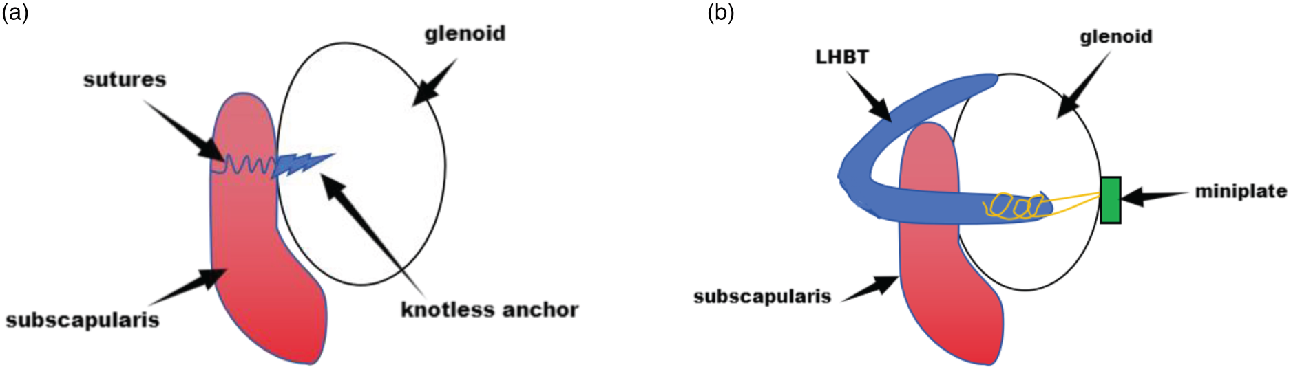
Schematic of bankart repair and subscapularis augmentation (bankart + ASA) (a) and arthroscopic subscapularis augmentation using the long head of the biceps tendon transposition (ASA-LHBT) (b).
This study was approved by Ethical Committee of our hospital and written informed consent was obtained from all patients.
Preoperative preparation
Recurrent anterior dislocation of the shoulder joint was diagnosed by referring to patients’ medical histories, and shoulder joint laxity was assessed by a physical examination of anterior, posterior, and inferior lax mobility. 19 Shoulder instability was determined using the apprehension test, load-and-shift test, and assessment of the muscles that press the humeral head toward the scapular glenoid. The anteriorly dislocated shoulder joint was relocated manually, following which radiography, 3D computed tomography (CT), and magnetic resonance imaging (MRI) were conducted to assess the extent of the Hill-Sachs defect of the scapular glenoid and humeral head. Eligible patients who did not fulfill the exclusion criteria were informed of their medical conditions, treatment options, surgical risks, complications, and the importance of postoperative rehabilitation before undergoing the ASA-LHBT.
Patient position, anesthesia, and portal creation
Following general anesthesia and a brachial plexus block, the patients were placed in the lateral decubitus position with the affected arms maintained at 30° abduction with 10 lb traction. Routine posterior, anterior, and anterosuperior portals were created, followed by an arthroscopic examination to detect all intracapsular pathologies.
Detachment, fixation, and braiding of the LHBT
The LHBT was identified between the distal bicipital groove and superior edge of the pectoralis major. An anterodistal portal was established at the superior edge of the pectoralis major muscle. The distal end of the LHBT was fixed with a 4.5 mm rivet at the superior edge of the pectoralis major, and the proximal end of the LHBT was cut off. The LHBT was subsequently pulled from the rotator cuff space and braided with two high-strength sutures in a whipstitch style for approximately 3 cm; the diameter of the braided tendon was approximately 4–5 mm (Figure 2(a)).
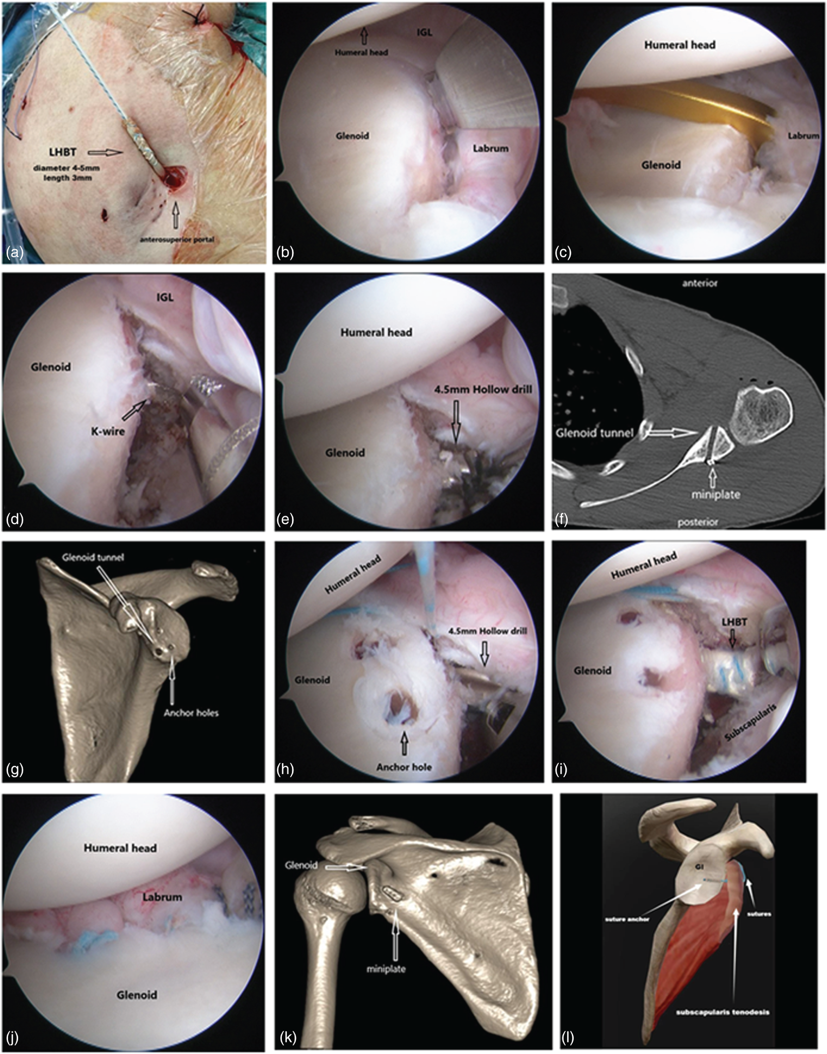
The procedure of arthroscopic long head of the biceps transposition + subscapularis augmentation (ASA-LHBT). (a) After transection, the long head of the biceps tendon is pulled from the anteroposterior portal and braided with high-strength sutures. (b) Loosening of the glenohumeral ligament complex of the joint capsule to the 6-o’clock position. (c) Insertion of anterior tibial guide. (d) The K-wire is drilled approximately 7 mm below the plane of the glenoid. (e) The K-wire is overridden with 4.5 mm cannulated drill. (f) Glenoid tunnel and miniplate. (g)The glenoid tunnel is established at approximately the 9 o’clock position in front of the glenoid and 7 mm below the articular surface. (h) The hollow drill is left in the tunnel and the anchor holes are drilled. (i) The long head of the biceps tendon is pulled into the scapular glenoid tunnel. (j) Glenoid labrum and glenohumeral ligament complex repair. (k) Miniplate located at the 3 o’clock position to the scapular glenoid. LHBT, long head of the biceps transposition. (l) Schematic illustration of ASA surgery.
Arthroscopy was performed using the anterolateral approach, and the glenohumeral ligament complex was released into a 6-point position (Figure 2(b)). The acromial glenoid defects were subsequently assessed.
Establishment of the glenoid tunnel
A tibial guide was placed into the joint with the tip of the guide positioned at 9 o'clock anterior to the glenoid (left shoulder), and the guide rod passing through the bare spot of the glenoid. A 2 mm K-wire was drilled posteroanteriorly into the glenoid, typically 7 mm below its surface. The posterior entry point of the K-wire was located at approximately 3 o'clock position (left shoulder). A glenoid tunnel measuring approximately 30–35 mm in length was created by overriding the K-wire with a 4.5 mm cannulated drill (Figure 2(c)–(g)).
Prefabricated glenoid labrum suture anchor tunnel
After establishing the bone tunnel, a 4.5 mm hollow drill was positioned in the tunnel, and anchor holes were drilled to ensure that subsequent placement of anchor nails would not interfere with the glenoid tunnel (Figure 2(h)).
Placement of guide suture with tendon introduction
A 3.0 mm rivet-guided cannula was passed through the subscapularis muscle anteroposteriorly via the upper third of the subscapularis at the myotendinous junction. The inner core was removed, leaving the cannula in place. The PDS sling was folded and inserted into the hollow cannula and passed through the glenoid tunnel posterior to anterior, using a suture retriever to pull out the suture across the subscapularis. The guide suture was identified anterior to the subscapularis muscle, and the braided LHBT was subsequently introduced. The distal end of the transposed LHBT passes through the subscapularis into the glenoid tunnel (Figure 2(i)), and the glenoid labrum and glenohumeral ligament complex were repaired (Figure 2(j)).
LHBT fixation
The posterior portal incision was manually enlarged, and the soft tissues of the tunnel were fully separated. The ends of the braided sutures were passed through the two holes of the microplate and pushed into the incision with a knot pusher along the braided suture to ensure firm attachment. The glenohumeral joint was maintained in a neutral position of external rotation and all braided sutures were tied to the miniplate (Figure 2(k)).
Treatment of the bankart + ASA group
Preoperative preparation, patient positioning, and anesthesia were identical to those in the ASA-LHBT group. Three glenoid labral anchor was placed at the 3–6 o'clock position in the right shoulder or at the 6–9 o'clock position in the left shoulder to repair the Bankart injury. In the neutral position, the middle and upper thirds of the subscapularis muscle were perforated with the ends of the tendon sutures, which were passed through a knotless anchor with the two ends forming a U-shaped parallel fixation into the scapular glenoid bone (Figure 2(l)).
Postoperative rehabilitation protocol
Immediately after surgery, the affected limb was immobilized using a brace with the arm abducted for 6 weeks. On postoperative day 1, active movement training of the interphalangeal joints of the elbow, wrist, and hand; isometric contraction training of the upper arm muscles; shoulder pendulum activity training; shoulder external rotation activity training; passive shoulder supination; and internal rotation exercises were performed. Active supination and internal rotation exercises were initiated in the 4th week and ice packs were applied to the affected shoulder at the end of the exercises. Full joint activities were resumed within 8 weeks, muscle strength training around the shoulder joint was initiated after 12 weeks, and confrontation training was performed after 6 months.
Outcomes
All patients were followed up to determine whether they had recurrent shoulder dislocation, LHBT fixation failure, joint stiffness, or infection. At follow-up, preoperative and postoperative 3D CT 20 and MRI scans of the shoulder joint were acquired to observe the position of the humeral head and the anatomical relationships between the proximal and subscapularis muscles. Furthermore, shoulder joint mobility (forward flexion, abduction, internal rotation, lateral external rotation, and abduction at a 90 °external rotation angle) was assessed before and 1 year after surgery. Shoulder joint pain and function were evaluated using the visual analog scale (VAS) score, Rowe classification, and Constant–Murley shoulder outcome scores.
Statistical analysis
SPSS software was used for all statistical analyses. Measurement data were expressed as mean ± standard deviation. Shapiro-Wilk test was used to test the normality of continuous variables. Continuous variables are presented as mean ± SD. Differences between the two groups were compared using t-tests if the variables conformed to a normal distribution, otherwise using Mann-Whitney U-tests to compare differences between the two groups. Statistical significance was set at P < 0.05 denoting statistical significance.
Results
Among the 50 patients included, 37 were male and 13 were female, with a mean age of 25 ± 3.1 years (range 19–43 years). The 50 patients were followed up for a mean of 20.1 ± 0.7 months (range 13–28 months). All procedures were performed by the same surgeon. None of the patients experienced postoperative complications, such as recurrent shoulder dislocation, LHBT fixation failure, or infection. Postoperative CT and MRI revealed that the shoulder joints were in position, and the transposed LHBTs in the ASA-LHBT group were located in the glenoid tunnel without signs of loosening.
As presented in Table 1, at the 1-year postoperative follow-up, the active mobility of the shoulder joint, such as forward flexion, abduction, internal rotation, external rotation at the side, and external rotation at 90° abduction, was significantly improved in both the groups compared to that in the preoperative period. The VAS scores at the 1-year follow-up decreased in both the groups compared with the preoperative scores. In addition, both the Rowe and Constant–Murley scores demonstrated significant improvements at the 1-year follow-up (Table 2). Although angular loss at the 1-year follow-up in both external rotation at the side and at 90° abduction was present in both the ASA-LHBT and ASA + Bankart groups; the former exhibited significantly less angular loss than the latter (Table 3).
Comparison of shoulder mobility between the 2 groups before and 1-year after surgery.
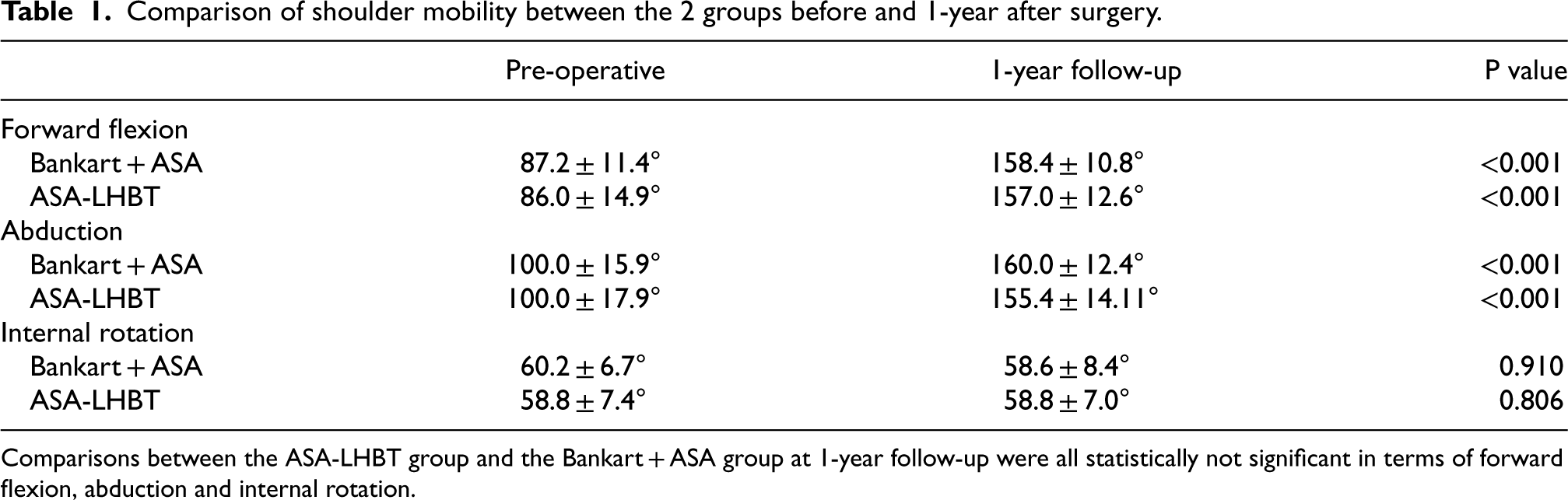
Comparison of shoulder mobility between the 2 groups before and 1-year after surgery.
Comparisons between the ASA-LHBT group and the Bankart + ASA group at 1-year follow-up were all statistically not significant in terms of forward flexion, abduction and internal rotation.
Comparison of the visual analog scale (VAS) score, rowe score and constant-murley score between the 2 groups before and 1-year after surgery.

Comparison of angular loss in terms of external rotation at side and external rotation at 90° abduction at 1-year follow-up.

Anterior shoulder instability is frequently caused by shoulder joint trauma during dislocation or subluxation. 21 The corresponding pathological changes include a Bankart injury in 87%, Hill-Sachs injury in 64%, and capsular tear and rotator cuff injury in 18% of the cases. 22 Recurrent anterior shoulder dislocations should be managed aggressively in young athletes engaging in contact sports, those with dislocations occurring on the dominant side, and military personnel with high training demands. 23
Unfortunately, the various treatments available for anterior recurrence are unreliable. Many studies have shown that simple arthroscopic Bankart repair has a high recurrence rate of dislocation, especially for patients with bone defect of 13.5–20% and high demand for motion.24,25 Subscapularis augmentation can effectively enhance the anterior stability of the shoulder joint and prevent the humeral head from dislocating forward. 8 According to Dimitri et al.'s analysis, 26 most patients had a significant difference in clinical scores after conventional Bankart repair, indicating that the shoulder joint function improved after surgery, but only about half of the patients could return to their preoperative level of motion, and about 14.8% of the patients had recurrent shoulder joint dislocation. Besides, in patients with >25% scaphoid glenoid defects, the risk of recurrent dislocations can be as high as 67% following arthroscopic Bankart repair. 5 Although the Latarjet procedure can achieve satisfactory stability by extending the scapular glenoid articular surface, it cannot override the Hill-Sachs lesions, which can be managed with the sling effect of the joint tendon to enhance the anterior stabilizing structures. Patients with a scapular glenoid defect of <13.5% achieved satisfactory outcomes and very low recurrence rates with arthroscopic Bankart repair and capsular shift of the glenohumeral ligament. Although satisfactory stability can be obtained for patients with a scapular glenoid defect of 13.5–20% with the Latarjet procedure, it is associated with several disadvantages, including destruction of the integrity of the coracoacromial arch, axillary nerve damage, and risk of bone graft failure. Moreover, the procedure requires specialized surgical instruments and entails a steep learning curve for surgeons. A previous study reported early postoperative complications of graft failure requiring reoperation in approximately two-thirds of cases and residual symptoms of nerve injury in half of the patients who underwent the Latarjet procedure. 27 Therefore, the Latarjet procedure is considered unsuitable for patients with <20% glenoid defects.
In a previous cadaveric study, Yamamoto 28 observed that the subscapularis sling effect is more important than the bone-blocking effect of the Latarjet procedure. Satisfactory surgical outcomes for recurrent anterior shoulder dislocation due to the sling effect of the subscapularis muscle produced by proximal transposition of the LHBT have also been previously reported. However, the sling effect of the LHBT on the lower part of the subscapularis tendon is weaker than that on the superior joint tendon, and is associated with a risk of vascular and nerve injury if the subscapularis muscle penetration site is too low.
Another retrospective study 17 demonstrated that arthroscopic Bankart repair combined with the ASA technique achieved very good results, with a low re-dislocation rate of only 3.2%. However, this technique may carry a potential risk of subscapularis muscle transection with high-strength sutures and reduced external rotational mobility of the shoulder joint. 29
Ensuring treatment efficacy while mitigating potential risks remains a significant clinical concern. Inspired by the promising attributes of these two procedures, we proposed the combined ASA-LHBT procedure. In the present study, we observed that this procedure yielded stable results through the transposition of the distal end of the LHBT and suspension of the upper third of the subscapularis tendon. The slip effect of the transposed LHBT can effectively prevent the risk of subscapularis muscle transection and external rotation limitation. In order to restore anterior shoulder stability in patients with 13.5–20% scapular glenoid defects, the effect of Bankart + ASA and ASA-LHBT on shoulder external rotation was compared to clarify whether the ASA-LHBT could restore anterior shoulder stability and to assess its effect on external rotation activity.
The ASA-LHBT and Bankart + ASA techniques were each performed in 25 patients each, and these patients were followed up for >1 year. None of the patients experienced recurrent shoulder dislocations after surgery. The active mobility of the shoulder, such as forward flexion, abduction, internal rotation, external rotation at the side, and external rotation at 90° abduction, significantly improved in both the groups compared to that in the preoperative period. The postoperative VAS score decreased, and the Rowe and Constant–Murley scores improved significantly in both the groups. The differences observed before and after surgery were statistically significant, indicating that both procedures were effective.
Nevertheless, patients in both the ASA-LHBT and Bankart + ASA groups experienced angular loss in external rotation at the side and at 90° abduction position, which was more significant in the Bankart + ASA group than in the ASA-LHBT group. This may be related to the different effects of the two surgical techniques on the subscapularis muscle. The traditional ASA technique involves fixing the upper third of the subscapularis muscle to the anterior glenoid with high-strength sutures, which limits the sliding of the subscapularis muscle relative to the glenoid when the shoulder joint performs external rotation, thus possibly causing external rotation limitation or subscapularis cutting. 29 The ASA-LHBT technique used in this study involves fixing the proximal end of the long head of the biceps tendon to the anterior glenoid, without affecting the attachment of the subscapularis muscle, which allows the subscapularis muscle to slide freely relative to the glenoid, theoretically reducing the restriction on the external rotation of the shoulder joint. These results indicate that the ASA-LHBT group maintained the external angle of the shoulder joint better than the Bankart + ASA group.
The technical advantages of the ASA-LHBT technique are as follows: First, the tendinous LHBT strengthens the upper third of the subscapularis. When the glenohumeral joint rotates, the subscapularis tendon slips in the direction of the muscle fibers, avoiding subscapularis muscle transection (Figure 3). Second, the scapular glenoid tunnel is drilled from the posterior to the anterior under arthroscopic surveillance, and the LHBT penetrates the upper third of the subscapularis tendon, thereby reducing the risk of axillary nerve injury. Third, a tibial guide is used to guide the bone tunnel establishment, which is easy to operate and does not require special instrumentation.
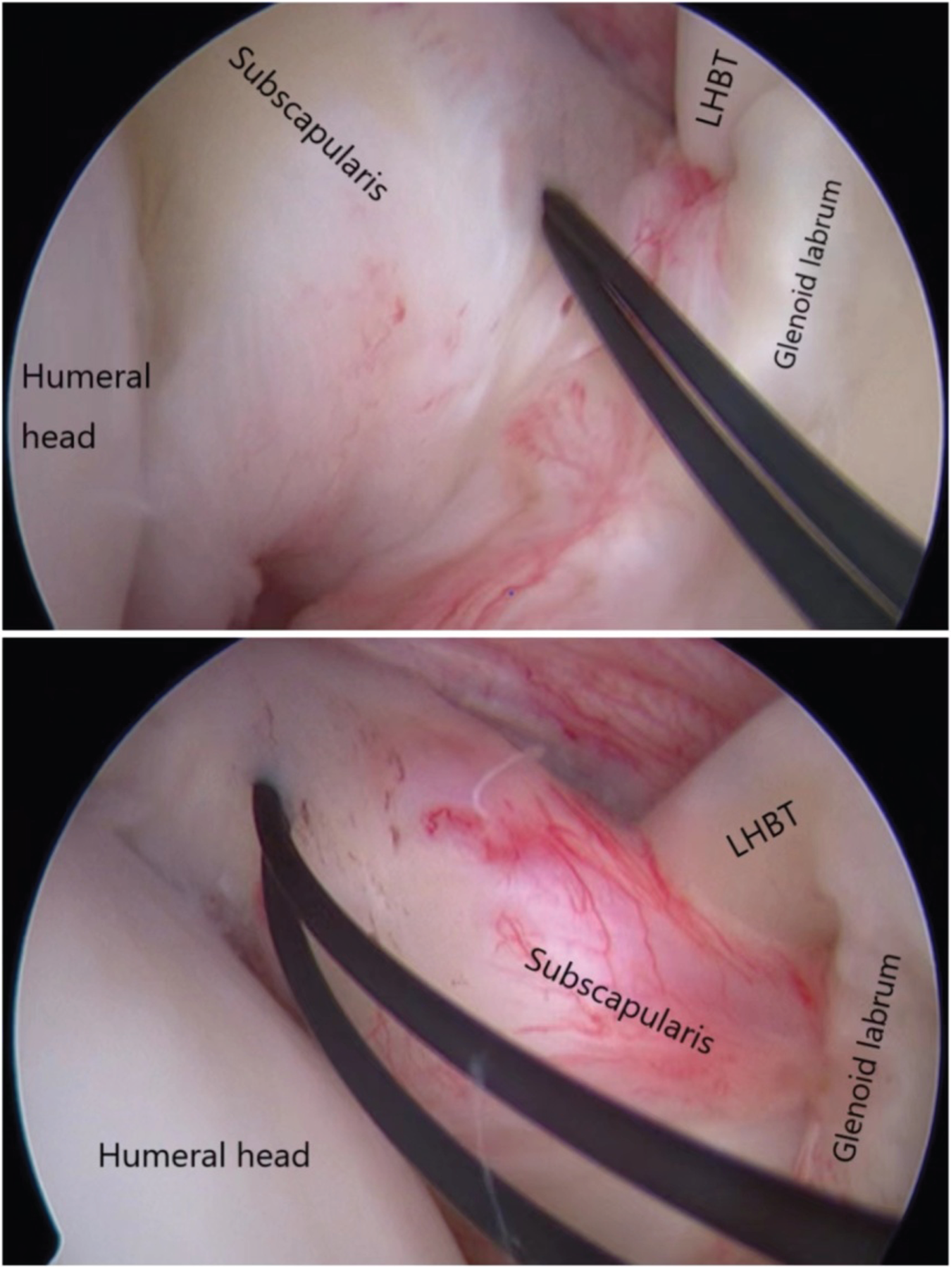
When the glenohumeral joint rotates, the subscapularis tendon slips in the direction oi the muscle fibers, avoiding subscapularis muscle transection. LHBT, long head of the biceps transposition.
However, the ASA-LHBT has several disadvantages: First, tension must be controlled when the LHBT is fixed to the posterior lead of the scapular glenoid. Excessive tension may restrict shoulder external rotation, whereas loose tension may incur the LHBT sling effect and the subscapularis blocking effect. Second, blind-knot fixation of the microplate carries a potential risk of error.
This study had several limitations that should be discussed. First, supportive biomechanical studies were not conducted. Second, the study conclusions were derived from a relatively small cohort, with potential risks of subjectivity and bias. Third, no comparisons were made between groups of patients with humeral head surface defects >20%, requiring the remplissage procedure. Fourth, the daily training activities of military patients, including throwing, horizontal and parallel bars, crawling, and hand-to-hand combat, should be equated to those of high-level competitive athletes, who have higher demands for shoulder joint stability. Finally, the follow-up time was relatively short, limited to only 1 year in most patients, with only one patient being followed-up for 5 years. Whether this will lead to accelerated joint degeneration and the development of osteoarthritis in the long term remains to be observed.
In conclusion, the results of this study revealed that the ASA-LHBT could effectively restore shoulder stability in patients with 13.5–20% scapular glenoid defects. Moreover, this technique reduced the risk of postoperative complications related to limited shoulder external rotation and axillary nerve injury. Therefore, we suggest that ASA-LHBT offers some advantages for young athletes engaging in contact or overhead sports, particularly military personnel with high training requirements, who experience dislocations on their dominant side.
Footnotes
Acknowledgment
None.

Ethical approval
This study was approved by Ethical Committee of our hospital and written informed consent was obtained from all patients.
Funding
The authors disclosed receipt of the following financial support for the research, authorship, and/or publication of this article: This study was supported by the Xiamen medical and health guiding project (No. 3502Z20224ZD1235), Xiamen Medical health science and technology project (No. 3502Z20194052), Xiamen Superior Sub-specialty construction project of Arthroscopic minimally invasive Orthopedics department (No. 2018296) and Xiamen Key Specialty construction project of Traumatic Orthopedics department (No. 2015347).
Declaration of conflicting interests
The authors declared no potential conflicts of interest with respect to the research, authorship, and/or publication of this article.