
Abstract
Background
Mesenchymal stem cells-derived exosomes, crucial in regenerative medicine, have been explored for their potential for the functional modification of bone scaffolds.
Objective
To design a functionally modified biomimetic nanohydroxyapatite using exosomes and explore its effects on bone regeneration.
Methods
A biomimetic nanohydroxyapatite (named as tHA) was fabricated as previous methods using a polydopamine (pDA) structure as a template, and exosomes (Exo) derived from periodontal ligament stem cells (PDLSCs) were used to functionally modify the tHA scaffold material through pDA. The effects of functional composite scaffold (tHA-Exo) on cells proliferation and osteogenic differentiation were investigated. Furthermore, their effect on bone regeneration was also evaluated in vivo.
Results
Exosomes can be loaded onto the tHA via pDA and the tHA-Exo releases exosomes in a sustained and stable manner. tHA-Exo showed improved cytocompatibility compared to controls. Additionally, tHA-Exo significantly enhanced the proliferation and osteogenic differentiation of PDLSCs. More importantly, animal experiments have shown that tHA-Exo could dramatically promote bone regeneration.
Conclusion
The tHA nanoparticles, functionally modified by the PDLSCs-Exo through pDA, significantly promoted bone regeneration by improving its cytocompatibility and osteogenic potential, which could serve as a promising material for promoting bone regeneration.
Introduction
Alveolar bone defects, with a high incidence, may be caused by e.g. periodontitis, 1 cysts, 2 inflammation, 3 or tumors,4,5 which lead to the loss of the supporting tissues of teeth, impair the occlusal function, and then have negative effects on the overall health. Autogenic bone grafts are usually regarded as the standard procedures for treating bone defects in clinics, but the limited donor source and the risk of donor site morbidity, immunologic rejection and cross infection restrict its extensive applications in the treatment of large-sized bone defects. 6 To repair the damaged alveolar bone, especially the periodontal bone tissue, alveolar tissue engineering, including bone graft scaffold materials, cells, and bioactive molecules, has been a research hotspot to develop bone graft scaffold with excellent bone regeneration and repair capabilities for serious bone defects. 7
Nano-hydroxyapatite, with excellent biological activity and mechanical properties due to its similar structure to the main mineral component of bone matrix, has been identified as excellent bone graft scaffold materials for bone tissue engineering. 8 An ideal biomimetic bone scaffold should be biocompatible, osteogenic, mechanically strong, and antimicrobial. To further enhance the functions of scaffold materials, recent research has attempted to modify these scaffolds in a variety of ways, mainly including the use of material composites, the loading of bioactive factors, and surface modification. 9 Polydopamine (pDA), as an efficient adhesion coating, adhere to almost all types of inorganic and organic material surfaces effectively and stably by forming covalent and noncovalent bonds (such as π bonds, van der Waals bonds, and hydrogen bonds) on material surfaces. Then, pDA can be oxidized in a weakly alkaline environment and rapidly crosslinked to form a polymer, which can be used as an intermediate medium to continue to efficiently and stably adsorb bioactive factors to achieve functional modification of scaffold materials. 10 Therefore, we fabricated a polydopamine (pDA)-templated nano-hydroxyapatite (tHA) in former studies inspired by mussels and found that tHA holds great potential with improved cytocompatibility and osteogenic capacity for bone tissue engineering, 11 and tHA can load bioactive molecules (some peptides, such as BFP-1, QK) via pDA though catechol chemistry with enhanced osteogenic effect in promoting bone regeneration.12,13
Exosomes (Exo), a cup-shaped structure with a size of 30–150 nm secreted by almost all cells, play an essential role in intercellular communication by transferring bioactive molecules to target cells, such as lipids, proteins, and nucleic acids, and show potential for treatment and diagnosis of many diseases. 14 Currently, numerous studies have shown that exosomes have potential applications in regenerative medicine, including bones, 15 cartilage, 16 muscle, 17 nerves, 18 dentin-pulp complex, 19 cardiac 20 and so on. Compared to mesenchymal stem cell (MSCs) transplantation, MSCs-Exo has been identified as more feasible for clinical application due to its higher safety and stronger plasticity. 21 As a cell-free biomaterial, exosomes offer several benefits, such as high stability, accessible storage, and low immunogenicity, and can partially solve the problems encountered in clinical applications of regenerative medicine, such as the source, quantity, and immune rejection of seed cells. Therefore, combining exosomes with tissue engineering scaffold materials can provide a new generation of scaffold biomaterials that are better suited for tissue repair.
Human periodontal ligament stem cells (PDLSCs), derived from the periodontal tissue scraped from extracted teeth due to orthodontic treatment or third molar, have emerged as an effective candidate for cell therapy due to their robust proliferation and multi-differentiation capabilities. 22 Significantly, PDLSCs-derived exosomes have been demonstrated to have the ability to regulate proliferation, migration, osteogenic differentiation, and angiogenesis, thereby significantly enhance bone regeneration.23–26 To enhance the osteogenic potential of tHA, we engineered PDLSC-exosome-coated tHA nanohydroxyapatite via pDA (tHA-Exo) and evaluated the osteogenic efficacy of tHA-Exo on bone formation in vitro and vivo. We believe that the design strategy with enhanced bioactivity holds a promising potential for applications in alveolar tissue engineering.
Materials and methods
PDLSCs culture
PDLSCs were isolated from healthy periodontal ligament tissues of premolars extracted from 16 patients aged 12–18 years due to orthodontic treatment, and the procedures were approved by the Experimental and Ethics Committees of Zunyi Medical University (LunSheng (2020)1-110). The culture method was similar to the previously described method. 27 Firstly, the periodontal ligament tissue was carefully scraped from the middle-third of the fresh extracted teeth and was digested in 3 mg/ml of type I collagenase at 37 °C for 30 min; after centrifugation, the periodontal ligament tissue was resuspended and seeded in T25 cell culture bottle cultured with complete culture medium. The complete culture medium was prepared with a-Minimum Essential Medium (a-MEM; HyClone, USA) supplemented with 10% fetal bovine serum (FBS; Gibco, USA), 1% Penicillin-Streptomycin Solution (100X; Solarbio, Beijing, China). Cell culture medium was replaced every 3 days. P3-P5 cells were used for further treatment.
A flow cytometry assay was applied to identify the cell immunophenotypes using the Human MSC Analysis Kit (BD Stemflow™ Human MSC Analysis Kit, San Diego, CA, USA). PDLSCs (P4) were cultured with osteogenic differentiation medium, adipogenic differentiation medium, or chondrogenic differentiation medium for a predetermined time, cells were fixed with 4% paraformaldehyde and then separately stained with Alizarin Red S staining (Solarbio, Beijing, China), Oil Red O staining (Solarbio, Beijing, China), and Alcian Blue staining (Solarbio, Beijing, China).
Isolation and identification of exosomes
PDLSCs (P5) were seeded to 10 cm plates (5 × 10⁶ cells/plate) and cultured with 10 ml culture medium prepared with exosome-free FBS when cells reached 80% confluency. Exosome-free FBS was achieved by centrifuged at 120000 g 4 °C for 18 h. Exosomes were isolated from the culture supernatant after incubation 48 h according to the instructions of the exosome extraction reagent (Invitrogen, Carlsbad, CA, USA). Briefly, the supernatant was centrifuged at 300 g for 5 min to pellet any contaminating cells, followed by centrifugation at 3000 g for 15 min to eliminate cell debris. The resultant supernatant was filtered through a 0.22 μm filter and then centrifuged at 4000 g for 40 min in Millipore (100KD). Exosome extraction reagent was mixed with supernatant at 2:1 ratio with gentle agitation and incubated at 4 °C overnight. The exosomes were resuspended in sterile PBS after centrifugation at 10, 000 g for 1 h. The exosomes stock solution was kept in storage at −80 °C.
The morphology of exosomes was detected by transmission electron microscopy (TEM) (Hitachi, HT-7700). The total protein concentrations of exosomes were measured by a BCA Protein Assay Kit (Solarbio, Beijing, China). Exosomal markers, including CD63 (1:2000; Abcam; ab134045) and CD81 (1:2000; Abcam; ab109201), and negative marker, Calnexin (1:2000; Abcam; ab133615), were assessed by Western blot. The particle size of exosomes was analyzed by nanoparticle tracking analysis (NTA) with an NS300 nanoparticle analyzer (Nanosight, Malvern, Worcestershire, UK).
Preparation and characterization of tHA-Exo
The procedure to synthesize tHA were the same as the previously described. 12 Firstly, 2 mg/ml dopamine (Sigma, St. Louis, MO, USA) was added to CaCl2 solution (Aladdin, Shanghai, China). Afterward, Na2HPO4 solution (Aladdin, Shanghai, China) was slowly dropped into the CaCl2−dopamine mixture with continuous stirring until the Ca/P ratio reached 1.67. The pH of solution was maintained at 8.5 through the addition of Tris-HCl (10 mM; Aladdin, Shanghai, China). The mixture was keep stirring for 12 h at 60 °C and then was kept for one day under room temperature after fully reaction. The precipitation was collected and dried in an oven at 60 °C. To synthesize tHA-Exo, PDLSCs-Exo (500 ug/ml) was added to tHA solution (100 ug/ml). The mixture was vigorously stirred for 24 h at 4 °C. After washing, the end products were lyophilized, ground down, and stored at −20 °C for subsequent research.
The morphology of tHA was detected by transmission electron microscopy (TEM, H-9000, Hitachi). PKH26-labeled PDLSCs-Exo were used to visualize conjugated exosomes using fluorescence microscope (Olympus, IX73 + DP73, Japan). PDLSCs were co-cultured with tHA-PKH26-labeled-Exo to detect cell uptake capacity for released exosomes. After 12 h of co-culture, cells were fixed with 4% paraformaldehyde and stained the nuclei with DAPI (Boster, Wuhan, China) after being washed three times with PBS, and images were taken using laser scanning confocal microscopy (LSCM, Zeiss LSM 900 Airyscan, Germany).
In vitro cytotoxicity of tHA-Exo
The cytotoxicity of tHA-Exo was evaluated using the CytoTox cytotoxicity LDH detection kit (Promega, Madison, WI, USA). After the cells were treated with the prepared materials for 24 h, the supernatant from each sample was collected and assayed the level of lactate dehydrogenase (LDH) (%) according to the manufacturer's instructions. Similarly, a calcein-AM (green fluorescence)/PI (red fluorescence) double staining kit (Solarbio, Beijing, China) was utilized to reveal the live or dead cells after co-culture with materials for 24 h. In addition, the cell counting kit–8 (CCK-8, Dojindo, Tokyo, Japan) was utilized to assess the cell numbers at 1, 3, 5, and 7 days at 450 nm for OD value using microplate reader (Multiskan Go, Thermo).
ALP activity analysis
Alkaline phosphatase (ALP) activity of PDLSCs was assessed using an assay kit (Nanjing Jiancheng, China). After co-culture with the prepared materials for 7 days, PBS was used to wash cells and then Triton X-100 (1%) was added into each well to lysate cells about 1 h at 4 °C. Then, cell lysate from each well was transferred to a new 96-well plate for further measurement of ALP activity according to the manufacturer's instructions. ALP staining was also performed according to the instructions of the BCIP/NBT Alkaline Phosphatase Color Development Kit (Beyotime, Wuhan, China). After 7 days of co-culture, PDLSCs were washed with PBS and fixed in 4% paraformaldehyde for 30 min. After three washes with PBS, the PDLSCs were incubated in alkaline solution for 30 min at room temperature, followed by three washes with PBS to terminate the reaction.
ARS staining
Calcium deposition was assessed by Alizarin Red S (ARS) staining. After 21 days of exposure to the prepared nanoparticles in osteogenic induction medium, PDLSCs were fixed with 4% paraformaldehyde. Afterward, cells were washed and stained with 1% ARS (pH 4.2; Solarbio, China) for observation. A cetylpyridinium chloride solution (10%, 1 h) was used to quantify the amount of calcium deposition according to the previous protocol.
Western blot
Cells were washed with PBS and lysed in RIPA (Beyotime, China) supplemented with a protease inhibitor cocktail (Solarbio, China) after 7 days of osteogenic induction. Protein simples were quantified using BCA assay kit (Solarbio, China), and protein simples were then separated on SDS-PAGE and transferred onto PVDF membranes (Millipore). The membranes were incubated with ALP (1:2000; HUABIO; SA40-00) and Runx2 (1:1000, Abcam; ab236639) antibodies overnight at 4 °C after blocked with 5% skim milk. The membranes were treated with HRP-conjugated secondary antibodies (Goat Anti-Rabbit IgG, 1:100000, Proteintech; Goat Anti-mouse IgG, 1:100000, BIOSS). The blotted membranes were visualized using ECL detection kit (Meilunbio, China) and quantified using the ImageJ software (National Institutes of Health, Bethesda, MD, USA). GAPDH (1:1000000, proteintech) antibody was used as internal control.
Animal experiment
The animal experiment was approved by the Animal Ethics Committee of Zunyi Medical University (approval no. Lunsheng (2020)2-217). To assess the effect of tHA-Exo on bone formation, male Sprague-Dawley rats (8 weeks) were utilized in our study. These animals were anesthetized with 5% chloral hydrate solution by intraperitoneal injection and then created two full-thickness bone defects (5 mm in diameter) in the calvaria using a dental ball drill. Saline irrigation was used to avoid heat-induced osteonecrosis, and a gentle operation was needed to avoid damage to the dura mater and brain. The critical-sized calvarial defects were then randomly assigned to the blank group, tHA group, and tHA-Exo group (n = 8). All animals were sacrificed after 8 weeks, and the specimens were harvested for further analysis. Samples were scanned using Micro-CT (VivaCT40, SCANCO Medical AG, Switzerland) to obtain 3D images of a new bone. For histological evaluation, decalcified sections were prepared and applied for hematoxylin−eosin (H&E) staining, Masson staining and immunohistochemical (IHC) staining of OCN expression.
Statistical analysis
All experiments were carried out at least three times, and the data were expressed as mean ± standard deviations. Statistical analyses were conducted using GraphPad Prism version 8.3.0 (GraphPad Software, USA). One-way analysis of variance (ANOVA) with the Scheffe multiple comparison test (post-hoc analysis) were used to determine the significant differences among the groups. P value < 0.05 was considered statistically significant.
Results
Characteristics of PDLSCs
PDLSCs were isolated from the periodontal tissue of premolars extracted for orthodontic treatment and were found to exhibit a uniform spindle-shaped morphology (Figure 1(A)). Alizarin red S, oil red O and alcian blue staining showed the presence of mineralized nodules, lipid droplets, and acid mucopolysaccharides, which indicated that PDLSCs are capable of osteogenic, adipogenic, and chondrogenic differentiation in vitro (Figure 1(B)-(D)). Flow cytometry analysis showed that PDLSCs were positive for surface markers of MSCs, including CD44 (100%), CD73 (100%), CD90 (100%) and CD105 (97.74%), and negative expression of CD45/19/34/11b (0.78%) (Figure 1(E)).
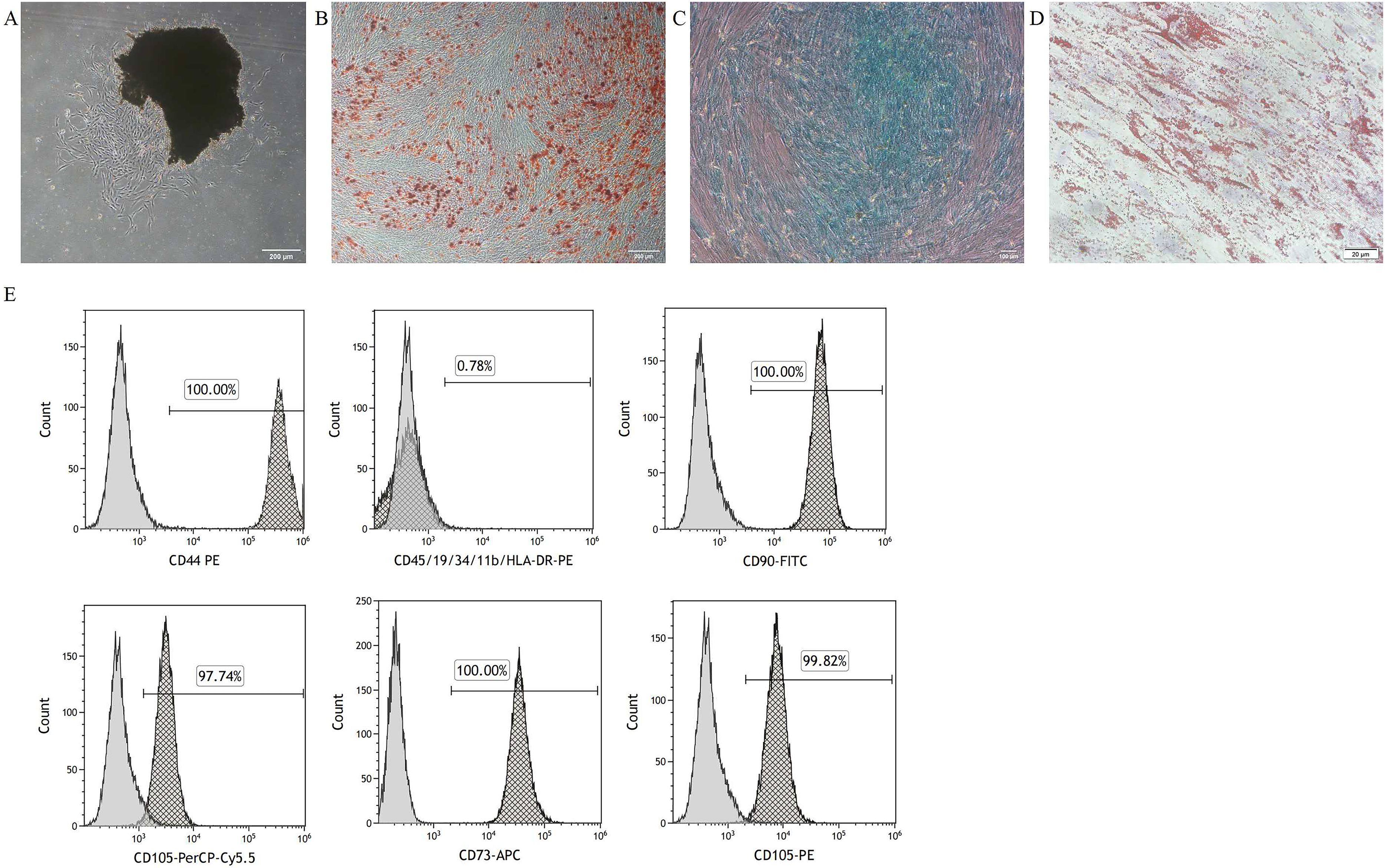
Identification of PDLSCs. (A) Representative images of PDLSCs observed by microscopy (scale bar, 200 μm). (B) Alizarin Red S staining for osteogenic differentiation (scale bar, 200 μm). (C) Alcian blue staining for chondrogenic differentiation (scale bar, 100 μm). (D) Oil Red O staining for adipogenic differentiation (scale bar, 20 μm). (E) Flow cytometry for PDLSCs.
Characterization of PDLSCs-Exo
TEM showed that PDLSCs-Exo exhibited double-membrane structures and cup-shaped morphology (Figure 2(A)). WB indicated that PDLSCs-Exo positively expressed the exosomal markers (CD 63 and CD 81), and negatively expressed Calnexin (Figure 2(B)). NTA analysis revealed that the particle size of PDLSCs-Exo was approximately 140 nm (Figure 2(C)).

Identification of Exo. (A) Morphology of Exo under transmission electron microscopy (scale bar, 100 nm). (B) Exo surface markers (CD 63 and CD 81) and negative marker (Calnexin) were detected by western blot. (C) The nanoparticle analysis of Exo.
Characterization of tHA-Exo
TEM images (Figure 3(A)) revealed that tHA exhibited a plate-shaped structure (approximately 15 nm in diameter and 80 nm in length), which had similar characteristics to natural HA in the bone tissue but were susceptible to aggregation due to the adhesion property of pDA, and the loading of exosomes did not alter the structure of tHA. To further confirm the successfully loading of Exo onto tHA, the fluorescence label assay was employed to visualize the presence of PHK-26 labeled Exo (red fluorescence light) on tHA-PHK-26 labeled Exo, as shown in the images (Figure 3(B)). The internalization of PDLSCs-Exo release from tHA-Exo was visualized using LSCM. The images showed that PKH26-labelled Exo (red dots) released from tHA-PKH26-labelled Exo and were internalized by cells and distributed in cytoplasm (Figure 3(C)). The previous study demonstrated that tHA had little toxicity to PDLSCs when the concentration exceeded 100 ug/ml. 28 Live and dead staining images showed that a small amount of red fluorescence was displayed in tHA group, while tHA-Exo group displayed a weaker red fluorescence signal (Figure 3(D)). Similar results were also found in CCK-8 assay, which demonstrated that tHA at 100 ug/ml had no obvious cell-killing effects, and tHA-Exo had a significant promotion effect on cell proliferation compared to control and tHA group (Figure 3(E)). LDH assay was performed to further assess the cytotoxic effect of tHA-Exo on the integrity of plasma membranes, and the resulted indicated that LDH leakage in the tHA group was significantly increased after 24 h of co-culture with PDLSCs and LDH releasing level was decreased from 9.72 to 5.79% when cells were subjected to tHA-Exo (Figure 3(F)). These results suggest that Exo endows tHA with superior cytocompatibility.

Characterization and cytocompatibility of tHA-Exo. (A) Morphology of tHA under TEM (scale bar, 500 nm and 20 nm). (B) Images of tHA and tHA-Exo using fluorescence microscope. Exo was labeled with PKH26 (red) (scale bar, 50 nm). (C) Internalization of tHA-Exo by PDLSCs using LSCM. Exo was labeled with PKH26 (red), and Nuclei were stained with DAPI (blue) (scale bar, 20 nm). (D) Live and dead staining of tHA and tHA-Exo (scale bar, 200 nm). (E) CCK-8 assay. (F) LDH assay. ns indicates not significant, *p < 0.05, **p < 0.01, ***p < 0.001, ****p < 0.0001.
The osteogenic effect of tHA-Exo on PDLSCs
As shown in Figure 4(B), a higher ALP level was observed in cells after co-cultured with tHA (P < 0.05), indicating that the positive effect of tHA on osteogenic differentiation, and the ALP level was significantly increased in the tHA−Exo group when compared to tHA (P < 0.05). These results were similar to ALP staining (Figure 4(A)). Concurrently, ARS staining proved the tHA group distinctly exhibited more calcium nodules than the control group (P < 0.05) and most of the amount of calcium nodules were found in the tHA−Exo (P < 0.05) (Figure 4(C) and (D)). WB analysis revealed an increase in the expression of ALP and RUNX2 in the tHA group, and tHA-Exo have significant enhanced osteogenic proteins (ALP and RUNX2) when compared to tHA group (Figure 4(E)).
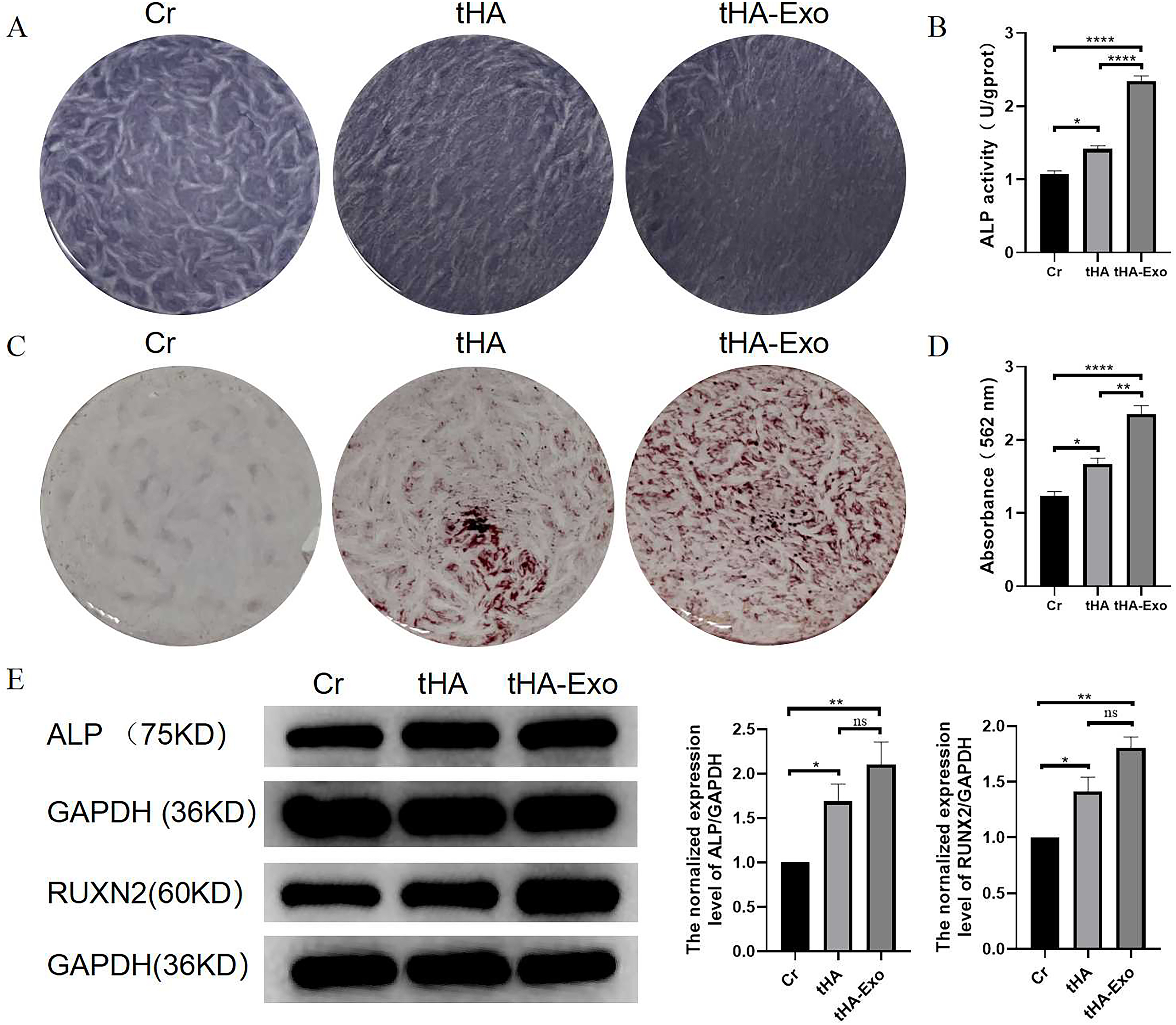
tHA-Exo promote the osteogenic differentiation of PDLSCs. (A) The ALP staining of PDLSCs after treated with tHA or tHA-Exo for 7 days (scale bar, 100 μm). (B) The ALP activity of PDLSCs after treated with tHA or tHA-Exo for 7 days (n = 3). (C) Photographs of alizarin red S staining for PDLSCs after treated with tHA or tHA-Exo for 21 days (scale bar, 100 μm). (D) The quantitative analysis of alizarin red S staining (n = 3). (E) WB analysis of ALP and RUNX2 in PDLSCs from different groups (n = 3). ns indicates not significant, *p < 0.05, **p < 0.01, ***p < 0.001, ****p < 0.0001.
tHA−Exo promote bone regeneration in bone defect model
After in vitro evaluation, the in vivo effect of tHA−Exo on bone regeneration was also assessed in our study. According to the observation of Micro-CT (Figure 5(A) and (B)), compared to the control group (16.1%), the bone regeneration is significantly accelerated in the tHA group (41.4%), indicating the osteogenic efficacy of tHA nanoparticle in vivo as well. After coated Exo on tHA, the amount of the newly formed bone tissues was further increased in the tHA−Exo group (58.9%). Obvious bone structures were detected in tHA and tHA−Exo group and fibrous tissues occupied the majority of the space in the control group at 8 weeks after surgery according to the results of H&E and Masson staining (Figure 5(C) and (D)). IHC staining of OCN expression (Figure 5(E) and (F)) revealed a similar trend similar to Micro-CT assessment. Overall, these in vivo results provided direct evidence that the combined immobilization of Exo could synergistically promote in vivo bone regeneration.
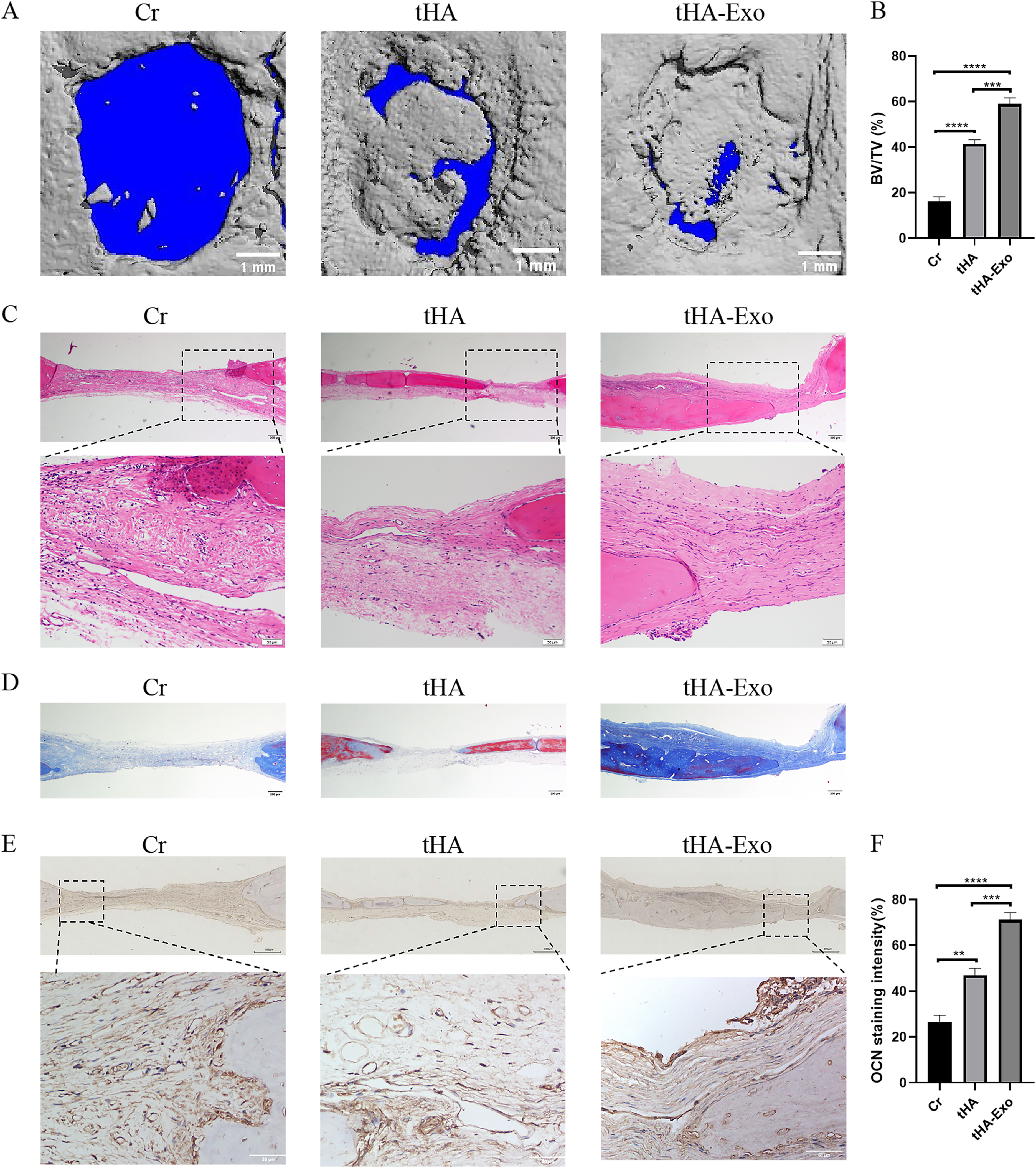
tHA-Exo promote bone regeneration in SD rat skull defect model. (A) Micro-CT images of alveolar bone loss (Scale bar: 1 mm). (B) Quantitative analysis of micro-CT images. (C) HE staining (Scale bar: 200 μm). (D) Masson staining (Scale bar: 200 μm). (E) Immunofluorescence staining for OCN (Scale bar: 200 μm). (F) Quantitative analysis of immunofluorescence staining for OCN. Data are presented as the mean ± SEM (n = 3). *p < 0.05, **p < 0.01, ***p < 0.001, ****p < 0.0001.
Discussion
This study explored the osteogenic effects of tHA coated with Exo through in vivo and in vitro experiments, and the results indicated that Exo can directly coat on tHA via pDA and significantly improve biocompatibility and reduce the cytotoxicity of tHA, and therefore remarkably enhanced the osteogenic ability of tHA and promoted its effect on new bone formation in vivo.
Hydroxyapatite in human bones exists in the form of nanoscale single crystals, uniformly distributed in the collagen matrix. 29 Therefore, artificial nano hydroxyapatite is often used as a bone substitute material for the repair and regeneration of bone tissue defects.8,30 Nano-hydroxyapatite can form strong bonding with natural bone tissue, and its calcium and phosphorus components play important regulatory roles in stem cell differentiation and bone remodeling processes. However, traditional nano hydroxyapatite has problems such as harsh synthesis conditions, irregular crystal size and morphology, and poor biocompatibility. 31 In recent years, the technology of synthesizing nano hydroxyapatite using biomimetic methods to simulate the formation process of biomineralization has become a research hotspot.
In the previous research, a polydopamine templated hydroxyapatite material, we named tHA, was biomimetically synthesized through the strong complexation between the catechol functional group in the pDA structure and calcium ions using polydopamine as a template. tHA has a calcium phosphorus ratio and crystallinity similar to natural skeletal apatite crystals, 32 which can effectively exert bone induction and bone conduction activity, thus exhibiting better osteogenic effects than traditional nano hydroxyapatite.11,33 However, further research has found that the osteogenic ability of tHA improves as the concentration increases, but the cytotoxicity also increases significantly, which limits the application of tHA materials in bone tissue engineering.11,28 In addition, studies have reported that hydroxyapatite materials are prone to inducing the expression of some cellular inflammatory factors such as TNF-a, IL-6, and IL-10, which disrupt the cellular microenvironment and lead to material implantation failure. 34 Similar studies have also found that high concentrations of hydroxyapatite materials can cause cell damage and death, with certain cytotoxicity. 31 Therefore, reducing the cytotoxicity of high concentrations of tHA is crucial for its clinical application.
Methods to modify bone scaffolds have been researched, mainly including optimization and innovation of scaffolds, the loading of bioactive factors and surface modification, to further improve the functions of scaffolds. Recently, pDA can adhere to almost all types of inorganic and organic material surfaces via forming covalent and noncovalent bonds, and can be an intermediate to continue to efficiently and stably adsorb bioactive factors, such as functional proteins, drugs or exosomes, to achieve functional modification of scaffold materials. 10 Zhou et al. conducted a study that loading exosomes onto a biomimetic scaffold to successfully functionally modify its osteogenic ability by coating pDA. 35 Therefore, our study utilized the pDA component in tHA to load exosomes into the scaffold material.
Exosomes, carrying virous proteins, lipids, genetic biomolecules, and metabolites, have attracted great attention in bone regeneration field. PDLSCs, as promising stem cells for cell therapy, have some superiority, such as easy to achieve, excellent ability to osteogenic, angiogenic and chondrogenic differentiation. 36 Compared to PDLSCs, exosomes derived from PDLSCs have also been demonstrated to have effect of promoting cell proliferation, 37 migration, 38 osteogenic differentiation, 39 angiogenesis, 40 osteoclastogenesis, 41 inflammation response, 42 thereby promoting bone regeneration. Therefore, we applied PDLSCs-Exo to coat tHA via pDA to functionally modify these biomimetic nano-hydroxyapatite material.
Recent studies have demonstrated that mesenchymal stem cell (MSC)-derived exosomes could be used as bio-mimetic tools to directly regulate osteoblast proliferation and activity, simultaneously induce stem cells into an osteogenic linage. Exosomes not only endow scaffold with abundant biofunction, such as regulating cell proliferation, migration, osteogenic differentiation, and angiogenesis, but also improve the excellent biocompatibility of scaffold. Kang et al. found that exosomes promoted the cell attachment and proliferation on the scaffold and enhanced the osteogenesis and vascularity regeneration in the scaffolds in vitro and in vivo. 43 Exosome make PLGA/Mg-GA scaffold with enhanced osteogenesis property. 44 Lu et al. demonstrate that the USCEXOs/GelMA-HAMA/nHAP composite hydrogel with controllable and biocompatible may effectively promote bone regeneration by coupling osteogenesis and angiogenesis. 45 Similar to these studies, our results revealed that PDLSCs-Exo significantly enhanced tHA ability to promote osteogenic differentiation and new bone regeneration.
Many signal pathways, such as calcium-dependent pathway, BMP/Smad pathway, Wnt/catenin pathway, are involved in the regulation of osteogenic process for MSCs. Therefore, the desired bone graft materials (tHA-Exo) should be delivering multiple biological cues to achieve maximum potential of target stem cells for precisc mimicry of the real bone formation condition. The PDLSCs-Exo functionlized tHA apatite nanocomposites maybe regulate MSCs function through two different pathways in a stable manner. On the one hand, tHA-Exo promote osteogenic differentiation of MSCs though calcium-dependent signaling due to the similar Ca/P ratio to the real bone structure. According to the previous literature, tHA could sustainably release Ca2+ towards surrounding environment and will provide nucleation sites during mineral deposition of osteogenic progenitor cells. On the other hand, PDLSCs-Exo have been found to have the ability to regulate proliferation, migration, osteogenic differentiation, and angiogenesis, thereby significantly enhance bone regeneration. Liu et al. found that PDLSCs-Exo promoted BMSCs osteogenic differentiation through altering exosomal microRNA profiles. 46 Lan et al. demonstrated that PDLSC-Exo are capable of promoting cell proliferation, migration and osteogenic differentiation, inhibiting H2O2-induced apoptosis, and activating the PI3K/AKT and MEK/ERK signaling pathways. 37 Kang et al. study indicated that the overexpression of miR-205-5p in PDLSCs-Exo inhibited inflammatory cell infiltration, decreased the expression of inflammatory factors, and regulated Treg/Th17 balance via targeting XBP1. Lu et al. found that PDLSCs-Exo inhibited osteoclast formation through the miR-31-5p/Enos signaling pathway. 41 PDLSCs-Exo enhanced BMSCs migration via AKT and ERK1/2 pathway, 38 and promoted BMSCs proliferation and osteogenic differentiation, and thus promoted bone regeneration. 39 Together, PDLSCs-Exo significantly improved tHA osteogenic ability, loading Exo via pDA in vitro and vivo through the synergistic effect of Ca2+ and PDLSCs-Exo.
Conclusion
In this study, we fabricated a bioactive functionalized nano-hydroxyapatite for bone regeneration. Our finding showed that improved cytocompatibility was achieved for tHA by coating PDLSCs-Exo through assistance of pDA and demonstrated that the tHA-Exo scaffold promoted PDLSCs proliferation and osteogenic differentiation in vitro and improved bone regeneration efficiency in vivo. Our work provides an instructive insight into the design of biomimetic apatite nanocomposites, which hold great potential for applications in bone regeneration. Therefore, incorporating exosomes with biomimetic bone scaffolds via pDA represents a promising approach to functionally modifying biological scaffolds.
Footnotes
Acknowledgements
We thank Prof. Yu (Zunyi Medical University) for sharing the BD Stemflow™ Human MSC Analysis Kit and making the flow cytometry assay for us.
Funding
This research was funded by The National Natural Science Foundation of China (grant number 82060205) and Zunyi Science and Technology Plan Project (grant numbers Zun Shi Ke He HZ Zi (2023) 82 and Zun Shi Ke He HZ Zi (2022) 423).
Declaration of conflicting interests
The authors declared no potential conflicts of interest with respect to the research, authorship, and/or publication of this article.