
Abstract
Three-dimensional (3D) (bio)printing has emerged as a relevant approach in bone tissue regeneration, enabling the precise fabrication of biomimetic scaffolds. The incorporation of extracellular vesicles (EVs) into 3D-(bio)printed constructs represents a promising cell-free strategy to enhance bone regeneration. EVs, as natural mediators of intercellular communication, contribute to osteogenesis, angiogenesis, and immune modulation. This review aims to evaluate current evidence on the use of EVs-enhanced 3D (bio)printing for bone regeneration. The literature search was conducted across different databases. In vitro and in vivo studies using EVs-containing (bio)printed constructs to assess osteogenic differentiation and/or bone regeneration were included. Out of 552 articles, 35 met the inclusion criteria. Most EVs were derived from bone marrow mesenchymal stem cells and were incorporated into scaffolds either before or after printing. Extrusion-based bioprinting was the most commonly used method. Nearly all studies reported enhanced osteogenic differentiation and bone formation in EV-treated groups, underscoring their therapeutic potential. EVs-based bioinks retain the regenerative benefits of stem cells while avoiding challenges associated to cell-based therapies. Despite encouraging results, standardization in EV isolation, storage, and delivery remains crucial for clinical translation. This review highlights the growing significance of EVs in regenerative medicine and identifies key areas for future research and development.
Impact Statement
This systematic review highlights the potential of extracellular vesicles (EVs)-based (bio)printed scaffolds in bone tissue engineering. By harnessing EVs’ regenerative properties, such as promoting osteogenesis, angiogenesis, and immunomodulation, this method presents a viable alternative to traditional cell-based techniques. When integrated with 3D (bio)printing, EVs facilitate the creation of bioactive scaffolds for bone regeneration, addressing challenges, such as cell viability and scalability. However, issues regarding the standardization of EV isolation, preservation, and integration must be addressed to fully realize the clinical potential of EV-enhanced (bio)printing technologies. Overall, EVs play a significant role in advancing personalized regenerative medicine solutions.
Introduction
Bone defects remain a major clinical challenge in modern medicine, resulting from various causes such as trauma, infections, tumors, and other bone-related conditions. 1 In recent years, the management of these defects has evolved, shifting from solely removing bone-related conditions, to prioritizing strategies that promote the complete restoration of defects left behind. 2 Bone regeneration has long been a topic of research interest due to the critical need to address critical bone loss and repair. Although autologous grafts, considered the gold standard for bone regeneration, offer advantages such as biocompatibility and osteoinductive properties,3,4 they are constrained by limitations including donor site morbidity, risk of infection, limited graft availability, additional surgical procedures, etc.5,6 In response, alternative approaches such as xenografts and synthetic bone substitutes have been explored. However, these methods often suffer from shortcomings such as poor mechanical properties, inadequate bone architecture, poor biointegration and unreliable degradation profiles.7,8
Recently, in parallel with more traditional three-dimensional (3D) printing approaches, the emergence of 3D bioprinting has provided new insight into regenerative medicine, with studies highlighting its success in the regeneration of both soft and hard tissues.9,10 By employing bioinks composed of living cells, bioactive molecules and/or biomaterials, 3D bioprinting allows for the high precision fabrication of structures that closely mimic native bone tissues.11–16 Inkjet bioprinting was the first technique of its kind, known for its accessibility and high cell viability (80–90%). It involves thermal or piezoelectric energy to generate and disperse bioink droplets.17,18 Extrusion-based bioprinting, a modification of inkjet bioprinting, uses continuous force to print cells. However, this method applies greater mechanical stress, which can reduce cell viability.18–20 Laser-assisted bioprinting is the latest advancement in the field. Its key advantage lies in being a noncontact, nozzle-free technique, which minimizes the risk of cross-contamination while ensuring high cell viability (>95%).18,21–24 Each bioprinting technique showed promising results, differing mainly in viscosity and optimal cell density for printing. 25
Bioinks play a central role in 3D bioprinting, serving as the foundational material for constructing engineered tissues. Their selection depends on factors such as the target tissue, the incorporated cells, and the printing technology. However, an ideal bioink should exhibit optimal mechanical, rheological, chemical and biological properties. 24 In bone tissue engineering, bioinks usually consist of biocompatible polymers that provide structural support for the printed construct, cells capable of osteogenic proliferation, and bioactive materials such as bioactive glass or calcium phosphate, which are known to induce osteogenic differentiation.12,13,26 Due to the limitations of cell-based approaches such as immune reaction, storage management and risk of tumor formation, there is a need to explore different strategies. 27
Recent research has increasingly focused on integrating extracellular vesicles (EVs) into bioinks, marking a promising advancement in cell-free regenerative strategies. 28 EVs, small cell-secreted vesicles enclosed in a lipid membrane, were initially thought to be cellular waste but are now recognized as key mediators of intercellular communication and tissue regeneration. 29 These vesicles can be classified into three main types based on their biogenesis, size and cargo: exosomes (40–100 nm), microvesicles (150 nm-1 μm), and apoptotic bodies (500 nm-2 μm). Exosomes are the most extensively studied type of EVs and are often referred to interchangeably with EVs. They are enclosed within a bilipid membrane similar to the cell membrane, reflecting their specific biogenesis pathway. Their cargo typically mirrors the molecular content of the cells from which they are secreted. Microvesicles, on the other hand, are primarily released by tumor cells, platelets and monocytes, often in response to cellular stress or apoptosis, through direct shedding of the plasma membrane. Apoptotic bodies, the largest of the three, are generated as a result of the apoptosis process and carry components derived from the dying cells.30–35
The application of EVs in tissue engineering holds great promise as a cell-free approach, addressing challenges associated with cell-based methods, such as immunogenicity and difficulties in cell isolation and expansion. By combining EVs with 3D (bio)printing, researchers aim to harness their regenerative potential in creating customized, biocompatible scaffolds for bone healing.36–40
This systematic review aimed to investigate the effectiveness of extracellular vesicles in combination with 3D (bio)printing strategies in promoting bone regeneration, providing insight into their potential to overcome current limitations and drive advancements in regenerative medicine.
Materials and Methods
Study protocol and registration
This systematic review was conducted according to the Preferred Reporting Items for Systematic Reviews and Meta-Analyses (PRISMA) guidelines using PICO methods to define the search strategy.41,42 The protocol for this review was registered with the International Prospective Register of Systematic Reviews (PROSPERO) with registration number CRD42024565925.
Focused question
The focused question was defined as follows: “In in vitro cell models or experimental animal models of bone defects, have 3D-(bio)printed scaffolds incorporating extracellular vesicles been demonstrated to effectively enhance osteogenic differentiation and/or promote bone regeneration?”
Eligibility criteria, information sources and search strategy
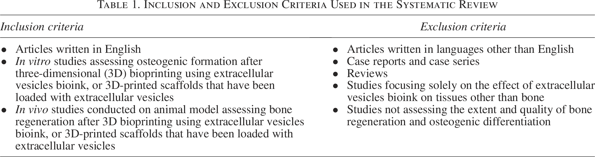
The eligibility criteria are listed in Table 1. The search strategy was developed based on the PICO reporting system for in vitro cell models or experimental animal models (P) using 3D-printed scaffolds enriched with EVs or 3D-bioprinted bioinks containing EVs (I) comparing with the scaffolds without EVs (C) to assess osteogenic differentiation and bone regeneration (O) (Supplementary Table S1).
Inclusion and Exclusion Criteria Used in the Systematic Review
A structured electronic search was conducted for articles published in English up to 12th of March 2025 across multiple electronic databases: Clarivate Analytics’ Web of Science (encompassing Web of Science Core Collection—WoS, Korean Citation Index—KCI, SciELO Citation Index—SCIELO, ProQuestTM Dissertations & Theses Citation Index, Grants Index), Scopus, and PubMed (including MEDLINE). The initial searches aimed to identify key terms, synonyms, and relevant controlled vocabulary (Medical Subject Headings—MeSH, available at https://www.ncbi.nlm.nih.gov/mesh/) and to evaluate various search strategies. A comprehensive search strategy, developed collaboratively by an experienced medical librarian (J.J.) and the review team, underwent peer review following the PRESS guidelines. 43 Additionally, efforts were made to identify relevant unpublished materials such as research reports, conference papers, doctoral dissertations, and other gray literature through sources such as OpenGrey (http://www.opengrey.eu), Google Scholar (first 100 results), and various digital repositories (e.g., Networked Digital Library of Theses and Dissertations—http://www.ndltd.org, Open Access Theses and Dissertations—https://oatd.org, DART-Europe E-theses Portal—DEEP—https://www.dart-europe.org/basic-search.php, and EThOS—Opening access to UK theses—https://ethos.bl.uk). Furthermore, to ensure comprehensiveness, backward and forward snowballing techniques were employed using citation indexes (WoS and Scopus) and Google Scholar. Searches were repeated during the final stages of article preparation until March 12, 2025, confirming no new relevant trials were published after the initial literature review.
Study selection and data extraction
The Rayyan platform facilitated 44 the importation of all literature search results to eliminate duplicates and start screening. Initially, two independent investigators (M.M. and D.M.) thoroughly screened titles and abstracts to identify studies that met predetermined inclusion criteria. Articles that did not meet these criteria were excluded, and full texts of initially selected studies were retrieved for comprehensive evaluation. Authors of inaccessible articles were contacted via email to request the full texts. In the subsequent stage, the same two investigators independently assessed the full texts to determine relevance. Any discrepancies were resolved through consensus or consultation with a third investigator (O.K.).
For eligible studies meeting the inclusion criteria, two independent reviewers (M.M. and D.M.) conducted the data extraction, with any discrepancies resolved by a third reviewer (O.K.), using a customized data extraction form (Microsoft® Excel, Microsoft Corporation, Redmond, WA, USA). The extracted data were: first author’s name, publication year, 3D (bio)printing technology, EV cell origin, EV incorporation method in 3D-(bio)printed scaffold, culture medium, and extraction protocol, 3D-(bio)printed scaffold composition, control group, methods, and main outcomes.
Science mapping analysis
Science mapping analysis of scientific domains was performed from the list of included articles using keyword co-occurrence networking on VOSviewer (free software, version 1.6.18, Centre for Science and Technology Studies, Leiden University, Leiden, The Netherlands, 2022). Network analysis of the keywords was generated from the matrix of retrieved articles (threshold value at 4). 45 The term document matrix allowed the measurement of document similarities between clusters of topics.
Quality assessment and risk of bias analysis
Two reviewers (M.M. and D.M.) independently evaluated the quality of the studies according to the risk of bias. In vitro studies were assessed for bias using the Quality Assessment Tool For In Vitro Studies (QUIN), 46 with the evaluation of 12 statements as adequately specified, inadequately specified, not specified, and not applicable. For the in vivo studies, the Systematic Review Centre for Laboratory Animal Experimentation (SYRCLE) 47 was used, which involves evaluating ten items with yes/no/unclear judgments to determine the risk of bias.
Studies were categorized as having a high risk of bias if they received a “no” rating for at least two items. On the other hand, studies were considered to have a minimal chance of bias if at least seven criteria were evaluated as “yes,” and none were evaluated as “no.” Otherwise, the studies were classified as having a moderate risk of bias.
Data synthesis and statistical analysis
Because of the heterogeneity in study protocols, methods of outcome assessment, and outcome measures, a meta-analysis could not be conducted. Therefore, qualitative data extracted from each study were compiled and synthesized into analytical tables.
Results
Systematic review following PRISMA guidelines
Figure 1 (PRISMA flow diagram) illustrates the study selection process. A comprehensive database search yielded 552 records. Following the removal of 281 duplicates, 271 articles were subjected to title and abstract screening. Of these, 54 full-text articles were evaluated for eligibility. Nineteen studies were excluded based on predefined criteria, with detailed reasons provided in (Supplementary Table S2). Consequently, 35 studies met the inclusion criteria and were incorporated into this systematic review.
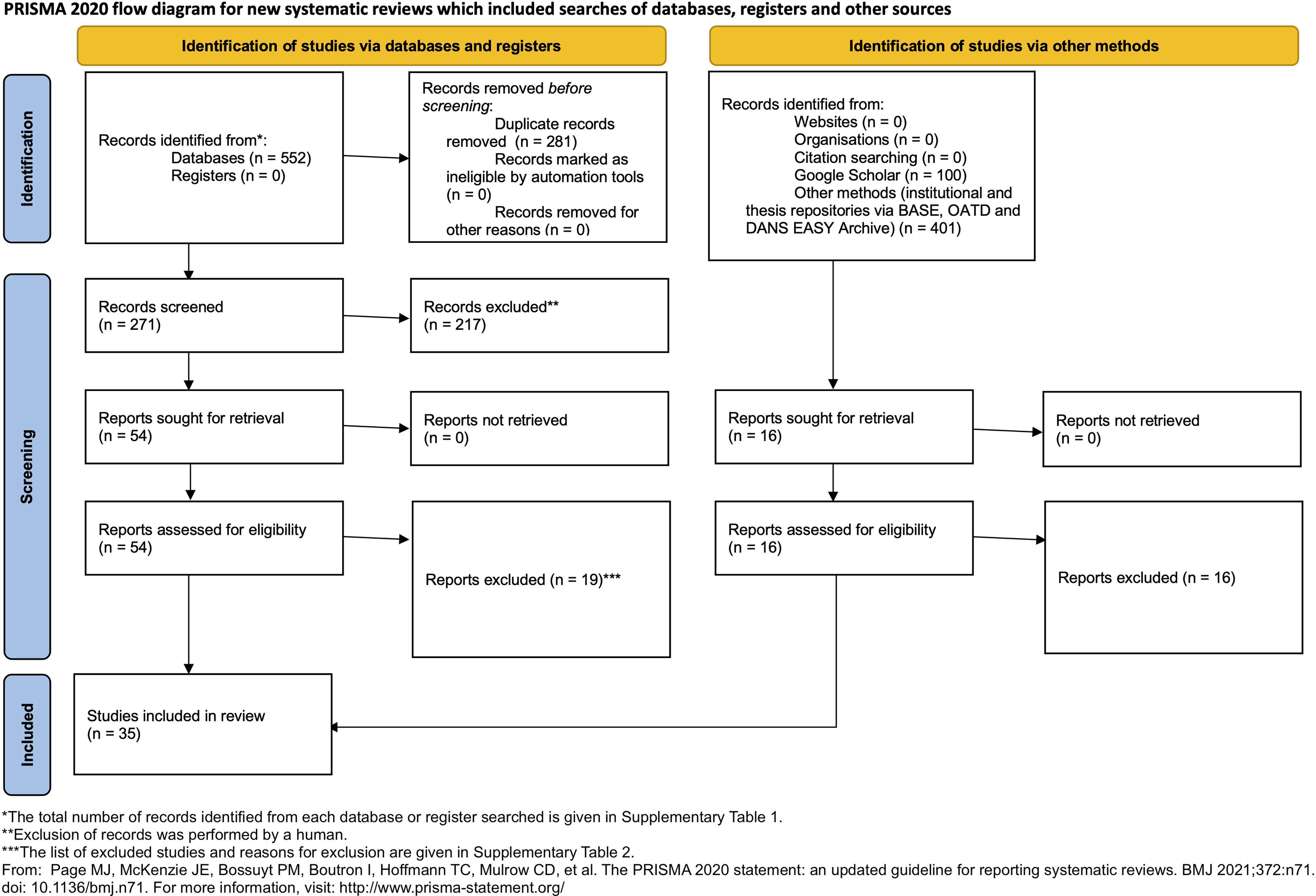
Flow diagram of studies selection according to PRISMA guidelines. PRISMA, Preferred Reporting Items for Systematic Reviews and Meta-Analyses.
A science map (Fig. 2) illustrated the spatial connections between the keywords. The most recurrent keywords were osteogenesis, bone regeneration, humans, animals, (3D) printing, and bone and bones.

Science mapping analysis of scientific domains: keyword co-occurrence networks among the included articles. Each node size is proportional to its degree and the link’s thickness represents the tie strength.
Study characteristics
Among the scoped articles, 8 studies48–55 (22.86%) were only in vitro, 16 studies56–71 (45.71%) were solely in vivo, and 11 studies72–82 (30.55%) combined in vitro and in vivo models. Of the in vivo studies, the animal model that was predominantly used was rats (55.55%) with calvarial defects (44.44%). The detailed information is presented in Figure 3 and Tables 2–4.
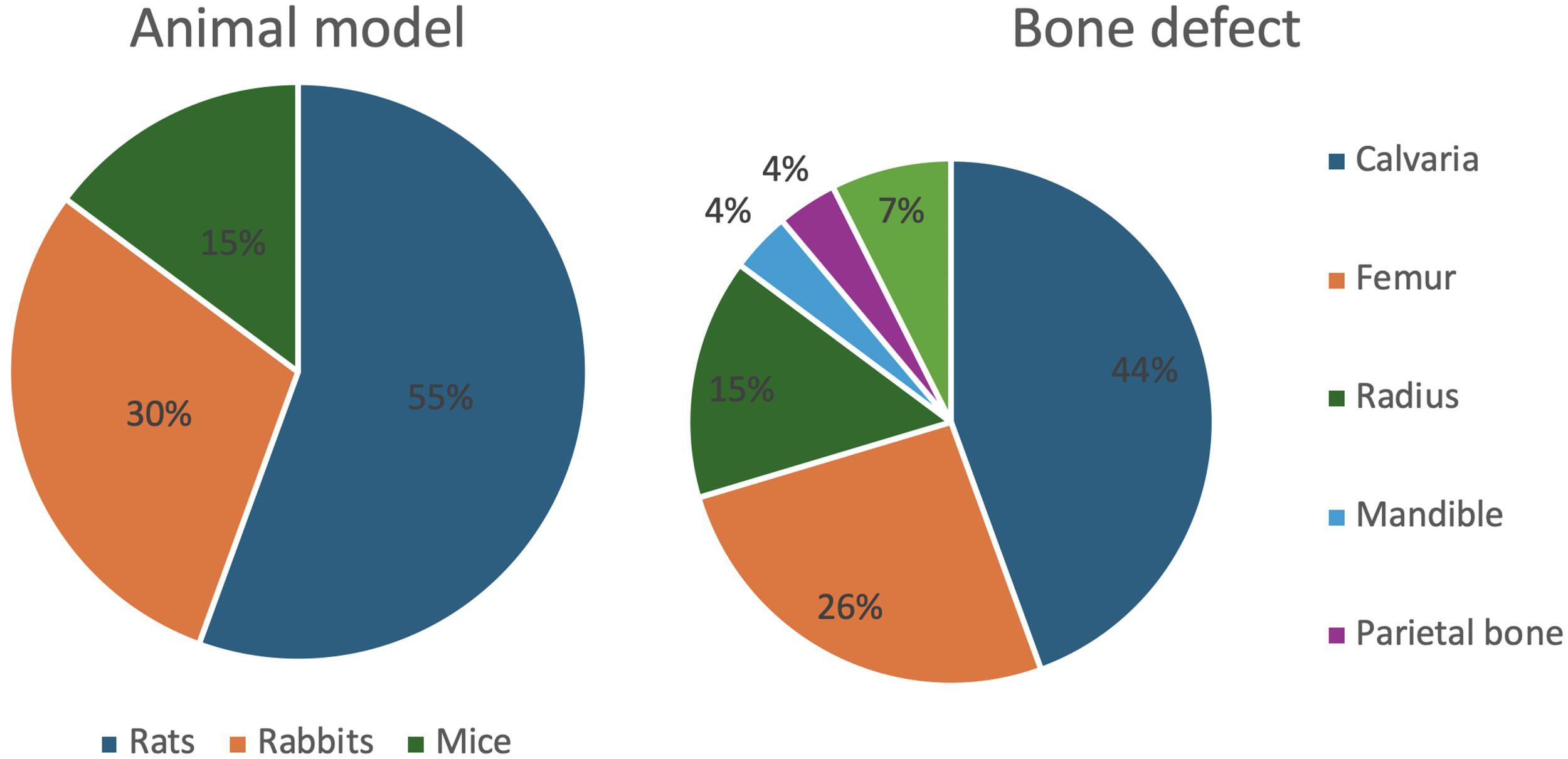
Study characteristics.
ECV Characteristics of in Vitro and in Vivo Studies
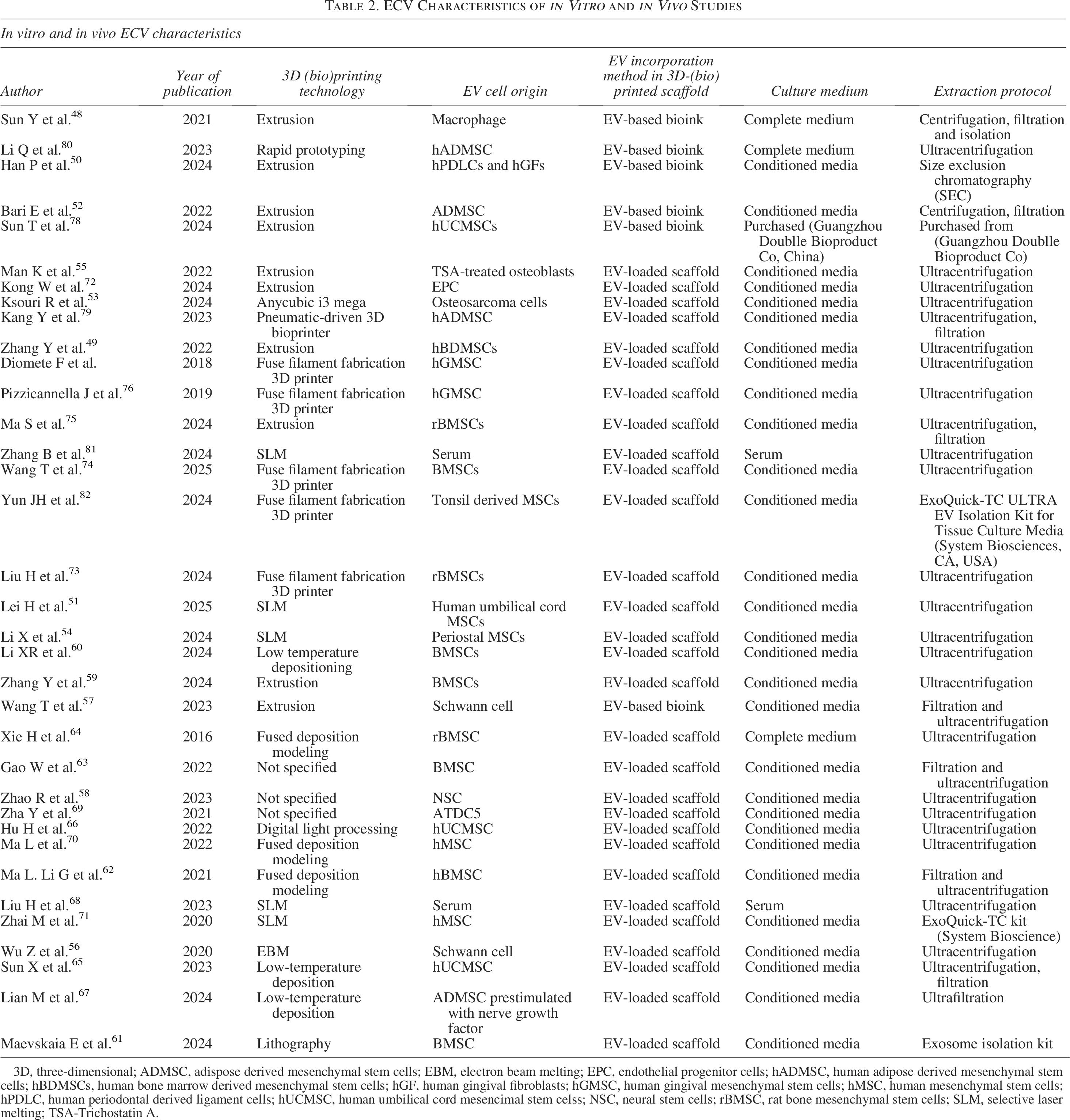
3D, three-dimensional; ADMSC, adispose derived mesenchymal stem cells; EBM, electron beam melting; EPC, endothelial progenitor cells; hADMSC, human adipose derived mesenchymal stem cells; hBDMSCs, human bone marrow derived mesenchymal stem cells; hGF, human gingival fibroblasts; hGMSC, human gingival mesenchymal stem cells; hMSC, human mesenchymal stem cells; hPDLC, human periodontal derived ligament cells; hUCMSC, human umbilical cord mesencimal stem celss; NSC, neural stem cells; rBMSC, rat bone mesenchymal stem cells; SLM, selective laser melting; TSA-Trichostatin A.
Analysis and Bioink Composition Spread for in Vitro Studies
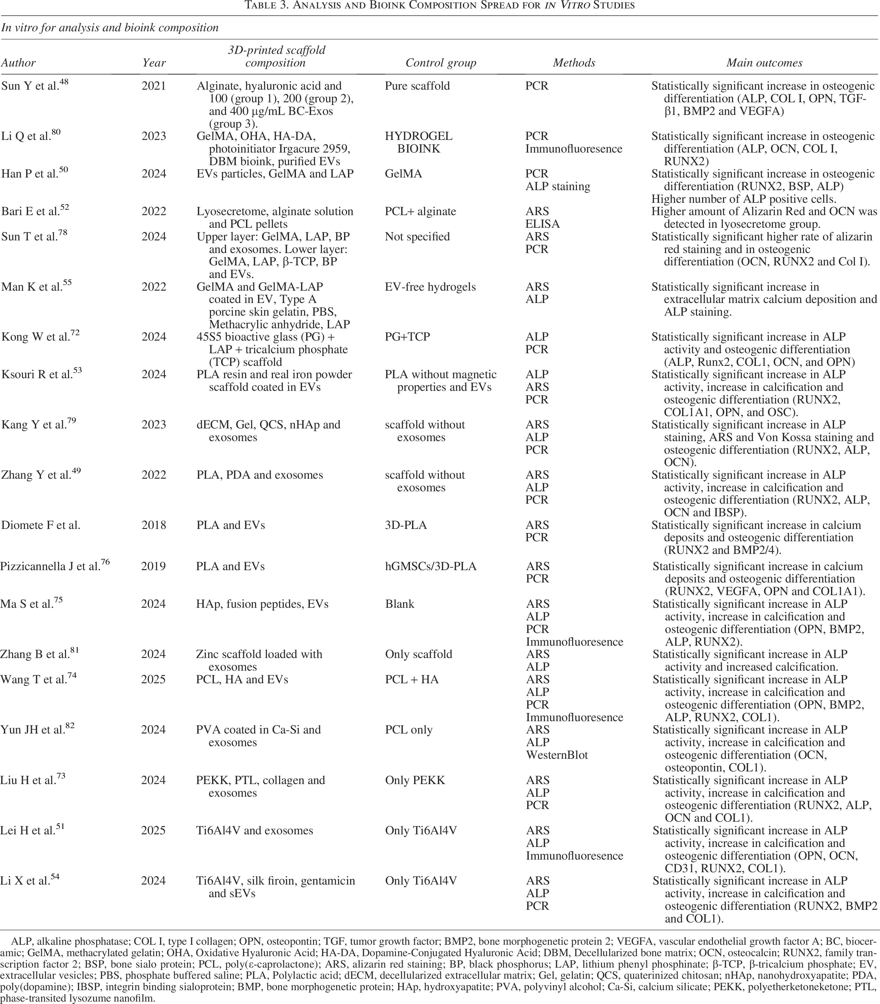
ALP, alkaline phosphatase; COL I, type I collagen; OPN, osteopontin; TGF, tumor growth factor; BMP2, bone morphogenetic protein 2; VEGFA, vascular endothelial growth factor A; BC, bioceramic; GelMA, methacrylated gelatin; OHA, Oxidative Hyaluronic Acid; HA-DA, Dopamine-Conjugated Hyaluronic Acid; DBM, Decellularized bone matrix; OCN, osteocalcin; RUNX2, family transcription factor 2; BSP, bone sialo protein; PCL, poly(ε-caprolactone); ARS, alizarin red staining; BP, black phosphorus; LAP, lithium phenyl phosphinate; β-TCP, β-tricalcium phosphate; EV, extracellular vesicles; PBS, phosphate buffered saline; PLA, Polylactic acid; dECM, decellularized extracellular matrix; Gel, gelatin; QCS, quaterinized chitosan; nHAp, nanohydroxyapatite; PDA, poly(dopamine); IBSP, integrin binding sialoprotein; BMP, bone morphogenetic protein; HAp, hydroxyapatite; PVA, polyvinyl alcohol; Ca-Si, calcium silicate; PEKK, polyetherketoneketone; PTL, phase-transited lysozume nanofilm.
Analysis and Bioink Composition Spread for in Vivo Studies
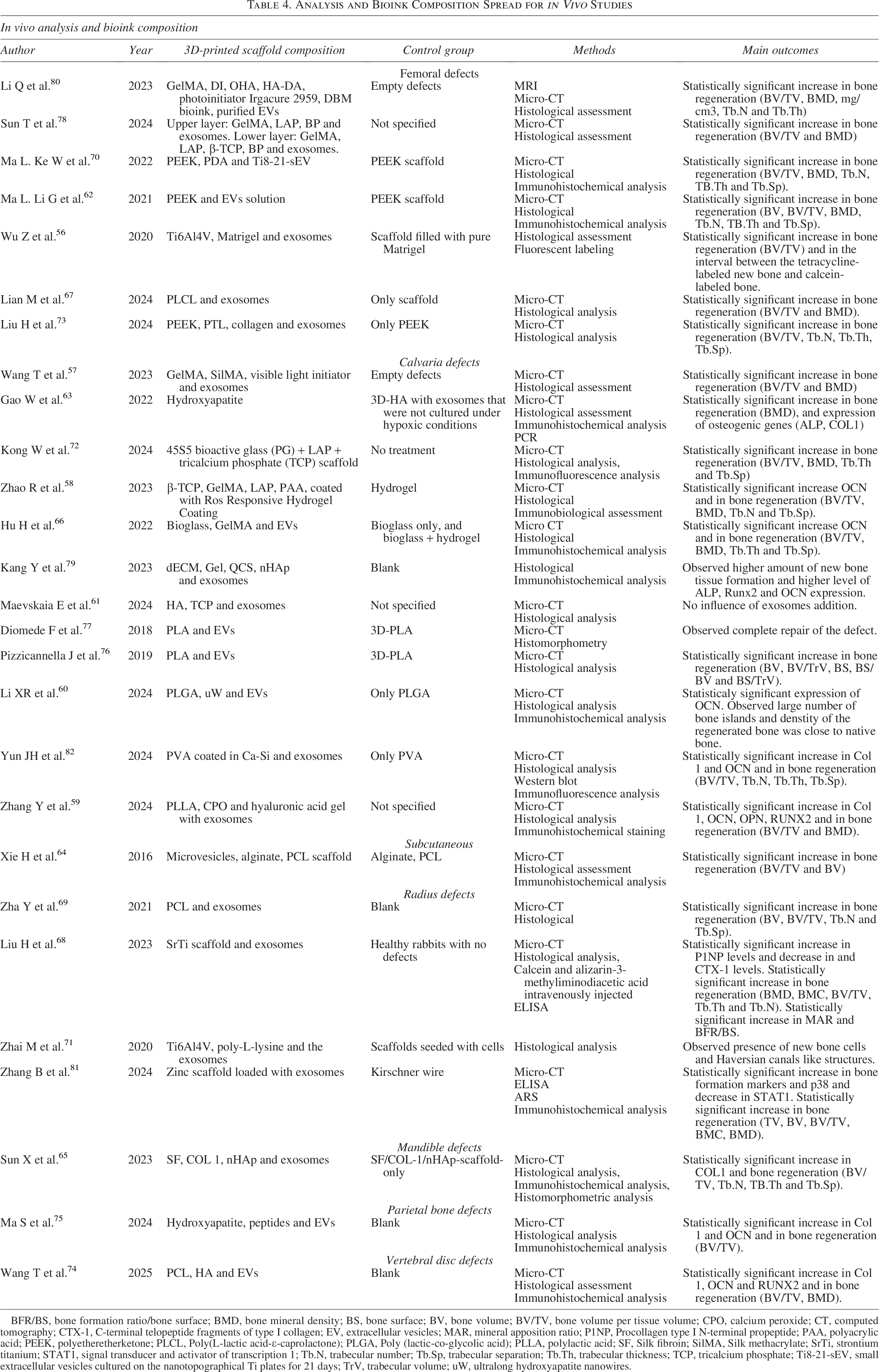
BFR/BS, bone formation ratio/bone surface; BMD, bone mineral density; BS, bone surface; BV, bone volume; BV/TV, bone volume per tissue volume; CPO, calcium peroxide; CT, computed tomography; CTX-1, C-terminal telopeptide fragments of type I collagen; EV, extracellular vesicles; MAR, mineral apposition ratio; P1NP, Procollagen type I N-terminal propeptide; PAA, polyacrylic acid; PEEK, polyetheretherketone; PLCL, Poly(L-lactic acid-ε-caprolactone); PLGA, Poly (lactic-co-glycolic acid); PLLA, polylactic acid; SF, Silk fibroin; SilMA, Silk methacrylate; SrTi, strontium titanium; STAT1, signal transducer and activator of transcription 1; Tb.N, trabecular number; Tb.Sp, trabecular separation; Tb.Th, trabecular thickness; TCP, tricalcium phosphate; Ti8-21-sEV, small extracellular vesicles cultured on the nanotopographical Ti plates for 21 days; TrV, trabecular volume; uW, ultralong hydroxyapatite nanowires.
The incorporation of EVs into (bio)printed scaffolds was achieved through two primary methods. The first involved integrating the EVs directly into the bioink before 3D printing, while the second method consisted of printing the scaffold first and subsequently coating or loading it with EVs. A total of 6 studies48,50,52,57,78,80 (17.14%) employed the first approach, whereas 29 studies49,51,53–56,58–65,67–77,79,81,82 (82.86%) adopted the latter.
Regarding (bio)printing technologies, extrusion-based bioprinting was the most frequently reported method, and it was used in 10 studies (28.57%). Eight studies (22.86%) employed fused filament deposition (FFD), five studies (14.29%) used selective laser melting, three studies (8.57%) used low-temperature deposition 3D printing. Other methods, including rapid prototyping, pneumatic 3D printing, digital light processing, electron beam melting, and lithography-based manufacturing, were each reported in one study (2.86%). Notably, four studies (11.43%) did not specify the manufacturing method used for the 3D scaffolds.
The origin of cells from which EVs were derived was also diverse, and results were grouped accordingly. EVs were obtained from bone marrow-derived mesenchymal stem cells in 10 studies49,59–64,73–75 (28.57%), adipose-derived mesenchymal stem cells in 4 studies52,67,79,80 (11.43%) same for human umbilical cord mesenchymal cells, mesenchymal stem cells in general in 6 studies54,55,69–71,82 (17.14%), gingival mesenchymal stem cells in 3 studies50,76,77 (8.57%), same as neural cells,56–58 and two68,81 (5.71%) extracted EVs from serum. Additionally, there was only one study for macrophages, 48 osteosarcoma cell line-derived EVs, 53 and endothelial progenitor cells 72 (2.86%).
Outcomes
EVs derived from mesenchymal stem cells (MSCs)
MSCs were frequently used as the source of EVs, with six studies not specifying the specific type of MSCs involved.
Regarding the in vitro studies, Man K et al. 55 used a human bone marrow-derived MSC (hBMSCs) in vitro model, demonstrating increased extracellular matrix calcium deposition (1.78 to 3.14-fold) and alkaline phosphatase (ALP) activity (1.21 to 1.94-fold) in the EV-treated groups compared to other groups across all time points. Two other investigators54,82 revealed that the osteogenic differentiation of rBMSCs and tonsile MSCs, respectively, was better in the group containing EVs.
Regarding the in vivo studies, Zha Y et al., 69 Ma L et al., 70 Zhai M et al. 71 and Yun JH et al. 82 performed in vivo studies without incorporating cells alongside extracellular vesicles. These studies reported significant improvements in new bone formation in the EVs-treated groups compared to groups without exosomes, with results comparable to groups treated with cells alone. While the first two studies reported statistically significant results, the third study performed subjective analysis without quantification.
Other studies reported the specific lineage of MSCs that they used.
EV derived from bone marrow-derived MSCs (BMSCs)
Ten studies explored BMSCs as the source of EVs: five in vivo, one in vitro and three combining both in vitro and in vivo models.
Regarding in vitro studies, Zhang Y et al. 49 and Ma S et al. 75 performed in vitro experiments with the addition of macrophages and hBMSC and significantly increased ALP activity, osteogenic gene expression, and calcium deposition in EV-treated groups. Liu H et al. 73 and Wang T et al. 74 both used BMSCs in their study design and reported better ostegenic differentiation in EVs containing groups.
Regarding in vivo studies,59,60,62,63,73–75 it was found that EVs significantly enhanced bone regeneration compared to control groups. Conversely, Maevskaia E et al. 61 reported no significant differences in bone ingrowth, bone-to-implant contact or bone augmentation with exosome treatment. Xie H et al. 64 observed improved bone regeneration in the BMSC-EV group compared to EV free groups.
EV derived from human umbilical cord MSCs (hUMSCs)
Some studies focused on hUMSCs.
Two studies51,78 used an in vitro approach to assess the effect of EVs on BMSCs and their osteogenic differentiation and concluded that their addition significantly improved the differentiation.
For the in vivo model, Sun T et al. 78 and Sun X et al. 65 used an approach without cells and reported superior bone formation and regeneration in EV-treated groups (61.86%) compared to other groups (41.90%). Hu H et al. 66 had a similar study design and results.
EV derived from gingival mesenchymal stem cells (GMSCs)
Three studies included GMSC-derived EVs: two used hGMSCs and one included periodontal ligament cells (hPDLCs) and gingival fibroblasts (hGFs).
For the in vitro model, Pizzicanella J et al. 76 and Diomede F et al. 77 demonstrated superior osteogenic differentiation in groups treated with cells and EVs. Han P et al. 50 found increased osteogenic protein expression and ALP activity in EVs-treated groups.
In vivo approach of Pizzicanella J et al. 76 reported higher bone volume per tissue volume (BV/TV) in the EVs and cell group (8.2 ± 0.3%) compared to other groups (3.9 ± 0.1%, 5.2 ± 0.2%, 4.1 ± 0.3%). Similarly, Diomede F et al. 77 showed significantly better bone regeneration in the 3D-PLA/PEI-EVs group (% of new bone = 12.2733915) compared to other groups.
EVs derived from adipose-derived mesenchymal stem cells (AD-MSC)
Four studies investigated AD-MSC-derived EVs.
Kang Y et al. 79 performed in vitro experiments with hBMSCs and HUVECs, and their results showed better osteogenic differentiation in the EVs group. Bari E et al. 52 reported higher levels of Alizarin Red and osteocalcin (OCN) in the EV-treated group (p < 0.001). Li Q et al. 80 performed in vitro experiments with rBMSCs and reported enhanced osteogenesis in vitro (p < 0.05).
For the in vivo model, Lian M et al. 67 used an approach without cells and reported significant bone regeneration with EVs treatment (p < 0.01). Kang Y et al. 79 experimented without cells and obtained a better formation of new bone (no quantification) in the EVs group. Li Q et al. 80 performed in vivo without cells and reported superior bone formation (p < 0.05).
EVs derived from neural cells
Three studies investigated EVs derived from neural cells (two from Schwann cells and one from neural stem cells), all using in vivo models without cells included in the bioinks or scaffolds.
Wu Z et al. 56 reported enhanced bone formation in the EVs-treated group based on micro-computed tomography (CT) and BV/TV analysis. Wang T et al. 57 observed similar results with significantly higher BV/TV in EV-treated groups (29.242% ± 7.681%) compared to controls (15.977% ± 3.577% and 8.329% ± 1.826%). Zhao R et al. 58 reported similar results with EV-treated groups showing significantly more new bone formation.
Other sources of EVs were found in the following articles:
In vitro studies used macrophages, endothelial progenitor cells (EPCs), and an osteosarcoma cell line (Saos2) as EV sources, and all reported that the addition of EVs significantly impacted osteogenic differentiation.48,53,72 One study 81 used serum-derived EVs and BMSCs for their in vitro testing and reported better osteogenic differentiation in the EVs group.
In vivo studies used EPCs and serum as a source of EVs, and concluded that EVs-containing groups had a higher rate of bone formation.68,72,81
Quality assessment and risk of bias
The assessment of study quality and risk of bias was conducted according to the QUIN method for in vitro studies, and the SYRCLE method for in vivo studies. The results from the quality assessment were graphically depicted using the display tool Robvis 83 (Figs. 4 and 5).
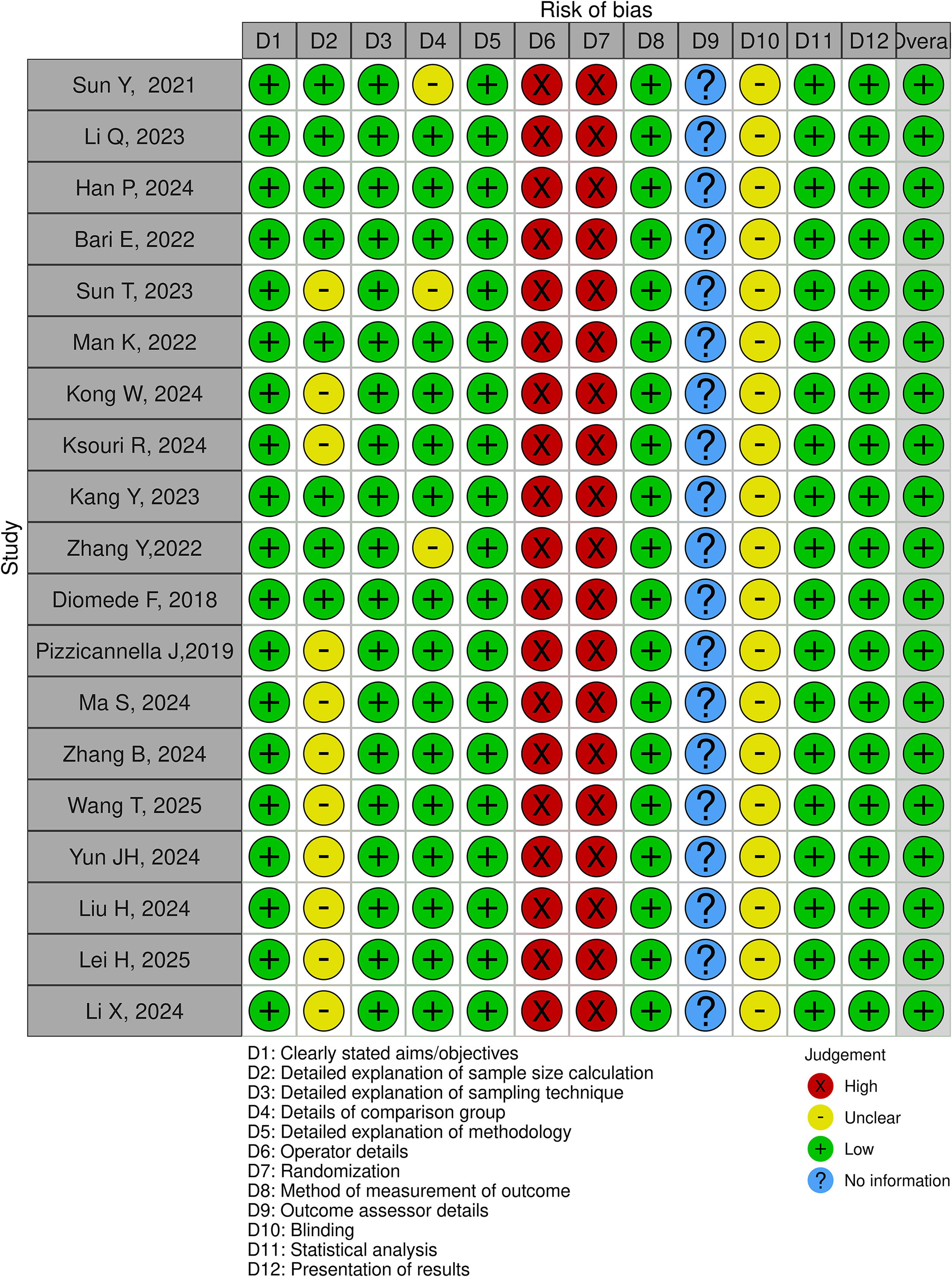
Quality assessment of selected studies using the QUIN method. QUIN, Quality Assessment Tool For In Vitro Studies.
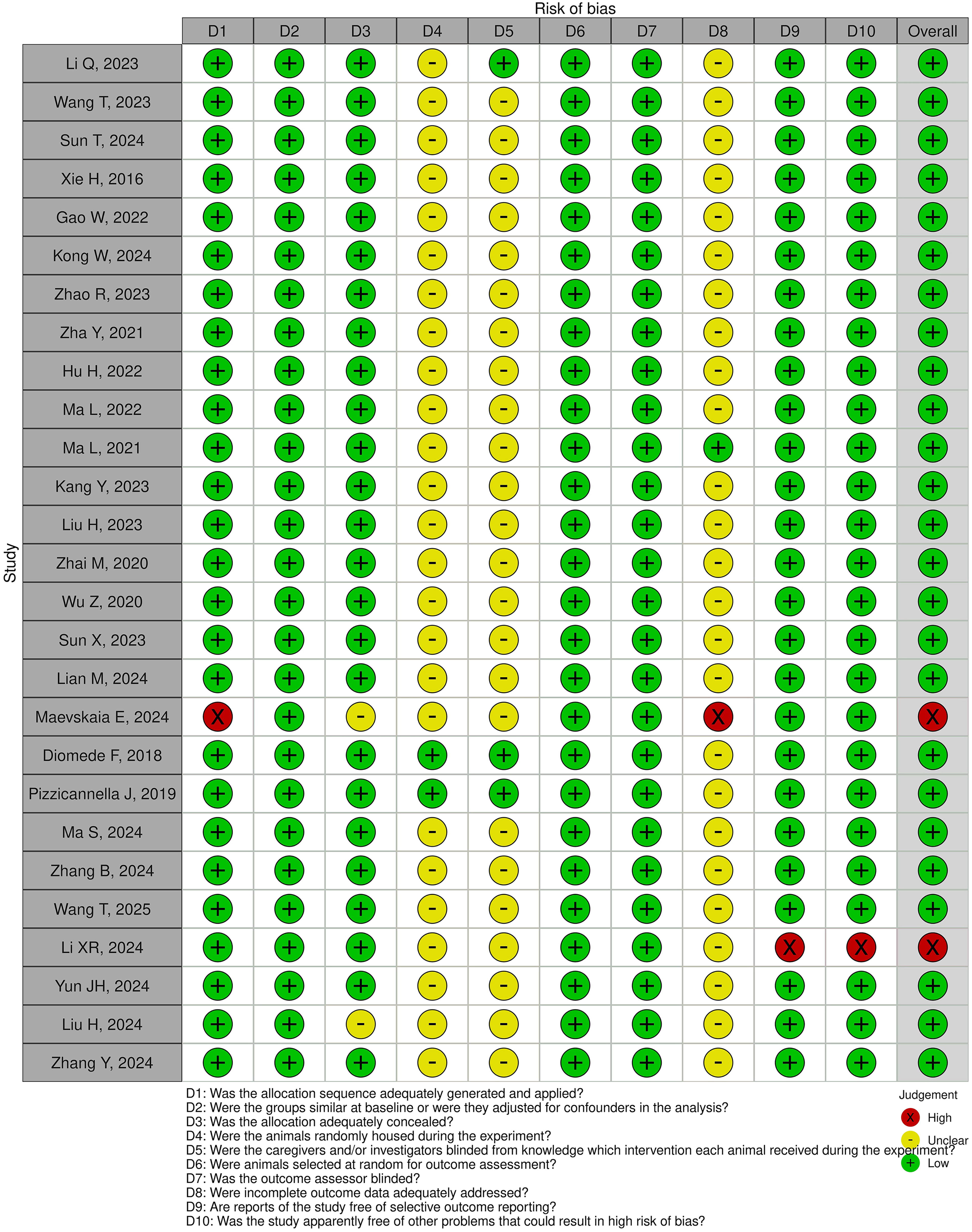
Quality assessment of selected studies using the SYRCLE method. SYRCLE, Systematic Review Center for Laboratory Animal Experimentation.
According to QUIN assessment, there were 56.94% of items in all the studies with low concern, 15.79% with some concern, 9.09% with no information (“outcome assessor details”), and 18.18% with high concern (“operator details” and “randomization”). Following the SYRCLE evaluation, there has been 70.37% of low concern, 28.15% for unclear, and 1.48% had high concern (for the studies performed by Maevskaia et al., 2024 and Li et al., 2024). Overall, the studies were found to be of satisfying quality for the risk of bias, except for two studies by Maevskaia et al., 2024 61 and Li et al., 2024, 60 which had a high concern overall score because of the inability to provide information regarding the allocation sequence applied, whether the incomplete outcome data was adequately addressed, selective outcomes reporting and problems related to high risk of bias.
Discussion
This review aimed to assess whether the incorporation of extracellular vesicles into 3D-(bio)printed constructs enhances bone regeneration in both in vitro and in vivo models. The majority of studies, with one exception, 61 reported that EVs-enriched scaffolds promoted higher osteogenic differentiation in vitro and enhanced bone formation in vivo compared to scaffolds without EVs.
Two primary EV incorporation strategies were identified: preprinting integration within the bioink and postprinting loading onto scaffolds. No differences were observed between these methods regarding osteogenic differentiation or bone regeneration. However, the lack of standardization in EVs sources, scaffold compositions, and experimental models hinders direct comparisons across studies.
BMSCs were the most common EV source, likely due to their established role in osteogenic differentiation and tissue regeneration.31,84 Due to the wide possibility of the differentiation of BMSCs into hematopoietic, osteogenic, or chondrogenic cells, many studies have been conducted with EVs derived from them. It has been demonstrated that EVs carrying similar cargo as their parent cells can have similar results in tissue regeneration. 33 Most studies concluded that adding EVs to the scaffolds promoted better bone regeneration. The sole exception, reported by Maekevska et al., 61 found no significant improvement in bone ingrowth, bone-to-implant contact, or bone augmentation with exosome addition. However, methodological differences may explain this discrepancy since the study assess mainly the osseointegration of implants. Other sources of EVs also demonstrated promising results. The choice of EVs source should be guided by the specific regenerative requirements of the target tissue, given that EVs cargo reflects the biological properties of their parent cells and can highly impact osteogenesis, immune modulation and vascular formation. 85
An interesting aspect of this review was the comparison of EVs alone versus their combination with cells in promoting bone regeneration. Of 35 studies, 18 did not include cells in their study design. In in vivo models, studies comparing bioinks containing EVs and cells to those with only one component found that the combination led to higher bone regeneration.64,76,77 For instance, Xie H et al. 64 reported that a bioink containing BMSCs and microvesicles (type of EVs) significantly enhanced bone regeneration compared to other groups. Similarly, Pizzicannella J et al. 76 incorporated hGMSCs into an EVs-containing bioink and observed an increase in Alizarin red staining, higher osteogenic gene expression in vitro, and improved bone formation in vivo. Diomede et al. 77 even reported full calvaria defect repair in all three tested groups: scaffold + cells, scaffold + EVs, and scaffold + cells + EVs. However, most studies focused on EVs alone rather than their combination with cells, warranting further investigation into their synergistic effects. It is hypothesized that the positive effect of EVs on bone regeneration stems from their specific bioactive cargo, released by the parent cells, which allows them to exert similar regenerative effects (Table 5).
Representative EV Cargo Profiles according to Cell Source and Environmental Conditions
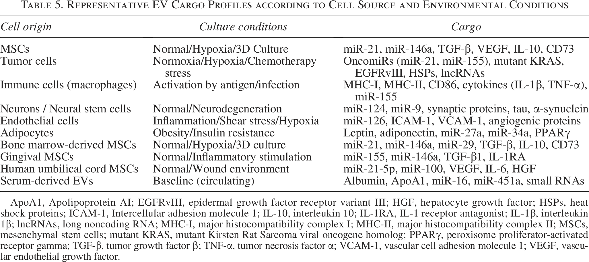
ApoA1, Apolipoprotein AI; EGFRvIII, epidermal growth factor receptor variant III; HGF, hepatocyte growth factor; HSPs, heat shock proteins; ICAM-1, Intercellular adhesion molecule 1; IL-10, interleukin 10; IL-1RA, IL-1 receptor antagonist; IL-1β, interleukin 1β; lncRNAs, long noncoding RNA; MHC-I, major histocompatibility complex I; MHC-II, major histocompatibility complex II; MSCs, mesenchymal stem cells; mutant KRAS, mutant Kirsten Rat Sarcoma viral oncogene homolog; PPARγ, peroxisome proliferator-activated receptor gamma; TGF-β, tumor growth factor β; TNF-α, tumor necrosis factor α; VCAM-1, vascular cell adhesion molecule 1; VEGF, vascular endothelial growth factor.
A substantial number of studies explored the incorporation of EVs into bone grafts and scaffolds outside the context of biofabrication.86–90 These findings demonstrate that EVs can support bone tissue engineering across a wide range of materials and experimental settings, highlighting their remarkable versatility. This broad applicability reinforces the growing interest in EVs as a key component in regenerative strategies, either as an adjunct to engineered constructs or as a standalone bioactive agent.
Building on this versatility, EVs also represent a promising alternative to cell-based therapies by enhancing tissue regeneration while avoiding immune compatibility issues and economic concerns. In bone tissue engineering, their proregenerative effects are mediated through multiple mechanisms, including angiogenesis promotion, 91 inflammation modulation, 92 stem cell recruitment, 93 and direct osteogenic stimulation (Fig. 6).
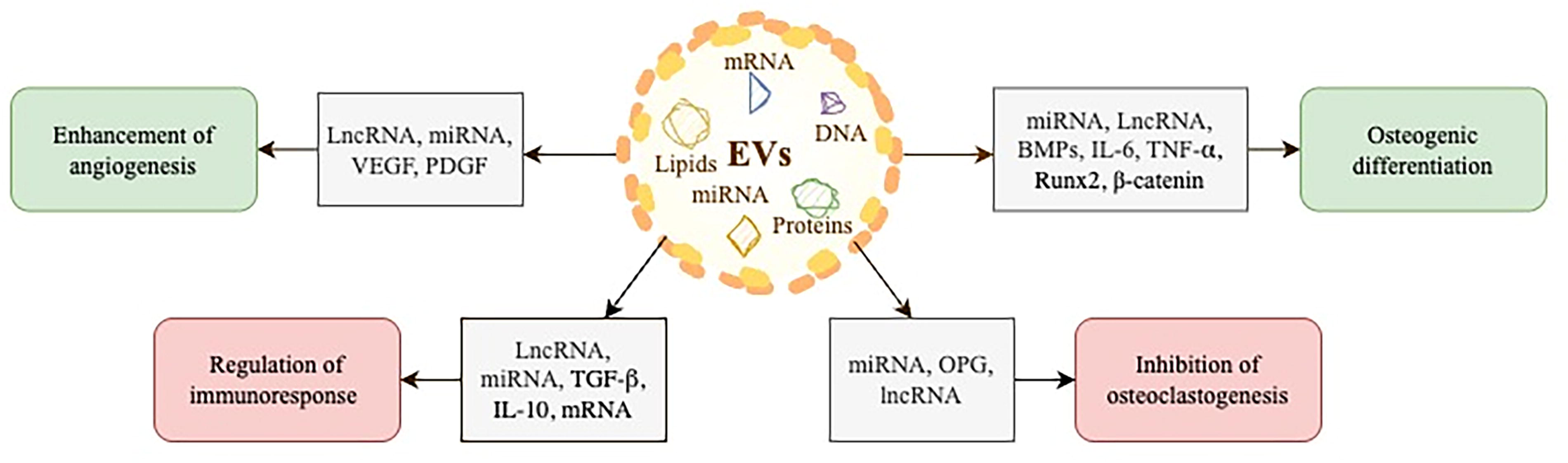
Schematic overview of the main regenerative mechanisms of EVs in bone repair. EVs, extracellular vesicles.
EVs-loaded 3D-printed scaffolds hold great promise for personalized bone regeneration by incorporating biological functionality with structural integrity. In a clinical setting, EVs provide a cell-free approach, evading complications that can arise from cell-based methods, 94 and providing scaffolds that can be adapted to each patient. EVs can be stored cryopreserved or lyophilized, facilitating long-term storage and transport, which is an advantage over live cell therapies. 95
The potential applications of EV-loaded scaffolds extend beyond just bone regeneration. The adaptability of 3D-printed scaffolds combined with the versatility of EVs enables their use in various fields of regenerative medicine, including cartilage repair,91,96 soft tissue healing such as skin 97 and tendons, 98 and neural regeneration. 99
To facilitate the integration of EVs into more studies and, ultimately, into human clinical trials, it is necessary to establish standardized protocols for their production, isolation, and characterization. First, EV production should be strictly defined, including cell culture conditions (cell type, culture medium composition, supplements, confluency, and passage number), along with any stimuli used to enhance EV secretion. Isolation procedures should be thoroughly documented, specifying the methods used and protocols for purity assessment. Finally, characterization protocols must be detailed and transparently reported, focusing on EVs identification, molecular markers and imaging techniques to assess size, concentration, and morphology. 100
The concentration of EVs loaded or incorporated into the bioinks is a crucial parameter to consider when considering the efficacy of these particles for tissue regeneration. Four studies in this review60,69,76,77 failed to report the exact concentration of EVs used, which should be a norm to compare results. Release profiles of EVs from the scaffold weren’t assessed in 18 out of 35 studies, which highlights the gap in research focusing on the appropriate reporting of EV-related experiments. To expand this field, we need transparent and thorough experiments that assess the characteristics of particles and how they act (release and internalization) when paired with other materials.
Standardized reporting of studies incorporating EVs is also crucial and has been proposed in the Minimal information for studies of extracellular vesicles guidelines. 101 Bridging the gap between preclinical findings and clinical applications requires well-designed translational studies. These should focus on determining the optimal EV dosage, evaluating long-term safety, and standardizing manufacturing processes. Additionally, combinatorial studies incorporating EVs with other bioactive molecules could further enhance their regenerative potential.
Despite the promising outcomes of EV-based strategies in tissue engineering, several challenges remain before their successful clinical translation can be achieved. A major limitation lies in the lack of uniformity regarding EV concentration and surface markers, which complicates cross-study comparisons. 102 Moreover, no consensus has been reached on the optimal method for incorporating EVs into bioinks or on the most suitable bioprinting strategy, and current protocols remain highly heterogeneous. These variations may significantly impact the biological activity of EVs and thus affect therapeutic outcomes.
Further obstacles concern scalability and reproducibility. EV yield and composition are strongly influenced by factors such as the cell source, culture conditions, and isolation methods, resulting in high variability and limited predictability of therapeutic efficacy.103,104 Storage conditions also pose a challenge, as EVs are sensitive to temperature fluctuations (e.g., freeze–thaw cycles) and may lose their bioactivity after prolonged storage exceeding one week.105,106 To enable the safe and efficient clinical use of EVs, future research must focus on establishing robust and standardized protocols for EV production, characterization, incorporation, and preservation.
When these challenges are adequately addressed, the integration of EVs with advanced biomaterials may offer a promising therapeutic solution for complex tissue regeneration, paving the way for a more personalized and effective approach to regenerative medicine.
Conclusion
This systematic review evaluated the use of extracellular vesicles alongside tissue engineering strategies based on (bio)printing to promote bone regeneration. In the in vitro cell model, 3D-(bio)printed scaffolds incorporating extracellular vesicles have demonstrated effectiveness in enhancing osteogenic differentiation, while in the experimental animal model, the addition of extracellular vesicles promoted bone regeneration. These results direct us toward broadening the use of EVs in tissue engineering by providing personalized bone regeneration while focusing on solving the challenges arising from implementing new strategies.
Authors’ Contributions
M.M.: Investigation, data curation, and writing—original draft. D.M.: Methodology, investigation, and data curation. J.J.: Methodology and writing—review and editing. R.D.: Writing—review and editing. A.J.: Supervision and writing—review and editing. O.K.: Conceptualization, supervision, and writing—review and editing.
Footnotes
Funding Information
This systematic review is part of Mina Medojevic’s doctoral thesis project. This international joint PhD program was made possible through the EIFFEL Excellence Scholarship (French Ministry for Europe and Foreign Affairs, N°P867856A). The project is also supported by funding from the Fondation des Gueules Cassées (N°35-2023), the Fondation de l’Avenir (N°AP-RM-24B-036), and the Department of Health Sciences and Technologies at the University of Bordeaux (N°R23061).
Disclosure Statement
The authors declare no conflict of interest.
Supplemental Material
References
Supplementary Material
Please find the following supplemental material available below.
For Open Access articles published under a Creative Commons License, all supplemental material carries the same license as the article it is associated with.
For non-Open Access articles published, all supplemental material carries a non-exclusive license, and permission requests for re-use of supplemental material or any part of supplemental material shall be sent directly to the copyright owner as specified in the copyright notice associated with the article.