
Abstract
Background
Cardiovascular diseases are the leading cause of mortality worldwide, with coronary artery bypass grafting being the most effective treatment for severe cases. While autografts are preferred, donor veins are often limited. Human umbilical arteries (hUAs) show promise as an alternative. However, to make vascular graft by decellularization, a traditional chemical method can damage tissue structure and function.
Objective
This study aims to evaluate the shortening of treatment time and the hUA decellularization efficiency of the perfusion bioreactor systems.
Methods
hUAs were perfused with 1% Triton X-100 for 6 h, followed by two different concentrations of (0.5% and 1%) SDS for 18 h, and subsequently subjected to a washing procedure. The decellularization process was evaluated using histological staining and DNA quantification, along with tests for cytotoxicity, cell adhesion and proliferation.
Results
The 0.5% SDS protocol proved most effective, reducing residual DNA to below 50 ng/mg of dry weight while preserving collagen structure. It showed no cytotoxicity to L929 cells, SEM analysis confirmed human umbilical vein endothelial cell (HUVEC) attachment and CCK-8 testing showed promoted HUVECs proliferation.
Conclusion
The decellularization protocol of perfusing through 1% TX for 6 h and 0.5% SDS for 18 h through the perfusion bioreactor system is efficient in the intact hUAs tissue. This sets the stage for future in vivo studies and potential clinical applications.
Keywords
Introduction
Cardiovascular diseases (CVDs) rank among the primary causes of mortality globally. 1 In severe cases, coronary artery bypass grafting (CABG) surgery is employed to reestablish blood supply to the ischemic myocardium, thus reinstating function and alleviating angina symptoms. 2 Thus, the demand for vascular grafts has also increased, particularly small-diameter vascular grafts (SDVGs) below 6 mm. The most optimal option is autograft grafts, particularly saphenous vein (SV) grafts, yet over 30% of patients do not have access to enough appropriate donor veins. Furthermore, synthetic small-diameter arteries have difficulties in endothelization and compliance mismatch due to poor biocompatibility, resulting in thrombosis, stenosis, and intimal hyperplasia.3,4 Consequently, the utilization of allograft derived from human umbilical arteries (hUAs) is suggested. The inner diameter of the hUAs is approximately 1–3 mm and can be easily collected without intrusive methods. Furthermore, they exhibit distinct vascular layers (tunica intima, media, and adventitia), analogous to those of the coronary arteries, facilitating surgical procedures.5,6
The hUA underwent elimination of immune-related elements via decellularization utilizing Triton X-100 (TX) and Sodium dodecyl sulfate (SDS). TX disrupted lipid-lipid and lipid-protein connections, while SDS solubilized cytoplasmic and nuclear membranes to extract cellular components.7,8
Frequently, SDS is individually used at a concentration of 1–2%. This showed efficiency in decellularization when eliminating cellular compartments, but it can damage the extracellular matrix.7,9,10 For example, Tuan-Mu et al. used 1% SDS for 48 h to decellularize the hUAs. The results showed the potential to remove cells with no nuclei in H&E staining, and the DNA content was below < 50 ng/mg dry weight. 11 However, the collagen density of decellularized hUAs was weaker than that of native tissue in the picrosirius red (PSR) image. Besides, the burst pressure was significantly reduced to approximately 450 mmHg, while native tissue was nearly 800 mmHg. 9 Furthermore, 3-((3-Cholamidopropyl) dimethylammonio)-1-propane sulfonate (CHAPS) was used to decrease the adverse effect of SDS in the research of P. Mallis et al. Particularly, the hUAs were decellularized by 0.5% CHAPS before treating with 0.5% SDS; the hUAs were incubated for 22 h for each detergent. Unfortunately, the results showed that nuclei existed in H&E staining, and the DNA content was higher than 50 ng/mg dry weight.11,12 Taken together, using Triton X-100 is considered.
While detergents can denature proteins due to prolonged exposure and high concentrations,7,10 physical methods can reduce exposure time by inducing cell lysis through temperature or pressure changes. 13 The perfusion bioreactor system enables continuous detergent flow through blood vessels, ensuring uniform tissue exposure and efficient washing of residual chemicals, which warrant attention due to their potential cytotoxicity. Thereby reducing the required chemical treatment time and completely removing residual detergents. 10
This article focuses on using perfusion bioreactor systems for decellularization with 1% Triton X-100 while reducing SDS concentrations to 0.5–1% in the short duration. The approach aims to minimize the adverse effects of prolonged detergent exposure on hUAs and residual detergents, achieving satisfactory decellularization by fully removing nuclear components and preserving the tissue structure that supports cell adhesion and proliferation for applications in vascular tissue engineering.
Materials and methods
The decellularization process of the human umbilical arteries
Human umbilical artery preparation
Human umbilical cords were obtained from Hung Vuong Hospital (HCMC) after acceptance of the volunteer. The cords were stored in cold 1X phosphate buffer saline (PBS 1X) (Merck, Germany) containing penicillin and streptomycin and transported to the laboratory. Arteries were isolated from human umbilical cords (20–30 cm long, 2–4 mm diameter) using sharp dissection. The Wharton's jelly surrounded the arteries, and the tissue was dissected from the arterial vessels. Two arteries were subsequently separated from the umbilical vein, which was then cut into segments of 5 cm in length and rinsed a few times with PBS 1X. Finally, umbilical arteries are preserved at −86 oC for analysis or were subjected to decellularization.
Decellularization of the human umbilical arteries
The arteries were perfused in the Biotek perfusion bioreactor (Sigma, USA) with 1% Triton X-100 (Merck, Germany) for 6 h, continued with SDS (Sigma-Aldrich, USA) at concentrations of 0.5% and 1% for 18 h, and finally perfused with distilled water for 24 h, all this procedure was performed at room temperature. Procedure was optimized through preliminary testing.
The evaluation of the decellularization efficiency
DNA quantification
Test samples were well minced and evaluated by quantifying the DNA followed by genomic DNA extraction using Dneasy Blood & Tissue Kit (QIAGEN, Germany) according to the manufacturer's instructions. DNA contents of test samples were determined by optical density (OD) at 260 nm measurements in a NanoDrop One Spectrophotometer (Thermo Scientific, USA). All measurements were performed in triplicate, and the amount of DNA was averaged and expressed as nanograms per milligram (ng/mg) of dry weight.
Histological analysis
The samples (length: 3 mm) were fixed in 10% formalin (Merck, Germany). Then, they were embedded in paraffin for cutting into slices. These were stained with Hematoxylin and Eosin (H&E). The results were recorded by an inverted microscope (Olympus, Japan).
The evaluation of the biological properties of acellular hUAs
The acellular hUA samples were freeze-dried, and gamma irradiated.
Cell line
Mouse fibroblasts – L929 cell line (ATCC, USA) were provided by The Laboratory of Tissue Engineering and Biomedical Materials, HCMC University of Science.
Human umbilical vein endothelial cells (HUVECs) (ATCC, USA) were provided by The Laboratory of Tissue Engineering and Biomedical Materials, HCMC University of Science.
In vitro cytotoxicity testing
The cytotoxicity of the hUAs was analyzed using an extract test by ISO 10993–5. L929 cells were used for the cytotoxicity assay. L929 cells were replated onto a 96-well plate (104 cells per well). The cells were cultured until reaching 80% confluence. Meanwhile, the hUAs were incubated in a culture medium (6 cm2 per mL) at 37 °C for 24 h to collect full liquid extract. In the cytotoxicity assay, the L929 cells were incubated with the liquid extract of the hUAs at 37 °C for 24 h. The culture medium and a liquid extract of latex gloves were used as blank and positive controls, respectively. After incubation for 24 h, the liquid extracts were replaced with MTT:culture medium (Sigma-Aldrich, USA) (ratio 1:9 v/v), and incubated for 4 h. After the medium was removed, 100 μL of dimethyl sulfoxide (DMSO) (Sigma-Aldrich, USA):absolute ethanol (Merck, Germany) (ratio 1:1 v/v) was added to each well to dissolve the MTT reagent. Absorbance at 570 nm was determined using OD Measurement EZ Read 400 (Biochrome, UK). Relative growth rate (RGR) was calculated on OD570 values according to the ISO 10993-5 protocol as RGR (%) = (OD test group/OD blank group) × 100. If the RGR value increased by more than 70%, the liquid extract would be considered to cause a noncytotoxic effect (ISO 10993-5).
Cell adhesion assay
The decellularized hUAs were cut into square strips (0.5 × 0.5 cm) and individually placed into 96-well plates. HUVECs were then seeded on the samples. The samples were then fixed in 5% glutaraldehyde at 4 oC for 2 h and processed to obtain dry samples. Finally, the attachment and morphology of the HUVECs on the samples were observed by scanning electron microscope (SEM).
Cell proliferation assay
The freeze-dried, and gamma irradiated samples were incubated in culture medium for 2 h and were minced to take into a 96-well plate. The HUVECs were then seeded into that 96-well plate. Finally, the assay was performed based on the instructions of the Cell Counting Kit-8 (CCK-8) (Sigma-Aldrich, USA). The 96-well plate reader with the OD 450 nm measurement EZ Read 400 (Biochrome, UK) was used to record the cell proliferation at days 2, 4, 6, 8, and 10 of the hUAs samples. All measurements were performed in triplicate, and the OD value was averaged.
Statistical analysis
The experiments were conducted three times, and the collected results were processed and graphed using GraphPad Prism 10 software. Each result was initially assessed for normality using the Shapiro-Wilk test and for homogeneity of variances via Levene's test at each time point. If the p-value exceeds 0.05, the data within that group are deemed to conform to a normal distribution, and the variances across the groups are regarded as equal (homogeneous). Given the absence of assumption violations, an ANOVA was conducted to evaluate the effects of treatment and time, followed by suitable post hoc tests for pairwise comparisons. One-way ANOVA was utilized for Figures 1 and 2, while two-way ANOVA was applied for Figure 3. A p-value < 0.05 indicates a statistically significant difference.
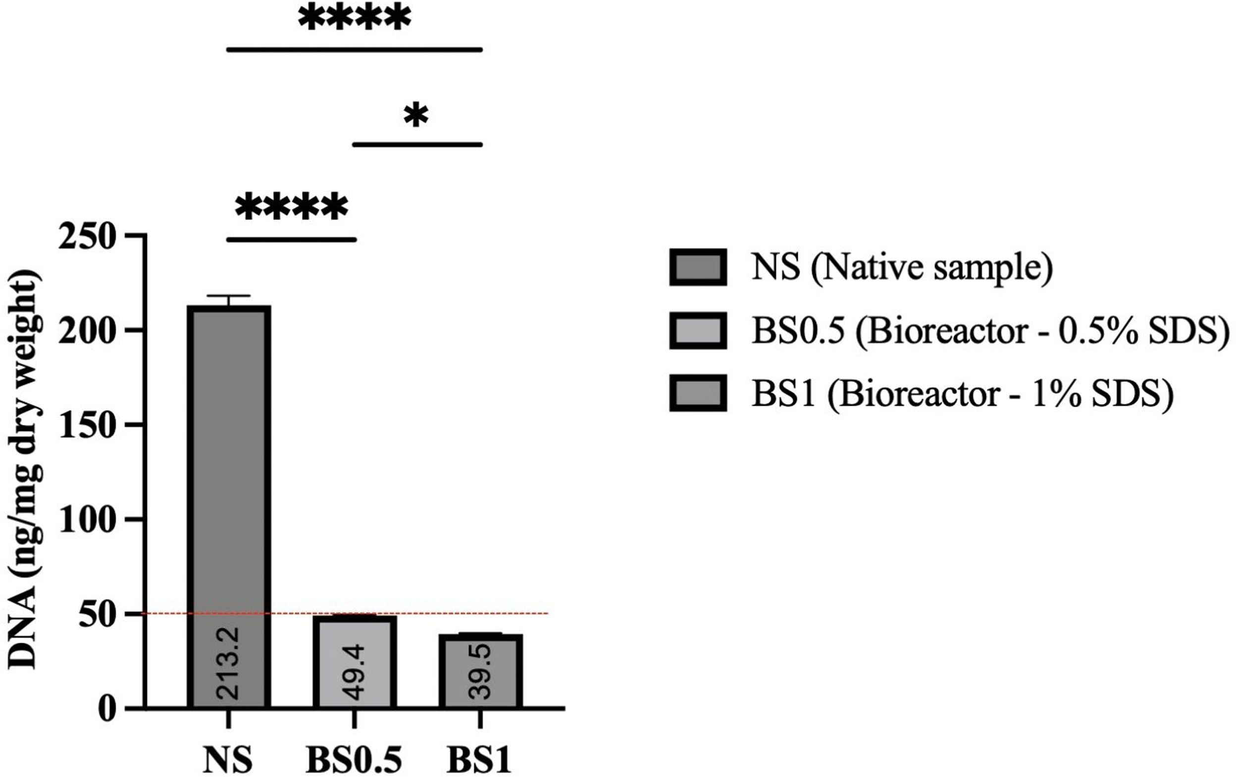
DNA quantification of the decellularized hUAs samples (ng DNA per mg dry weight). DNA content was calculated as mean ± SD (n = 3). *: P < 0.05; ****: P < 0.0001.
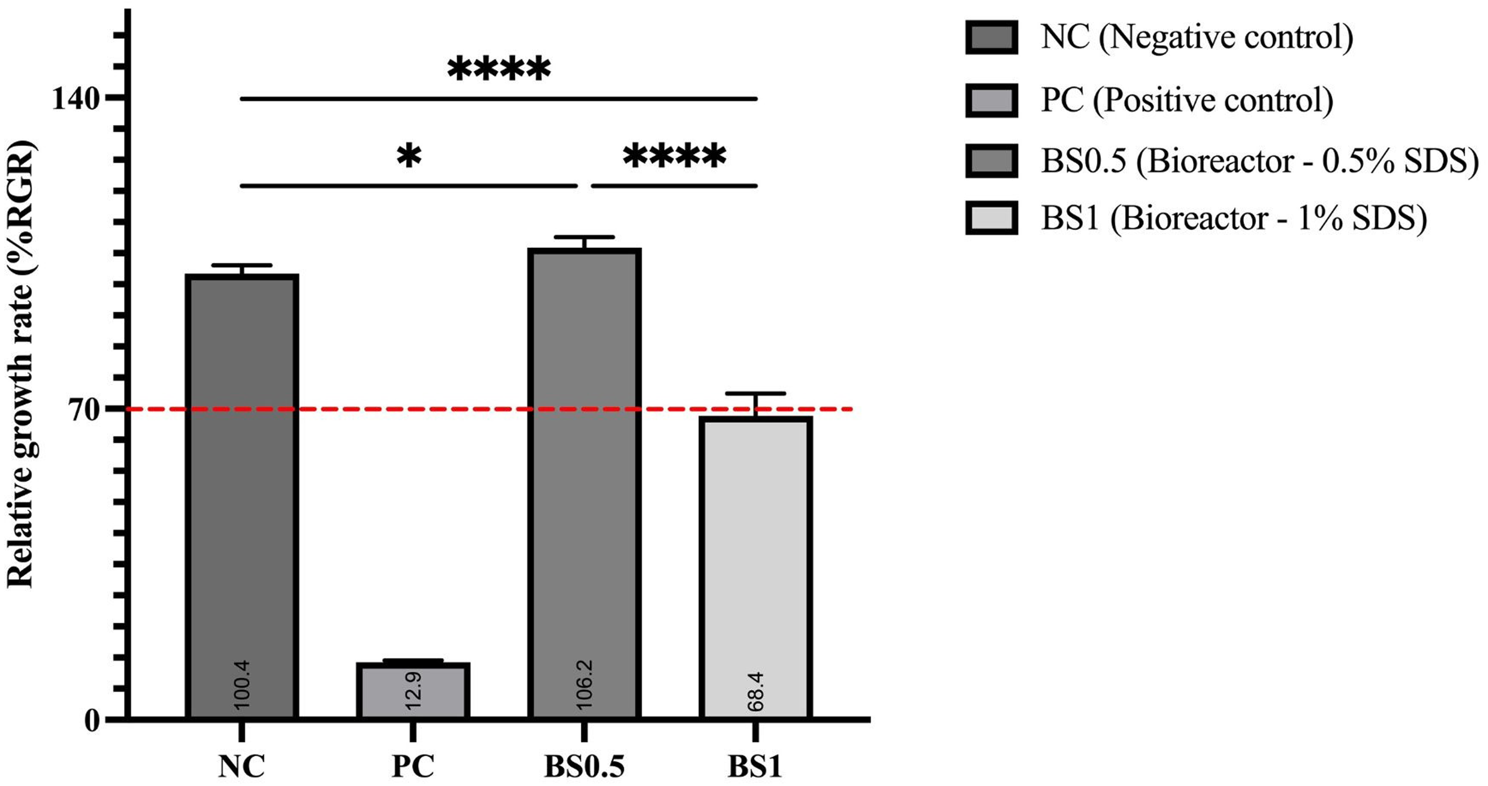
The relative growth rate of the decellularized hUAs. (–). Culture medium; (+). Latex extract; BS0.5, BS1. experiment groups. *: p < 0.05; ****: P < 0.0001.

Cell proliferation of the HUVECs after 2, 4,6,8, and 10 days.
Results
DNA residual quantification
Native hUAs exhibited a DNA concentration of up to 213.2 ± 5.1 ng DNA/ mg dry weight, whereas hUAs treated with 0.5% and 1% SDS in conjunction with a bioreactor displayed a significant reduction to below 50 ng/mg, with 49.4 ± 0.3 ng/mg and 39.5 ± 0.3 ng/mg, respectively (Figure 1).
Histological analysis
Hematoxylin and Eosin (H&E) staining revealed the structural characteristics of native hUA tissues. The extracellular fibers appeared pink, while the nuclei were stained purple, indicating the remaining cellular content (Figure 4(A), arrows). In samples treated with 0.5% SDS, no nuclei were detected (Figure 4(B)). The same outcome was seen in the 1% SDS group (Figure 4(C)). Moreover, the extracellular fibers of both treated groups preserved the tubular structure with three distinct layers, which was not observed the disintegration, similar to native tissues. However, there is evidence that the collagen structure of the BS1 was looser than that of the BS0.5, which is identical to the structure of the native tissue.

H&E staining of the hUAs samples. A. Native samples; B. BS0.5; C. BS1. Cell configuration in native tissue (black arrow). 40× magnification.
In vitro cytotoxicity testing
The morphology of L929 mouse fibroblasts treated with liquid extracts is shown in Figure 5. The fibroblasts have elongated shapes, which remained unchanged after 24 h of incubation in the culture medium (Figure 5(A)). In the BS0.5 group, the liquid extract did not impact the cells, as their morphology remained normal after 24 h of incubation (Figure 5(C)). After incubating with the MTT reagent, the BS0.5 group was observed to have a dense density of formazan crystal. The RGR value was also encouraged, which was found to be as high as the RGR value of the negative group. By contrast, the cell morphology was shown dead and exhibited cellular shrinkage in the BS1 group after 24 h (Figure 5(D)), which was similar to the positive group (Figure 5(B)). The corresponding result had been observed in the MTT result; there is an extremely lower amount of formazan crystal (Figure 5(H)). Together, the RGR value (Figure 2) was 68.4 ± 4.5, which is not adapted to the ISO standard (< 70%).

Morphological appearance of mouse fibroblast L929 cell line in a cytotoxicity assay. A. Cell morphology when incubated for 24 h in the culture medium. B. Cell morphology when incubated for 24 h in the latex extract. C. Cell morphology when incubated for 24 h in the BS0,5 extract. D. Cell morphology when incubated for 24 h in the BS1 extract. E. Cell morphology after MTT in the culture medium. F. Cell morphology after MTT in the latex extract. G. Cell morphology after MTT in the BS0,5 extract. H. Cell morphology after MTT in the BS1 extract. 10× magnification.
Cell adhesion assay
The HUVECs were seeded onto decellularized hUAs samples and observed using SEM. The SEM images revealed the morphology of the HUVECs, which is spread into a polygonal-rounded appearance after being cultured for 48 h (Figure 6(A), (B), black arrows). The results showed that cells adhered well to the surface of the blood vessels in the BS0.5 sample. Moreover, the cell density in the BS0.5 group was significantly high (Figure 6(A)). Additionally, the cells in the BS0.5 group appeared clearer and adhered closely to the blood vessel surface. Figure 6(C) shows the surface of the blood vessels of the control sample. Figure 6(D) shows the morphology of dead cells on the surface of samples.
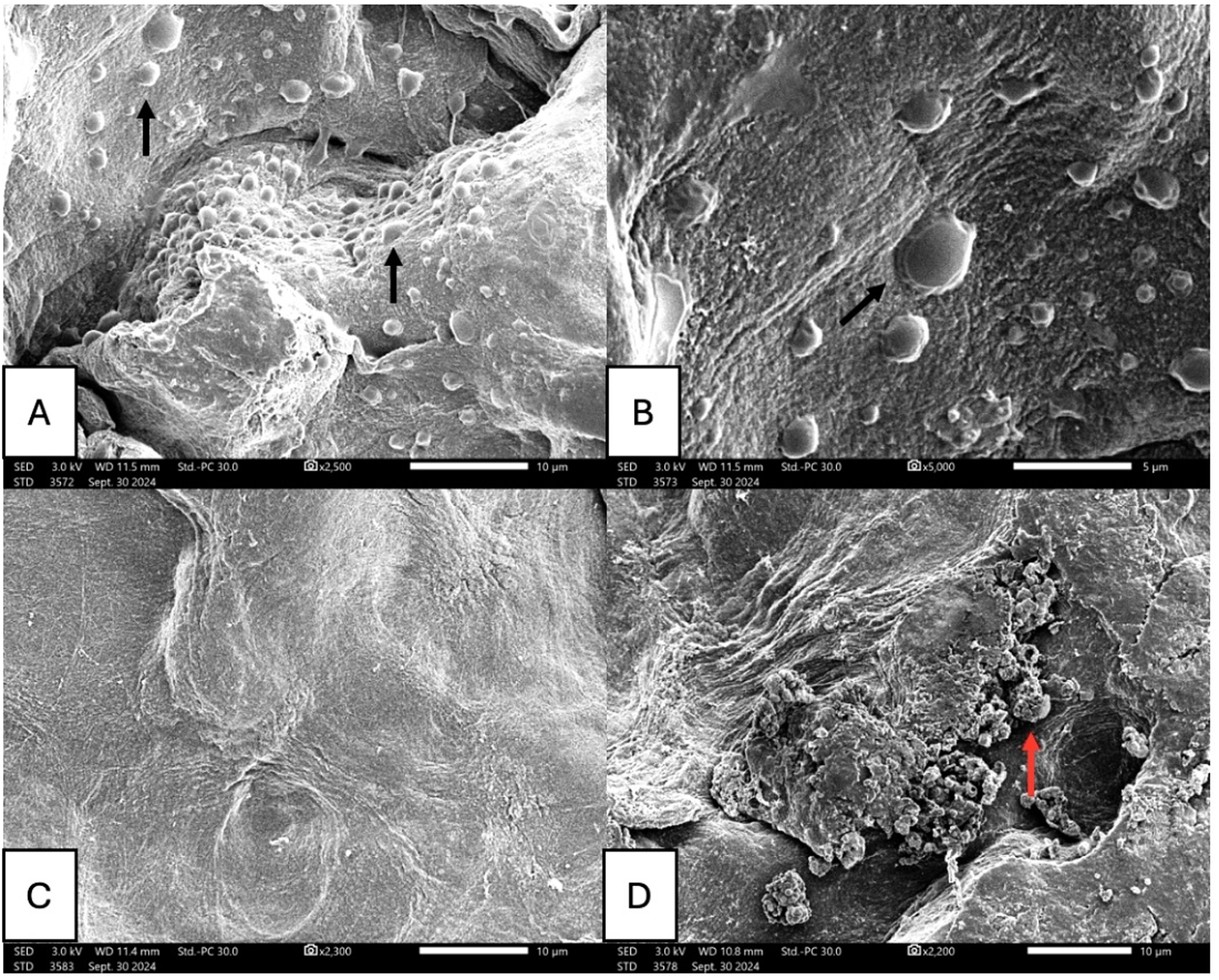
The SEM image of the HUVECs attachment (black arrows) on the hUAs samples. A, B. BS0.5 group; C. control group; D. dead cells. The scale bar corresponds to 5, 10 µm.
Cell proliferation assay
The cell proliferation of the HUVECs was observed in Figure 3. On day 2, the OD value of the BS0.5 group was lower than the OD value of the control group. Significantly, day 4 observed a remarkable increase in OD value in the BS0.5 group, specifically from 0.36 ± 0.05 to 0.99 ± 0.098. The OD value of the BS0.5 group remained stable from day 4 to day 8. At the end, day 10 observed the reduction of OD value in the control and the BS0.5 groups, from 1.62 ± 0.05 to 1.02 ± 0.07 and from 1.18 ± 0.12 to 0.95 ± 0.05, respectively.
Discussion
The hUAs were perfused in 1% Triton X-100 for 6 h, followed by SDS at two concentrations (0.5%, 1%) for 18 h in the perfusion bioreactor system. The histological structures and biological properties of the treated samples were examined to determine the optimal decellularization protocol for developing allograft-derived SDVGs, for potential use in CABG.
H&E staining and DNA quantification confirmed that the decellularization protocol with 0.5% SDS effectively removed cellular components while preserving the integrity of the ECM architecture and no cytotoxicity effect on L929 cells, which were absent in the decellularization protocol of 1% SDS concentration.
In a previous study, P. Mallis et al. decellularized the hUAs through agitation in 0.5% CHAPS, followed by 0.5% SDS for 22 h per detergent. However, this method showed no potential since the residual DNA contents exceeded 50 ng/mg. Indicating poor decellularization efficiency of the agitation. 14 Similarly, Tuan-Mu et al. also reported incomplete removal of cellular components when agitating the hUAs tissue with 0.5% SDS. They also suggested that the hUAs were completely decellularized when using 1% SDS for 48 h. 9 On the other hand, our study demonstrated that the perfusion bioreactor system significantly enhanced the efficiency of decellularization, in which the residual DNA levels of the 0.5% SDS protocol were lower than 50 ng/mg while the whole duration was spent only 24 h. The dynamic flow prevents the accumulation of cell debris, thereby reducing the risk of residual DNA or immunogenic material. Additionally, utilizing the transmural pressure to ensure uniform penetration increases the effects of detergents while using low concentration. In line with these findings, Fang et al. encouraged the important role of the perfusion bioreactor system in decellularization. Their research emphasized the potential results of completely removing cell components. 15 Furthermore, the advantage of the perfusion bioreactor system was shown in reducing SDS treatment duration and washing residual SDS, which has been concluded to have cytotoxicity effects on humans.15,16 The BS0.5 group showed no cytotoxicity through extract cytotoxicity assay, with the %RGR higher than 70%, as ISO 10993-5 recommended. These findings also suggested that the BS0.5 samples may support cell adhesion and proliferation, making them potentially suitable for tissue engineering applications. This was consistent with the result of the cell proliferation assay. HUVECs seeded onto the BS0.5 samples demonstrated growth patterns similar to the control group. The cells underwent main stages of growth, stability, and decline.
Besides these outcomes, SEM images illustrated the good attachment of HUVECs to the decellularized hUAs surface. Indicating the ECM scaffold is preserving well. This is because the perfusion bioreactor system minimizes the mechanical damage to the ECM structure, along with utilizing a low concentration of SDS (lower than 0.5%), which suggests no side effect on ECM integrity. 17 Also, Fang et al. established the attachment of HUVECs on the decellularized hUAs through bioreactors. 15 Moreover, the growth of HUVECs also suggested preventing graft thrombosis, the main cause of CVDs, since it promotes endothelization. 18
After forming a scaffold with adequate endothelialization and no immune-triggering stimuli from heterologous cells and scaffolds, the next step is to culture HUVECs and smooth muscle cells to test endothelialization and reestablish the scaffold in the bioreactor like a vascular conduit. After constructing entire blood vessels, the mechanical characteristics and biocompatibility will be assessed. Blood arteries are strengthened by smooth muscle cells, not collagen or elastin. 19 Thus, mechanical strength will be assessed following cell seeding on the scaffold. After HUVEC endothelialization, the functional cells contact the bloodstream, allowing more accurate biocompatibility testing via vascular grafting. CD31 and CD68 indicate inflammation and healing, CD41 and fibrinogen assess coagulation, and Von Kossa staining evaluates calcification. 20
Conclusion
This study suggested that the decellularization protocol of perfusing through 1% TX for 6 h and 0.5% SDS for 18 h through the perfusion bioreactor system was efficient in the intact hUAs tissue. The research also succeeded in reducing SDS concentration and detergent treatment duration in the corporation of the perfusion method. The acellular hUAs remained non-cytotoxic, allowing cells to adhere and proliferate effectively.
Footnotes
Author contributions
Conceptualization: Nho Thuan Nguyen, Tung Ngoc Hoang Pham, Hoang Minh Lam, Tuyet Thi Diem Hoang, Thang Quoc Bui and Ha Le Bao Tran; design of the work, the acquisition, analysis and interpretation of data for the work: Nho Thuan Nguyen, Tung Ngoc Hoang Pham, Hoang Minh Lam; drafting the work and revising it critically for important intellectual content: Tuyet Thi Diem Hoang, Thang Quoc Bui and Ha Le Bao Tran; final approval of the version to be published: all authors.
Funding
The authors disclosed receipt of the following financial support for the research, authorship, and/or publication of this article: This research is funded by the Ministry of Science and Technology under grant number ĐTĐL.CN-49/22.
Declaration of conflicting interests
The authors declared no potential conflicts of interest with respect to the research, authorship, and/or publication of this article.