



Abstract
John Hatton, LSA MRCS FRCS MD (1817–1871), was apprenticed from 1833 to Joseph Jordan, MRCS FRCS (1787–1873), a well-known Manchester surgeon. Jordan, who had been teaching anatomy since 1814, closed his Mount Street Medical School in 1834 and was elected as surgeon to the Manchester Royal Infirmary in 1835. He continued to lecture on surgery and surgical pathology at the Infirmary, and sometimes at the Pine Street Medical School run by Thomas Turner, LSA FRCS (1793–1873). During 1837–38 Hatton transcribed and illustrated these lectures in a bound manuscript and also added notes and drawings in his personal copy of The Dublin Dissector. He gained his Licentiate of the Society of Apothecaries (LSA) in 1836 and Membership of the Royal College of Surgeons (MRCS) in 1839 and set up in Manchester as surgeon from around 1840. This paper is based on three previously unrelated documents in the University of Manchester Archives: a handwritten catalogue of specimens in Jordan’s Anatomy Museum, Hatton’s annotated copy of The Dublin Dissector and his manuscript record of Jordan’s lectures. These documents provide a valuable insight into medical education during the 1830s.
Keywords
Joseph Jordan, MRCS FRCS and his Manchester School of Anatomy
Medical training underwent extensive reform between 1800 and the 1820s. After the Apothecaries Act (1815) came into effect, 1 a medical practitioner in England was required to hold a Licence from the Society of Apothecaries (LSA) and Membership of the Royal College of Surgeons (MRCS), obtained by examination and known colloquially as ‘Hall and College’. Before 1817 most young men entering medicine were apprenticed to a physician or surgeon for five years. A student could spend most of his apprenticeship in any provincial town; however, at some point, he would have to travel to London to complete courses of certified lectures given by staff at the London teaching hospitals before sitting the exams.
Manchester surgeon Joseph Jordan founded a School of Anatomy and gave organised courses of lectures in anatomy and surgery from 1814. He is recognised as ‘the founder of provincial medical education in England’ 2 because his lectures gained certificates of approval for teaching from the Apothecaries in 1817 and the London College of Surgeons in 1821. 3 He was an excellent surgeon and teacher; his life and work have previously been well documented 4 and this section is mainly related to his work as a medical educator.
Joseph’s family were established in the Manchester cotton printing business. He was the youngest of six siblings and at the age of 15, he was apprenticed to Mr John Bill (1756–1847), 5 a Manchester Infirmary surgeon, however, this was an unhappy experience; he was treated badly and mainly confined to a ‘gloomy cellar making pills and plasters’. Fortunately, his pupillage was transferred to another surgeon, William Simmons (1762–1830), 6 ‘a good and kind master’, who allowed him to ‘all departments of the Infirmary’. 7 Jordan acted as his ‘amanuensis and assistant’ until he was 19, spending most of his time at the Manchester Infirmary ‘where he dissected and engaged in pathological research’, laying the foundation of his career in anatomy and surgery. After spending a further year in Edinburgh with anatomist Alexander Munro tertius (1773–1859) and surgeon Charles Bell (1774–1842) he joined the Royal Lancashire Regiment as surgeon from 1807 to 1812. When he returned to Manchester, he joined an established practice but after two years he resigned. Perhaps his experience in Edinburgh had inspired him with a desire to teach anatomy; in 1814 he advertised his School of Anatomy in Wheeler’s Chronicle. He rented three houses on Bridge Street, the upper floors of which were used as a dissecting room and anatomical museum. He reasoned that a local school would save students the exorbitant cost of studying in London and save them from the moral danger of the Capital, and also provide a supply of locally qualified doctors. His classes became increasingly popular; the course ran from October to April and included lectures and demonstrations designed to comply with the examination rules of the Apothecaries and the College of Surgeons. The pass rate was high, and Jordan stated that he’d ‘never had a student rejected in anatomy’. 8 Before Jordan’s Medical School there were no opportunities for dissection or organised lecture courses outside of Oxbridge, London, Edinburgh and Dublin. The success of Jordan’s system can be measured by the number of similar medical schools that subsequently were founded in the major provincial cities during the 1820s–30s. 9
In November 1825, another Manchester surgeon, Thomas Turner, LSA FRCS (1793–1873) opened a rival medical school in Pine Street near the Manchester Infirmary. 10 He had studied medical education in France and had given courses of medical lectures at the Manchester Literary and Philosophical Society from 1822. His new School taught a wider range of medical subjects including physiology, midwifery, botany, chemistry and anatomy although there was no dissection until after 1825. 11 Jordan responded by opening a new purpose-built Medical School on Mount Street (1826) and expanded the teaching to include medicine and chemistry. A full account of the rivalry can be found in Brockbank’s Foundation; however, it is generally conceded that Pine Street was better-run, nearer to the Infirmary and became more popular with the students. In 1835 Jordan achieved his life-long ambition when he was elected as Surgeon to the Manchester Royal Infirmary. 12 He decided to close Mount Street and agreed with Turner to transfer all his museum specimens to the Pine Street School. 13 Turner’s School was well organised and maintained a high standard; it underwent further expansion and later acquired two others small schools that had opened in Marsden Street (1829) and later Chatham Street (1850). 14 In 1836 the Pine Street School became the ‘Manchester Royal School of Medicine’ and in 1872 was incorporated into Owens College (the forerunner of the Manchester Victoria University (1880) and the present day University of Manchester Medical School). 15
Jordan was essentially an anatomist and dissection was the bedrock of his teaching. In order to comply with the examination regulations for the LSA and MRCS, he gave a course of systematic lectures: ‘140 in each course, with demonstrations and dissection From October 1st to the end of April’. 16 Although there is a record of his museum specimens, 17 there is no surviving record of his early anatomical lectures, however after the closure of the Mount Street School he continued to give lectures on surgery and surgical pathology at the Infirmary and sometimes at the Pine Street School, and it is these lectures that were transcribed and illustrated by John Hatton in 1837–38. 18
The Catalogue of Jordan’s Museum Collection (1830s–80s)
Jordan’s museum collection of anatomical and pathological specimens was an important and valuable part of his medical teaching and, together with the facility for dissection, was a major attraction for potential students. The collection was transferred to the Pine Street Medical School around 1835 and later moved to the Owens College Medical School in 1873. 19 Fortunately, a manuscript catalogue of Jordan’s museum specimens has survived in the University of Manchester archives, although in poor condition. 20 It was probably originally written in the 1830s but later signed and dated ‘16 June 1883’ by one of his nephews, James Stephens, LSA FRCS (1835–1887), and titled a ‘Catalogue of Preparations in the Mount Street Museum belonging to Joseph Jordan’. It is 136 pages long and lists 1056 specimens, each one numbered and accompanied by hand-written notes. They are grouped into nervous system, eye, ear, respiratory, alimentary canal, hernia, urinary and genital, heart and blood vessels, skin tumours and comparative anatomy. For example, number 272 was a posterior brain abscess from a patient with headache and vomiting and 522 was a vas deferens filled with mercury. Jordan’s special interest in hernias and vascular disease was reflected in the collection by several examples of hernia, aneurysms and ‘ossified’ arteries including a complete obliteration of the aorta. Several specimens had injected blood vessels and other exhibits were experimental such as the effects of ligation of the aorta in a dog (721). All these would have been available to Hatton to help with his illustrations of Jordan’s lectures.
John Hatton, LSA MRCS FRCS MD (1817–1871)
John was the son of Susannah and John Hatton (senior), a book binder. His exact date of birth is unknown, but he was baptised in the Parish Church of Manchester on 9 April 1817 21 and his death certificate says he was 54 when he died in 1871. 22 Hatton’s Certificate from Apothecaries’ Hall (LSA 1836) 23 provides a valuable insight into his apprenticeship. It states that his sole parent was his mother, ‘Mrs Susannah Hatton of Manchester, widow’ and under ‘age’, his date of baptism is given incorrectly as 9 April 1815. It seems likely that his father died around 1820 and that because of difficult circumstances his mother arranged his indenture when he was 13. The Certificate notes that he was indentured to Walter Barton Stott, LSA MRCS (1799–1878), of Manchester for five years from 14 March 1831 and that he had attended approved lectures during 1832. Stott had been taught by Jordan and they were close friends, he demonstrated in anatomy at the Mount Street School and was surgeon to the Manchester Dispensary for Diseases of Children. 24
Hatton’s apprenticeship was exemplary; apart from anatomy and dissection he attended lectures in chemistry, materia medica, botany, anatomy, dissection, medical practice, midwifery, forensic medicine, all given by Manchester doctors including Jordan, and he also attended the Royal Infirmary for 12 months. He was apprenticed to Jordan in 1833 and gained his LSE in 1836. He underwent further training at University College Hospital in London and passed his MRCS in 1839. 25 It is not known exactly when or how long Hatton spent in London, however he is noted in the Royal Infirmary Board minutes as Jordan’s ‘surgical pupil’ in 1837. 26 Volume 1 of his Dublin Dissector is dated ‘1833’ and volume 2 ‘1837’ and indicate he was doing regular dissection. Although the Mount Street Medical School had closed in 1835, Jordan continued to teach dissection at his house in Bridge Street, assisted by his nephew, Edward Stephens, LSA FRCS MD (1804–1863), and presumably helped by Hatton during the final period of his apprenticeship when he also started to transcribe Jordan’s lectures during 1837–38. Stephens had been apprenticed to Jordan in 1819. He later reminisced about his experiences, describing the living conditions at Bridge Street as primitive, but ‘the five years were hard worked and happy’. He went on to gain MDs from Leyden and Berlin and was surgeon to St Mary’s Lying-in Hospital in Manchester and later lectured at Pine Street. 27
In Jordan’s biography, ‘Dr Hatton’ is referred to as ‘one of his assistants’ (trained apprentice sometime continued as an ‘assistant’). Jordan had extensive correspondence but did not write or publish much himself, so it was part of Hatton’s duties to keep Jordan’s papers in order and write replies to his letters etc. in addition to assisting with patients, teaching and dissection. 28 Generally, apprenticeships to a surgeon were for five years and could cost three hundred guineas or more. They were taught basic pharmacy, prescriptions (in Latin), osteology, midwifery, vaccination, pathology, anatomy and surgery. Part of the work was routine bottle washing, cleaning and domestic duties. Not all teachers were as enlightened as Jordan and some apprentices lived in poor conditions and their ‘training’ was no more than a routine of drudgery preparing pills and plasters. 29
By 1841 Hatton was living on Oxford Street in Manchester and working as Medical Officer to the Chorlton Union Workhouse, and Surgeon to the nearby Chorlton-upon-Medlock Dispensary on Cavendish Street, which had opened in 1826 following a fever epidemic. 30 The river Medlock was heavily polluted and Chorlton was an extremely poor and unsanitary area, largely inhabited by Irish immigrants living in dilapidated back-to-back houses. Outbreaks of measles, dysentery, whooping cough with a high mortality were common. The Dispensary was based in the Town Hall and grew rapidly during the 1830s dealing with frequent epidemics, acute medical and accident cases as well as a home-visit service. 31 Hatton would have dealt with all types of illnesses and accidents and also had a special interest in midwifery; he had been taught by obstetrician Thomas Radford MD (1793–1881) and reported two difficult obstetric cases. 32 In 1847 he put himself forward for a surgical post at the Royal Infirmary; he was supported by Sir James Bardsley MD FRCP (1801–1876), physician at the Infirmary and others, though Jordan’s name was absent. 33 His application failed in the face of stiff competition, which included a former Royal Infirmary house surgeon, a nephew of Thomas Turner, and a former apprentice of Jordan. 34
Hatton was heavily involved in the Public Health movement and after an outbreak of cholera in 1849 he kept detailed epidemiological records and started a campaign of sanitary improvements; he produced a map of cholera and fever cases in the area and presented his report, Sanitary Conditions of Chorlton-upon-Medlock, to the Manchester and Salford Sanitary Association in 1854. 35 He was now well established, he had married Jane Burton (1807–1883) in 1845 36 and after passing his FRCS (1854) he styled himself as ‘Consultant Surgeon to the Dispensary’.
In addition to his medical work he was for 15 years the Regional Secretary to the Lancashire and Cheshire Branch of the Provincial Medical Surgical Association. The PMSA became the ‘British Medical Association’ (BMA) from 1854 and that year the Association’s 22nd Annual Meeting was held in Manchester and was organised by Hatton. Dr James Wilson, MRCS (1792–1855), 37 president of the Meeting, presented Hatton with a ‘valuable piece of plate’ for conducting the Meeting with his ‘talent and zeal’. 38
Ill health forced him to resign in 1857; a testimonial in the BMJ described him as ‘one of the most zealous and efficient officers’ respected ‘for his professional acquirements and social qualities’. 39 He was presented with a ‘valuable timepiece’ by the BMA and a ‘handsome service of plate’ from colleagues in his local Branch. 40 His resignation was due to the onset of Bright’s disease 41 (chronic nephritis) and around 1860 he gave up his post at the Dispensary. He is described in the Medical Directory as ‘travelling’ and probably spent some time in Scotland; certainly, the University of St. Andrews Biographical Register of Medical Candidates records that he gained his ‘M.D 2.1 by examination’ in 1861. 42 From 1863 he is living quietly in Belvedere in Kent but still attended occasional BMA meetings; he died suddenly during a visit to a friend in Manchester on 26 January 1871 and was buried at Bowden Parish Church. 43
The Dublin Dissector
The Dublin Dissector, first published in 1829, was a popular dissecting manual written by Robert Harrison, BA MB MD FRCS (1796–1858), Professor of Anatomy in Dublin. 44 It was published in two volumes and subtitled: Manual of Anatomy, comprising a concise description of the bones, muscles, vessels, nerves and viscera, also the relative anatomy of the different regions of the human body, for the use of students in the dissecting room. It ran through several editions in Ireland and was the favourite students manual in most American medical schools. 45 It generally received good reviews, 46 though Harrison’s successor, Professor Alexander Macalister, MB MD FRS (1844–1919), described the book as ‘dreary’ and compiled from a French anatomy book! 47 Prior to the 1840s there were several anatomy textbooks, but very few that were intended as an aid to dissection. 48 Unusual for a dissection manual, The Dublin Dissector had no illustrations until after the 1847 edition.
John Hatton had both volumes of the third edition (1831). Volume 1 is signed and dated ‘26 January 1833’ and marks the start of his apprenticeship, while volume 2 is dated ‘1837’ and inscribed ‘9 Bridge Street, Manchester’ – a time when he would have been studying surgery and anatomy for his MRCS.
49
His leather-bound copy has interspersed blank pages bound in with the printed pages which he used to illustrate various anatomical structures with pen and ink drawings, sometimes finished with watercolour. In Volume 1 he did 10 illustrations; the early ones of some muscles were poor, but he quickly improved and his coloured sketches of stomach, gall bladder (Figure 1), pelvis and testis can be rated as ‘very good’. Volume 2 includes a section on skull and bones. There are over 20 drawings including some good illustrations of the vertebrae, temporo-mandibular joint and pelvis. Some are accompanied by informative notes, for example, a drawing of a dislocated hip is annotated ‘the thigh bone dislocates upward upon the dorsum iliac’. On another page he lists 17 muscles attached to the scapula and signs the page ‘JH’. Overall, his illustrations range from poor pencil drawings to nice ink and colour pictures. The illustrations do not always match the subject discussed in the printed text and not all the blank pages were used.
Sketch of the gall bladder and bile duct, Dublin Dissector, vol. 1, p. 164. (© University of Manchester)
John Hatton’s manuscript record of Jordan’s lectures
During 1837–38 Hatton made a hand-written copy of a series of surgical lectures given by Jordan at the Infirmary and the Pine Street Medical School. The manuscript consists of 229 folios in good condition, but the spine and front board are loose. 50 It is part of the University of Manchester Medical Manuscripts Collection and was donated by Dr Ernest Bosdin Leech MD FRCP (1875–1950) 51 who had obtained it from Dr J Douglas Boyd MB BCh (d.1954). 52 A letter inside the front board, dated 10 July 1932, explains that it had been given to Boyd by Mr Notman, a local school teacher 53 ; however, there is no record of previous ownership.
The course of lectures was the final part of Hatton’s training before he took the MRCS examination in 1839 and he intended that his record of the lectures should be a gift for Mr Jordan. Hatton’s handwriting is excellent and legible throughout. He signed and inscribed it: ‘presented to Joseph Jordan Esq., as a slight testimony of esteem and regard, by his obedient servant, John Hatton, 10 February 1838’. He transcribed 19 lectures, starting from 16 October 1837 and included 49 illustrations by his own hand and two other prints. The lectures are listed at the beginning of the manuscript and some loose lecture notes on ‘inflammation of the spinal membrane’ are probably part of lecture 20. Apparently, he did not record all the lectures as he was unable to attend the full course.
List of Jordan’s lectures in Hatton’s manuscript
Phenomena of circulation and inflammation Terminations of inflammation Formation of abscess, treatment of abscess Ulceration and granulation Mortification from arteries, veins, or nerves, in children and from blisters Mortification from gangrene, Lock Hospital gangrene, and separation of limb… Constitution affected by local causes and irritability of an organ, Treatment of inflammation Diseases of arteries Aneurysm of artery, aneurysm of aorta, pathology and natural cure of aneurysm Coagulation of blood in, symptoms of, diagnosis and natural cure of aneurysm Cure of aneurysm by ligature, medical treatment and anastomoses of vessels Aneurysm varix, hydrocele, passage of testicle into scrotum and stricture of chord Diagnosis of chord and hydrocele of chord Cure of hydrocele and hernia Common circumstances to hernia and strangulation Hernia of omentum Angular curvature of the spine, kyphosis Lateral curvature of the spine, scoliosis Inflammation of the spinal membrane (incomplete loose notes)
The first eight were introductory lectures on the basic pathological processes – inflammation, infection and ischaemia – essential knowledge to understand the cause of disease, similar to any modern text on surgical pathology. Lectures 9 to 13 are concerned with vascular disease, which was a special interest of Jordan. He reported a case of ‘obliteration of the aorta’ in 1830 54 and the diseased aorta was part of the pathological collection (no. 601); it was illustrated by two professional prints of the obliterated aorta ‘drawn on stone by HG James…from a preparation of Mr Jordan’ 55 – the only illustrations in the manuscript not done by Hatton. In 1858 Jordan presented a second case of this rare condition to Sociètè de Biologie in Paris. 56 Lectures 13 to 17 are concerned with testes and hernias – another special interest, which Jordan shared with his friend Sir Astley Cooper FRS (1768–1841) 57 who tried to persuade Jordan to publish his work on these subjects. 58
Hatton’s pictures are on separate pages inserted near to the relevant text. Some are pencil sketches, others are black ink or in watercolour and occasionally signed ‘JH’ or ‘copied from Dr Jones’.
59
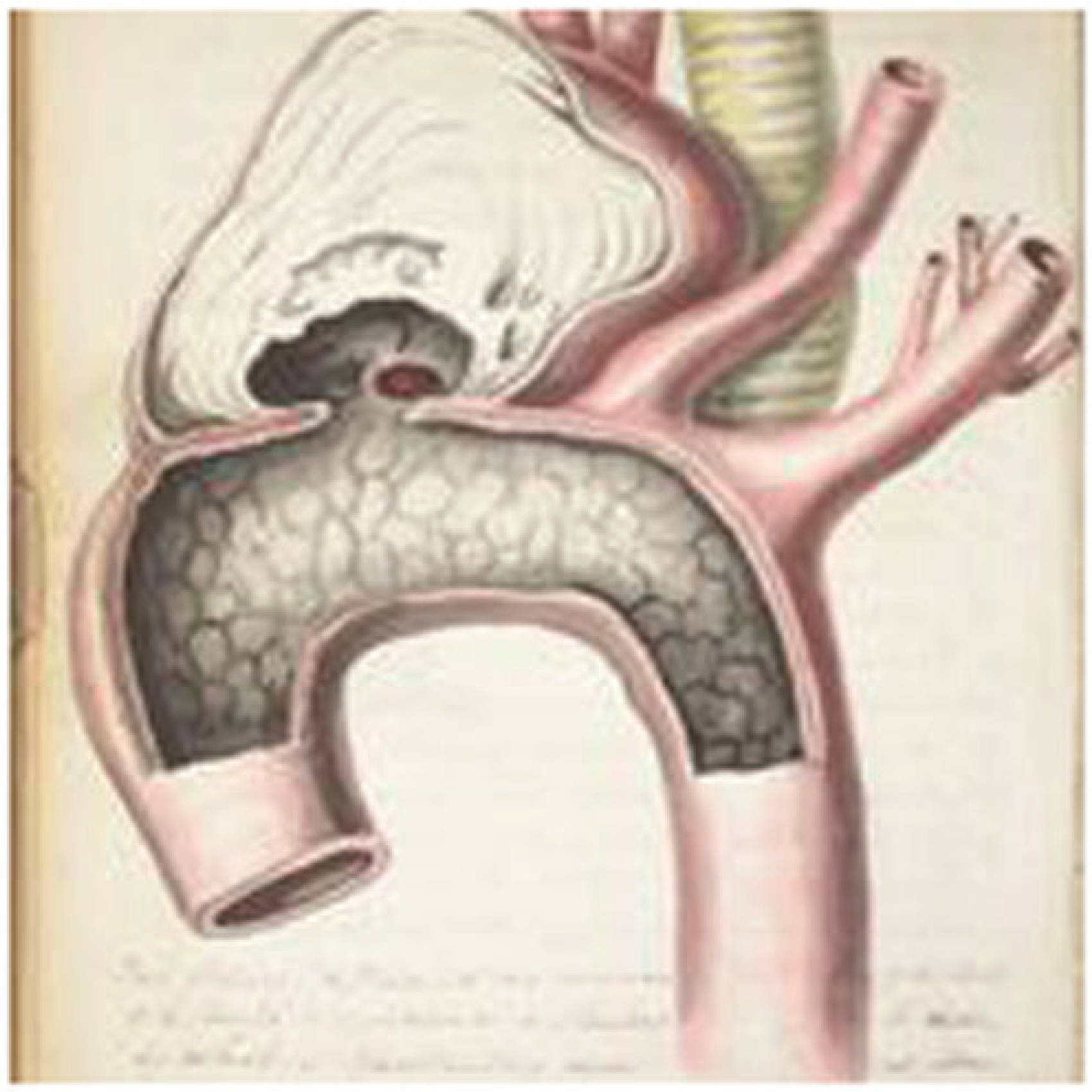
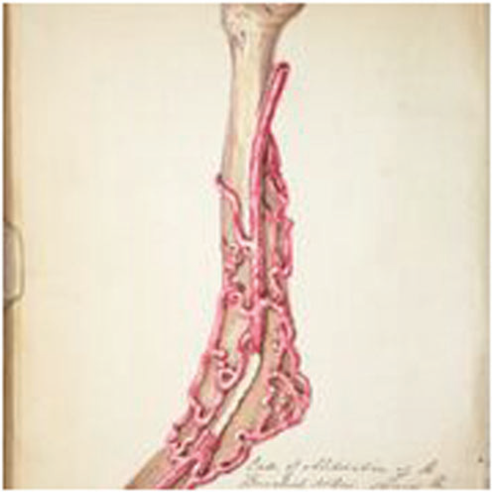
They clearly reflect the special interests of Jordan; of the 49 pictures, 22 illustrate vascular pathology and 24 relate to testes and hernias. His vascular series include some good drawings of the major vessels in the dog and horse, recording the effects of haemorrhage and ligature of the femoral and carotid arteries (Figure 2). These are related to discussion in the text about the risk of haemorrhage after surgical ligation and the formation of clot proximal to the ligature. Other illustrations include excellent drawings of an aortic arch aneurysm (Figure 3), and an obliterated brachial artery bypassed by an anastomosis (Figure 4). The later section includes good illustrations of hernia, testes and different types of hydroceles (Figure 5).
Two sketches showing the effects a ligature on the carotid artery in a dog after 12 days. On the right there is residual clot above and below the occlusion. On the left the ligature has completely divided the artery and the end of the lower half has cicatrized. Hatton ms. p. 87. (© University of Manchester) Watercolour of a large aortic aneurysm. Hatton ms. p. 114. (© University of Manchester) Watercolour of occlusion of the brachial artery bypassed by massive anastomosis. Hatton ms. p. 117. (© University of Manchester) Hatton's table of Jordan's differential diagnosis of hydroceles. (© University of Manchester)

Discussion
The history of medical education is complex. 60 Briefly, before 1800 only a few practitioners were sufficiently educated to gain registration with the Apothecaries or College of Surgeons. A wealthy student could be apprenticed to ‘walk the wards’ at a London teaching hospital or study for a degree at Edinburgh, Dublin, Oxbridge or at a Continental medical school. Most just paid to be apprenticed to an established practitioner although not all placements were satisfactory. Jordan provides a good example; he was apprenticed in 1802, but his first attachment was disastrous, and he was lucky to be transferred to William Simmons and by the time he was 19 he had laid the foundation of his career in anatomy and surgery. He did spend a year in Edinburgh but never bothered with any formal qualification until his MRCS in 1826, which he obtained only to strengthen his campaign for a post at the Infirmary and later, in 1843, he was awarded FRCS under the new Charter for the Royal College of Surgeons, England.
In part, the call for medical reform from 1806 came from ‘general practitioners’ who wanted a clear definition between their profession and irregular practitioners, druggists and midwives. Also, the need to improve the quality of training, especially with the growth of medical and scientific knowledge, was generally recognised. When Hatton gained his LSA in 1836 he was following a well-trodden route established by the 1815 Apothecaries Act, which called for the regulation of apprenticeships, medical training and certificates of examination. The LSA was soon linked to the MRCS and was such a success that, ‘young men flocked to take the examination, egged on by ambitious parents with visions of their boy growing rich, doing his rounds in a carriage and pair’. 61 Indeed, around Manchester, Jordan was known for his yellow carriage 62 and later, Hatton for his ‘blue brougham’. 63
Hatton transcription of Jordan’s lectures is of historical importance. His manuscript provides a record of topics of interest in surgical pathology during the 1830s. Several of the lectures refer to experimental surgery on animals relating to vascular occlusion and aneurysms, and others to techniques for hernias and hydroceles. Hatton’s illustrations add greatly to understanding the text; overall, his pictures can be described as ‘fair to good’ and his technique improved over time. He also had access to Jordan’s anatomical museum and the catalogue of the specimens, some of which can be identified in the text of Jordan’s lecturers. Hatton’s copy of The Dublin Dissector was the most up-to-date manual available. The absence of illustrations may seem strange but was probably related to cost of printing and clearly the idea was that the student added his own illustrations on the blank pages.
John Hatton provides a good example of the training of a medical student in the 1830s. As a qualified surgeon his career was typical for his time; slowly building up his private practice while at the same time forging a link with a local voluntary dispensary. His concerns with public health and sanitary reform were shared with many Manchester medical reformers and his work on cholera was of importance. He also supported the status and wider governance of the profession through his work for the BMA. He valued the importance of medical education and qualifications, especially his FRCS and MD and possibly may have eventually gained a position at the Manchester Royal Infirmary, but sadly his ambitions were cut short by serious illness.
Jordan’s School broke the monopoly of the London medical establishment’s control of teaching and examination and provided an example for other major cities to follow. It is arguable that Thomas Turner’s Manchester Royal School of Medicine was the ‘first proper’ medical school in Manchester with an advanced curriculum, library, laboratories, etc., but Jordan had laid the foundations and even provided the museum. Both men died in 1873 knowing that they had set in motion a great institution, what was to undergo a continuous development from the Manchester Owens College Medical School to the present-day University of Manchester Faculty of Medicine, Biology and Health. 64
Footnotes
Acknowledgements
The author is grateful to University of Manchester Library for permission to use the illustrations. The author also owes thanks to archivist Charlotte Hoare for advice on Hatton’s manuscript and his copy of The Dublin Dissector and arranging for their digitisation; archivist James Peters for access to the University of Manchester Archives and heritage officer Stephanie Seville for access to Jordan’s items in the Manchester Medical School Museum of Medicine and Health.
Declaration of conflicting interests
The author(s) declared no potential conflicts of interest with respect to the research, authorship, and/or publication of this article.
Funding
The author(s) received no financial support for the research, authorship, and/or publication of this article.
