



Abstract
Chimeric antigen receptor T (CAR-T) cells, created by gene editing systems along with recombinant adeno-associated virus (rAAV), provide a promising strategy for treating leukemia. rAAVs serve as a safe and effective donor template for homology-directed repair because they can avoid integrating into the host genome. However, only a few AAV serotypes can efficiently transduce human primary T cells at low multiplicities of infection (MOIs) with high packaging efficiency. To address this problem, variants derived from an AAV2 peptide library were screened in Jurkat cells and later validated in primary T cells. A high-ranking sequence identified outside the VR-VIII region, NNSKLTV, was discovered after three rounds of selection and was named Tot3. Tot3 demonstrated transduction efficiency similar to AAV2, but at a 27-fold lower MOI. In addition, Tot3 exhibited greater packaging efficiency and reduced thermal stability. Simultaneously, programmed cell death protein 1 (PD-1) knockout and CAR overexpression were achieved in human primary T cells using Tot3, with knockout and knock-in efficiencies reaching up to 70% and 55%, respectively. These CAR-T cells demonstrated significantly enhanced antitumor activity and increased survival times in a mouse model of diffuse B cell lymphoma.
Keywords
INTRODUCTION
Chimeric antigen receptor T (CAR-T) cell immunotherapy is a major advancement in clinical practice, with CD19 as a key target for treating acute lymphoblastic leukemia (ALL) and non-Hodgkin’s lymphoma.1–3 However, in a long-term clinical follow-up of CAR-T therapy targeting CD19, the 3-year duration of response was only 51%. 4 One reason is that the decreased expression of the CD19 antigen on the tumor cell surface allows it to escape immune detection.5,6 Another reason is that the increased expression of inhibitory immune checkpoint receptors (ICRs), such as PD-1, in CAR-T cells leads to their dysfunction and exhaustion.7,8 Notably, cross talk between CAR and ICRs could offer significant potential for disease treatments via synergistic effects. The PD-1/PD-L1 axis has been suggested as a key immunosuppressive pathway that enables CAR-T cells to evade a sustained effective response in cancer cells. In addition, the downregulation of PD-1 in CAR-T cells specifically contributes to their cytotoxic activity and persistence in vivo.9–12 Related clinical trials are currently ongoing in patients with multiple myeloma or solid tumors.13,14
CAR-T cells are produced by retroviral vectors, lentiviral vectors, or ribonucleoprotein (RNP). However, retroviral and lentiviral vectors can integrate into the genome randomly, risking abnormal transcription and translation of essential genes, which can lead to insertional oncogenesis and gene silencing.15,16 Massive cell death occurs when the RNP complex is transformed into primary T cells by electroporation. The cyclic guanosine monophosphate–adenosine monophosphate synthase–stimulator of interferon (IFN) genes pathway is responsible for the toxicity caused by electroporation. 17 Further investigation of RNP electroporation conditions is necessary for CAR-T cell production.
To minimize the toxicity from dsDNA during CAR-T cell engineering, an rAAV vector was used in the nucleofection of the gene editing system. The AAV6 vectors were used as donors for homology-directed repair (HDR) templates (HDRTs) and transduced into primary human T cells along with LbCpf1, 18 ZFN, 19 or Cas9 to achieve efficient single- or dual-CAR knock-in.20–22 In addition, AAV-DJ mediated CAR against CD4 directly achieves tumor control in mice with T cell leukemia. 23 For large-fragment integration, rAAV vectors containing an HDRT have been demonstrated to be the most reliable method for knock-in efficiency, demonstrating high recombination efficiency while maintaining minimal toxicity. 24 So rAAV vectors delivering HDRT were used for T cell editing in this study. The efficiency of this scarless integration mediated by rAAV is influenced by several factors, including AAV capsid serotype, AAV backbone format, the selection of genome-editing tools, and more.25,26
Although high transduction efficiency of rAAVs has been successfully demonstrated in hepatocytes, 27 neural cells, 28 and retinal cells, 29 it remains low in primary human T cells. To the best of our knowledge, AAV serotypes 6 and 2 are the two natural capsid serotypes that are more efficient than other serotypes; however, a high dose of these vectors is required to achieve meaningful infection. To improve rAAV transduction efficiency in primary T cells, the AAV capsid can be engineered using various methods, such as rational design, directed evolution, and in silico design. 30 For example, the AAV6 capsid was modified to enhance the transduction efficiency in T cells,24,31 although the yields were significantly reduced due to packaging issues after the capsid was modified. 32
In this study, to enhance transduction efficiency and minimize integration risk in human primary T cells for CAR-T therapy, AAV2 capsid variants were selected using a peptide display library and were used to knock-in the anti-CD19 CAR and knockout the PD-1 gene simultaneously.
MATERIALS AND METHODS
Plasmids
The sequence information for wild-type AAV capsids, including pRC1, pRC2, and others, was obtained from the NCBI database and synthesized by General Biology (Anhui, China). pAd is a helper plasmid that contains the adenovirus genes required for rAAV production. Psub201 comprises the complete wild-type AAV2 genome, including inverted terminal repeats, replication protein (Rep), and capsid protein (Cap). For the subsequent saturation mutagenesis of the AAV capsid library and the characterization of mutant performance, Psub201-Shuttle and pRC2-Shuttle were cloned by introducing an AflII (NEB, R0520L) restriction endonuclease site downstream of N587 in the VP1 sequence. Based on pRC2-Shuttle, DNA fragments expressing novel serotypes were digested with BsiWI (NEB, R3553L) and AflII, followed by homologous recombination to generate Tot1-14 (pRC2-Tot1-14).
For reporter gene EGFP and luciferase expression, scAAV-EGFP and ssAAV-luciferase were used as previously described. 33 To compare the editing efficiency of single-stranded AAV (ssAAV) and self-complementary AAV (scAAV), the U6 promoter-driven PD-1-targeting gRNA and CMV promoter-driven EGFP were cloned into the expression backbones of either ssAAV or scAAV, named ssAAV-sgRNA (ssAAV-U6-gPD-1-CMV-EGFP) or scAAV-sgRNA (scAAV-U6-gPD-1-CMV-EGFP), respectively. To accurately integrate the CAR sequence into the PD-1 locus, the ssAAV-sgRNA vector was modified by replacing CMV-EGFP with an HDR cassette. FLAG-Tag was fused to the N-terminus of the CAR against CD19 (FMC63 clone) or CD22 (m971 clone) to detect CAR expression. The EF promoter-driven CAR sequence is flanked by the left homologous arm (LHA) and right homologous arm, which target the first exon of PD-1, and is named ssAAV-F19 or ssAAV-F1922.
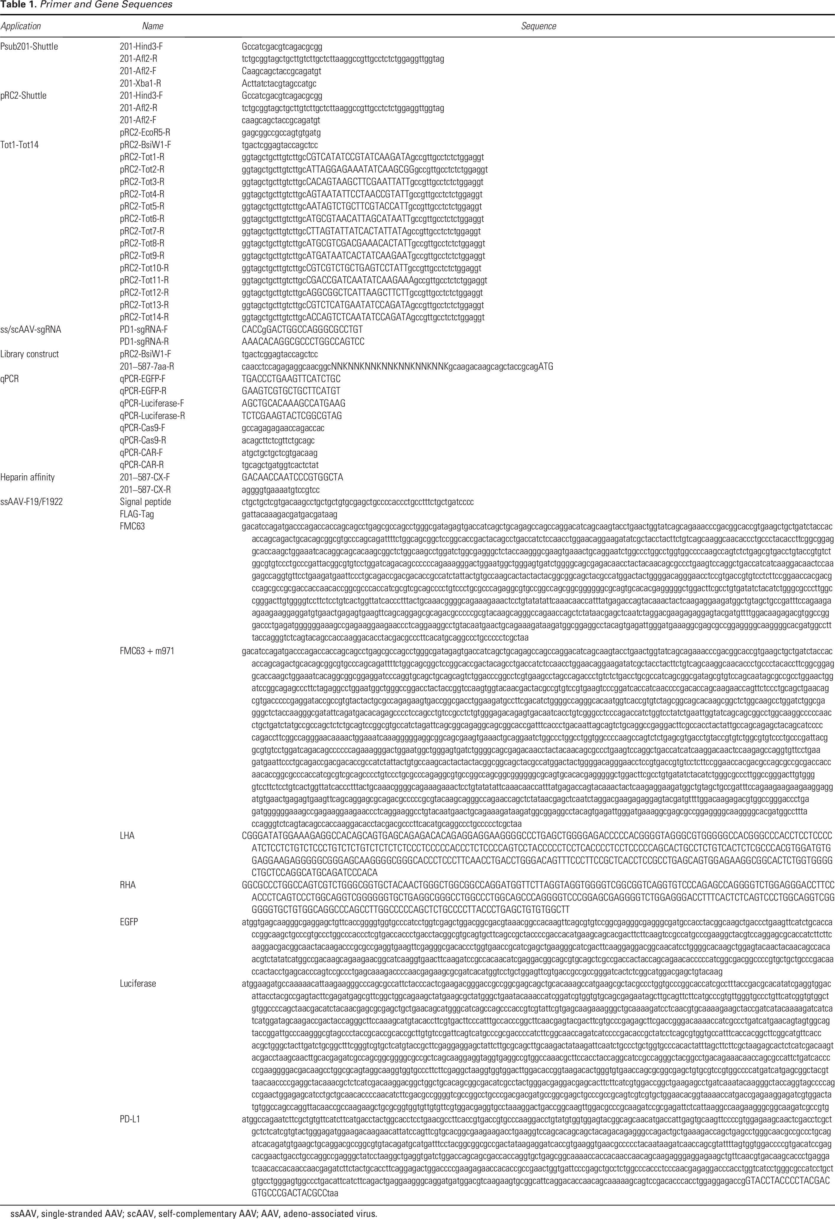
For co-expression of luciferase and EGFP, the CMV promoter-driven luciferase and EGFP co-expression via P2A, which was cloned into the lentivirus (LV) expression vector PLVX-Puro, is called LV-GL (CMV-luciferase-P2A-EGFP). In addition, PGK promoter-driven PD-L1 was fused to the C-terminus of EGFP, called LV-PD-L1(CMV-Luciferase-P2A-EGFP-PGK-PD-L1), with EGFP serving as a marker for fluorescence-activated cell sorting (FACS)-based selection. All primers used are listed in Table 1.
Primer and Gene Sequences
ssAAV, single-stranded AAV; scAAV, self-complementary AAV; AAV, adeno-associated virus.
Cell lines
HEK293 and HEK293T cells were purchased from American Type Culture Collection. Jurkat cells were kindly provided by the Stem Cell Bank (Chinese Academy of Sciences). Nalm6 cells were purchased from Cellcook. For stable Nalm6-GL or Nalm6-PDL1 cell construction, Nalm6 cells were transduced with LV-GL (called Nalm6-GL) or LV-PD-L1 (called Nalm6-PD-L1). EGFP-positive populations were obtained by FACS using a BD FACSAria III. Adherent HEK293 and HEK293T cells were cultured in Dulbecco’s modified Eagle’s medium (Gibco, C11995500BT) supplemented with 10% fetal bovine serum (FBS) (NEWZERUM, FBS-E500) and 1% antibiotics (penicillin 100 U/mL and streptomycin 10 mg/mL) (Solarbio, P1400). Other suspension cells were cultured in RPMI 1640 medium (Gibco, C11875500BT) with the same supplements. The incubator was maintained at 37°C with 5% CO2.
Primary human T cells
Healthy volunteers provided fresh blood. Volunteer recruitment was reviewed and approved by the Ethics Committee of West China Hospital (2018, No. 463). PBMCs were isolated by Ficoll (Solarbio, P8900) gradient centrifugation. CD3+T cells were further isolated using a commercial kit (Miltenyi Biotec, 130-097-043). CD3+T cells were cultured in X-VIVO 15 (Lonza, 04-418Q) medium supplemented with 200 U/mL IL-2 (Peprotech, 130-095-367) and 10% heat-inactivated FBS (Gibco, 10091148). Human T-Activator CD3/CD28 Dynabeads (Gibco, 11131 D) were used for T cell activation and proliferation for 2 to 3 days.
Mouse work
Female NCG mice (6 weeks of age) were all purchased from GemPharmatech (Nanjing, China). Mice were housed under specific pathogen-free conditions with a 12-h light/12-h dark cycle, following the WestChina-Frontier PharmaTech (WCFP, Chengdu, China) animal operation procedures. For in vivo bioluminescence imaging, the mice received D-luciferin potassium salt (Abcam, ab143655) by intraperitoneal injection at a final concentration of 150 mg/kg and were maintained under isoflurane anesthesia for 5–10 min before imaging. Bioluminescent activity was presented as photons using an in vivo imaging system Lumina III instrument (PerkinElmer).
Nucleofection
After 2 to 3 days of activation, the beads were removed, and the supernatant was discarded, followed by centrifugation (200 g, 10 min). The operations were conducted according to the protocol of the kit (Lonza, V4XP-3032). In brief, 1 × 10⁶ cells were resuspended in 16.4 μL of solution buffer and added to the RNP complex, which contained 3.6 μL of supplement mix with 60 pmol of Cas9 nuclease protein (IDT, 1081060). The mastermixes were transferred into the nucleocuvette, and the EH-115 program was selected, immediately followed by adding prewarmed fresh medium. For gene editing, rAAVs were then added to the culture after electroporation until analysis.
Flow cytometry
Flow cytometry analysis for EGFP expression was performed 2 days after rAAV transduction of the target cells, whereas other flow cytometry analyses for the T cell editing experiments were conducted 5 days post-transduction. For EGFP detection, the cells were harvested by centrifugation (300 g, 5 min) and washed thrice with ice-cold PBS. The cells were subsequently resuspended in PBS and then directly tested for intrinsic EGFP. For detection of CAR or other protein expression on the surface of the cells, the prewashed cells were suspended in 100 μL of cold PBS and kept on ice with Human TruStain FcX (BioLegend, 422302) for 20 min. After blocking, the cells were incubated with antibodies for an additional 30 min. Following three washes with ice-cold PBS, the samples were analyzed using a FACSAria™ III Flow Cytometer (BD). The following antibodies were used: Brilliant Violet 510™ anti-human CD3 (BioLegend, 300448), PerCP-Cyanine5.5 anti-human CD4 (BioLegend, 357414), APC anti-human CD4 (BioLegend, 317416), PE-Cyanine7 anti-human CD8a (BioLegend, 300914), PE anti-human CD279 (PD-1) (BioLegend, 329906), Brilliant Violet 421™ anti-human TIGIT (VSTM3) (BioLegend, 372710), APC anti-human CD366 (Tim-3) (BioLegend, 345011), APC anti-DYKDDDDK Tag (BioLegend, 637308), FITC anti-human CD45RA (BioLegend, 304106), and PerCP-Cyanine5.5 anti-human CD62L (BioLegend, 304105). The data were analyzed with FlowJo.
AAV capsid library production
For the generation of the primary library, primers containing 21 random nucleotides were designed and synthesized. PCR was performed to amplify a capsid fragment containing a random sequence. At the same time, Psub201-shuttle was digested with BsiWI (NEB, R3553L) and AflII (NEB, R0520S), generating a 7.6 kb backbone fragment. Both the insert and vector, resulting in linearized DNA fragments, were purified using a gel extraction kit (Omega, D2500-02) and assembled together with Gibson assembly recombinase (NEB, E2621L). Library plasmid measuring 50 ng and 12.5 μg of helper plasmid pAd were co-transfected into 1.5 × 10⁷ HEK293 cells, and the cells along with supernatants were harvested 48 h later for further purification. The iterative library sequences were recovered from the cell genome during the last cycle of selection by PCR. Importantly, the PCR cycle must be kept below 25 during genome rescue to minimize amplification bias. The frequency and enrichment of each randomized sequence were analyzed as previously described. 24 All primers used are listed in Table 1.
rAAV production and titration
rAAVs were produced using a protocol that was previously described. 34 In brief, three plasmids (pAd, pRC, and rAAV transgene plasmids) were transfected into HEK293 cells at a mass ratio of 1:1:1, and cells were harvested 72 h post-transfection. Purification was performed using cesium chloride gradient ultracentrifugation. rAAV genomes were quantified by qPCR using transgene plasmids as controls.
qPCR
For rAAV titer detection, DNaseI (TransGen, GD201-01) and proteinase K (TransGen, GD201-01) were used to remove external DNA and capsid proteins, respectively, to expose the genomic DNA of rAAV. For heparin affinity and rAAV binding/internalization assay, processed rAAV samples and extracted genomes were used for subsequent qPCR. qPCR was performed using an ABI QuantStudio3 and SYBR mix (ABI, A25742) with the corresponding primers. rAAV transgene plasmids and rAAV genomic DNA were detected simultaneously by qPCR, and the genome copy numbers were calculated based on the standard curve. All primers used are listed in Table 1.
ELISA for cytokines
For cytokine detection, T cells were cocultured with tumor cells at an E:T ratio of 1:1 without IL-2 for 24 h. The supernatants were collected for subsequent experiments. The next step was performed according to the manufacturer’s instructions using the following kits: TNF-α (Dakewe Bio, 1117202) and IFN-γ (Dakewe Bio, 1110002). Briefly, 100 μL of a 100-fold diluted sample and 50 μL of biotinylated antibody were added to a precoated plate and incubated for 1 h at room temperature. After washing the plates with PBS (3 × 5 min), 100 μL of streptavidin-HRP was added, and the mixture was incubated at room temperature for another 20 min. The plates were then washed with PBS (3 × 5 min), 100 μL of TMB was added, and the mixture was incubated at room temperature for 10–20 min. The reaction was terminated with 100 μL of stop solution. The measurements were taken at 450 nm and 630 nm wavelengths using a microplate reader. Cytokine concentrations were calculated from the standard curve generated by the standard material.
Cytotoxicity assay
To evaluate the cytotoxic activity of T cells treated with rAAVs for 5 days, Nalm6-GL or Nalm6-PD-L1 cells were cocultured with T cells in 96-well plates at the indicated E:T ratios for 24 h. Next, according to the instructions of the luciferase assay kit (Beyotime, RG006), the cell lysates were incubated with D-luciferin substrates for 5 min, and relative light units (RLUs) were recorded using a microplate reader. The RLUs of the no T cell group were used as control, and the cytotoxicity was calculated according to the following formula:

Capsid thermal stability
The thermal stability of the AAV vector capsid was evaluated as previously described. Briefly, AAV vectors (2 × 10¹⁰ vg in 20 μL of PBS) were exposed to a series of temperatures (54°C, 56°C, 58°C, 60°C, 62°C, 64°C, and 66°C) for 15 min. A measure of 2.5 μL of each sample was transferred onto a nitrocellulose membrane (Servicebio, G6014) and blocked with 5% nonfat milk (Sangon Biotech, A600669) for 1 h. A20 antibody (ARP, 61055) and B1 antibody (ARP, 03-61058) were used to detect intact capsid and denatured capsid proteins, respectively. HRP-conjugated anti-mouse secondary antibody (Abbkine, a21010) was used for signal detection.
Heparin affinity
To compare the affinity of variants to heparin agarose, scAAV-EGFP vectors (1 × 10¹¹ vg in 100 μL of PBS) were incubated with heparin agarose (Yeasen, 20493ES08) for 1 h at room temperature. Unbound rAAV vectors were removed by low-speed centrifugation (300 g, 5 min), followed by three washes with PBS. Stepwise elution was performed using a series of NaCl solutions (0.2 mol/L, 0.3 mol/L, 0.4 mol/L). The viral genomes of the flow-through, wash, and elution fractions were then quantified by qPCR.
rAAV binding and internalization
rAAV binding and internalization were measured as described. Briefly, Jurkat cells and activated CD3+T were kept on ice for 30 min to arrest internalization. Cells were treated with rAAV2 or Tot3 at a multiplicity of infection (MOI) of 1 × 10⁴ (Jurkat) or 2 × 10⁴ (CD3+T). Cells were then placed on ice for an additional hour. Then, the cells were washed thrice with precooled PBS to remove unbound rAAV, and the genomes were quantitated using qPCR. For rAAV internalization assay, after binding, the cells were placed at 37°C for 1 h to enable rAAV uptake. Following washing with PBS buffer containing 0.05% trypsin, genomes were extracted and quantitated using qPCR.
Heparin competition
For the heparin competition assay, primary human T cells or Jurkat cells were seeded in 24-well plates at a density of 1 × 10⁵ cells/well. Sequentially diluted heparin sodium solution (MCE, HY-17567A) was preincubated with the cells for 2 h before challenging them with rAAV in an incubator. Primary human T cells (AAV2: 2 × 10⁵ vg/cell, Tot3: 2 × 10⁴ vg/cell) and Jurkat cells (AAV2: 1 × 10³ vg/cell, Tot3: 40 vg/cell) were then transduced with scAAV-EGFP. The percentage of EGFP-positive cells was determined by flow cytometry 48 h later (FACSCalibur, BD Biosciences).
Indel analysis
The genomic DNA of human T cells was isolated through proteinase K (TransGen Biotech, GE201-01) digestion and isopropanol precipitation. PCR was performed on the genome of PD-1 with designed primers (PD1-F: 5′-CCACGTGGATGT GGAGGAAG-3′, PD1-R: 5′-CCACACAGCTCAGGGTAAGG-3′). A 445 bp band containing the indel region was separated by agarose gel electrophoresis and purified using an Omega Gel Extraction Kit (Omega Biotek, D2500-02). The indel mutagenesis efficiency was quantitatively assessed by Sanger sequencing and analyzed with Synthego (https://ice.synthego.com/#/).
Western blot analysis
Jurkat cells and human primary T cells were harvested and lysed using RIPA lysis buffer (Beyotime, P0013) supplemented with 1% protease inhibitor cocktail (Bimake, B14001). The lysates were centrifuged at 13,000 g for 5 min at 4°C, and protein concentrations were quantified using the Enhanced BCA Protein Assay Kit (Beyotime, P0009). Total protein samples (10 μg) were loaded and separated by SDS-PAGE, then transferred to PVDF membranes (Millipore, IPVH00010) after electrophoresis. The membrane was incubated with 5% nonfat milk (Sangon Biotech, A600669) buffer at room temperature for 1 h. Then, the membrane was incubated with an anti-CRISPR-Cas9 antibody (Abcam, ab191468) overnight at 4°C. The membrane was washed with TBST (3 × 5 min) and then incubated with an HRP-conjugated goat anti-mouse IgG antibody (Abbkine, A21010) at room temperature for 1 h. For reference gene detection, the membrane was washed with TBST (3 × 5 min) and then incubated with HRP-conjugated GAPDH Mouse McAb (Proteintech, HRP-60004) at room temperature for 1 h. Substrate (Abbkine, BMU102-CN) was added, and the signal was captured using a Bio-Rad ChemiDoc Imaging System device.
Statistical analyses
Statistical analyses were performed using GraphPad Prism Version 10. Intergroup comparisons were conducted by one-way ANOVA, two-way ANOVA, or the log-rank Mantel–Cox test as indicated. Details are provided in the corresponding figure legends. All data are presented as the mean or mean ± SEM, with significance levels indicated as *p < 0.05, **p < 0.01, ***p < 0.001, and ****p < 0.0001.
RESULTS
Screening AAV2 variants for enhanced transduction efficiency in human T cells
To identify a parental serotype that enables efficient transduction in human T cells, Jurkat cells were transduced with natural AAV serotypes carrying the luciferase transgene (Supplementary Fig. S1A). It was found that AAV2 exhibited superior transgene expression in Jurkat cells based on the results of the luciferase activity assay (Supplementary Fig. S1B). Therefore, saturation mutagenesis was performed by assembling random heptamer sequences at the residue N587 of the AAV2 capsid protein, which is the critical binding site of heparan sulfate proteoglycan (HSPG). 35
To estimate the variant count of the AAV2-derived capsid library, the diversity of parental and evolved libraries was characterized by next-generation sequencing (NGS) (Fig. 1A). Viral genome DNA was extracted, and NGS was performed using Illumina-based amplicon sequencing. Based on NGS data, we estimated the amount to be around 1 × 10⁷ with a lower limit of 1.7 × 10⁶. After three rounds of screening, the number of library sequences dropped to 9 × 10⁴, indicating enrichment of dominant sequences. Comparative analysis revealed that the third-round library showed remarkable evolution of dominant sequences, with the most prevalent sequence exhibiting a normalized fold change of over 11,000-fold enrichment (Fig. 1B). The percentages of asparagine (N), leucine (L), and negatively charged aspartic acid (D) increased further in the third round of selection, representing approximately 45% of the total residues at the corresponding position (Supplementary Fig. S2A, B). In addition, a unique motif (Y/WLD) might play a crucial role in T cell infection with AAV (Fig. 1C). To assess the changes in heparin binding capacity caused by the random insertion of a heptamer sequence at N587, we performed qPCR on individual populations after binding to heparin agarose. Compared with the about 30% of heparin agarose-unbound virus particles (flow-through + wash) in the parental and second-round library, this proportion rose to 46% in the third-round library (flow-through + wash) (Fig. 1D).
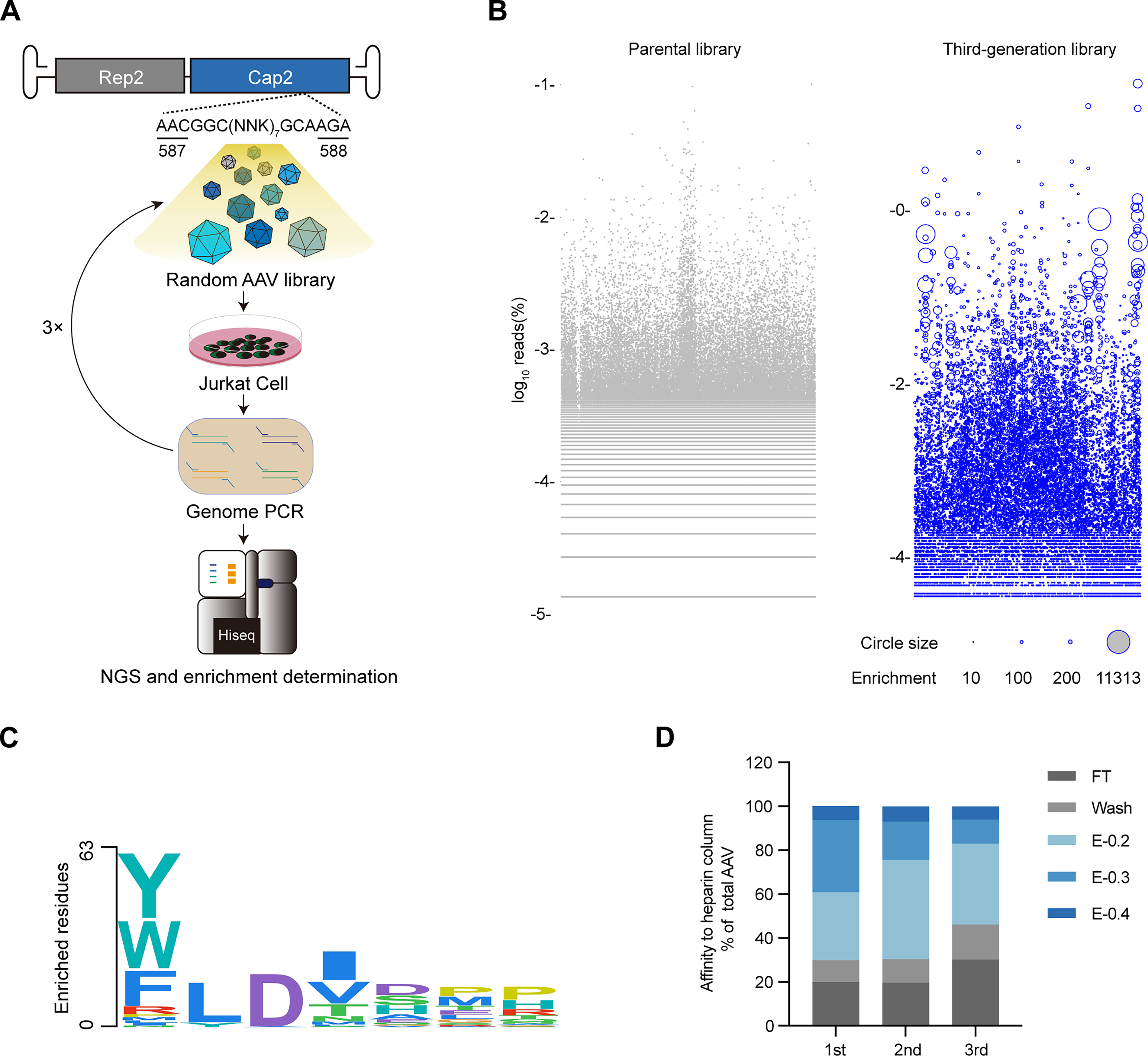
Directed evolution analysis of AAV2 peptide display library in Jurkat cells.
Characterization of the evolved AAV2 variants
To thoroughly evaluate how yield and transduction efficiency together affect transgene expression capacity, 14 variants from the second- and third-round evolved library were chosen for initial testing. A series of scAAV vectors encoding EGFP under a chicken beta-actin promoter in either AAV2 or variant capsids were packaged and used to infect Jurkat cells (Supplementary Fig. S3A). Superior EGFP expression was observed in 13 mutants, except for Tot12, indicating a successful selection (Supplementary Fig. S3B-D). Among the candidate variants, Tot3 (insertion NNSKLTV) showed the greatest increase in EGFP expression and was chosen for further validation.
In Jurkat cells, at an MOI of 9 × 10², over 95% of cells were EGFP positive after using Tot3, which signifies a 27-fold increase in transduction efficiency compared with AAV2 (Fig. 2A). These results were recapitulated in primary human T cells, including CD3+, CD4+, and CD8+T cells (Supplementary Fig. S4). To achieve the same suboptimal transduction efficiency, Tot3 can be used with 27 times fewer viral particles, and ≥93% of EGFP-positive cells can be obtained using Tot3 at an MOI ≥ 2.43 × 10⁵ (Fig. 2B). Compared with AAV2, enhanced binding and internalization efficiencies in Jurkat and primary T cells were observed by Tot3 (Fig. 2C–F). In particular, the yield of Tot3 tripled compared with AAV2 (Fig. 2G). The thermal stability assay showed that AAV2 capsids deteriorated at 66°C, whereas Tot3 began to degrade at 62°C (Fig. 2H). The lower thermal stability of the capsid could be related to more effective transgene expression due to improved uncoating efficiency after internalization.36,37 These data collectively demonstrate that Tot3 simultaneously enhanced packaging efficiency and transduction efficiency in human T cells.

Characterization of evolved Tot3 in vitro.
The current understanding is that of intracellular trafficking pathways involved in rAAVs attachment to target cells and receptor-mediated cellular trafficking.38,39 Given the decreased heparin affinity during capsid evolution (Fig. 1D), we aimed to determine whether AAV2 and Tot3 vectors enter human T cells in an HSPG-dependent manner. In Jurkat cells, low doses of heparin sodium significantly increased rAAV-Jurkat attachment, mainly for the AAV2 vector (<5 μg/mL), with fewer effects observed for Tot3 (<0.5 μg/mL). However, additional heparin sodium reduced the expression levels of AAV2 and Tot3 vectors as the doses increased, suggesting that both vectors depend on HSPG interactions to infect Jurkat cells (Fig. 2I). In primary T cells, EGFP expression decreased significantly in the presence of heparin sodium for the AAV2 vector, while the effect was much weaker for the Tot3 vector (Fig. 2J).
Effective PD-1 knockout in human T cells with the selected Tot3
Since Tot3 significantly improves transduction efficiency, it was used in the rAAV-CRISPR system for gene editing in human T cells. The gene editing efficiency of ssAAV or scAAV was compared for delivering sgRNAs. It is known that scAAV can bypass the rate-limiting second-strand synthesis and results in faster, more efficient transgene transcription, although its packaging capacity is half that of ssAAV (2.3 kb vs 4.7 kb).40,41 To assess the delivery efficiency of the sgRNA, EGFP was co-expressed in the same vector (Fig. 3A). Cas9 was delivered into T cells either by electrophoresis or by ssAAV-SpCas9, which was driven by a short, universal EF promoter (Fig. 3B, Supplementary Fig. S5A, B). Western blot analysis showed that Cas9 protein expression was higher in Tot3 compared with AAV2 (Supplementary Fig. S4C). Due to the low expression levels of Cas9 proteins in primary CD3+T cells, Cas9 was delivered via electroporation before ssAAV-sgRNA or scAAV-sgRNA transduction (Fig. 3B). Western blot analysis confirmed that the electroporated Cas9 protein persisted for 2 days (Fig. 3C). Flow cytometry analysis revealed that EGFP expression levels increased in a dose-dependent manner in both Jurkat cells and primary human T cells 2 days after transduction, with significantly lower EGFP levels in the ssAAV-sgRNA group compared with the scAAV-sgRNA group. Furthermore, Tot3 achieved significantly higher EGFP expression levels compared with AAV2 (Fig. 3D, Supplementary Fig. S5D).

The Tot3-CRISPR system enables efficient PD-1 knockout in primary human T cells.
To evaluate the genome editing efficiency of ss/scAAV2- and ss/scTot3-sgRNA, Jurkat cells were coinfected with different combinations of AAV vectors (Supplementary Fig. S5B). Both ssAAV-SpCas9 and scAAV-sgRNA packaged in Tot3 resulted in a 74% indel frequency, indicating more efficient genome editing than the AAV2 vector (Supplementary Fig. S5E). Five days after transduction, Sanger sequencing was performed on PD-1 exon 1 in primary T cells, and indel frequencies were analyzed by inference of CRISPR edits. The scTot3-sgRNA-transduced cells showed the highest indel frequency of PD-1 (60%) at an MOI of 5 × 10⁵ (Fig. 3E). Moreover, PD-1 expression decreased in a dose-dependent manner for both the ssAAV- and scAAV-expressing sgRNA. PD-1 expression was reduced by approximately 75% in scTot3-sgRNA-treated cells at a high MOI of 2.5 × 10⁶ (Fig. 3F). In line with CD3+T cells, Tot3 exhibited higher gene editing efficiency in CD4+T subtypes than parental AAV2 (Supplementary Fig. S6A, B).
It has been reported that LV- or RNP-mediated PD-1 knockout in T cells can trigger the expression of alternative ICRs, such as TIGIT/LAG-3/TIM-3, and others. 18 However, rAAV-mediated PD-1 knockout did not alter the expression levels of TIGIT or TIM-3 in the CD3+, CD4+, or CD8+T cell populations (Supplementary Fig. S6C, D). Nevertheless, the antitumor activity of PD-1 knockout T cells against Nalm6-PD-L1 cells was slightly improved (Supplementary Fig. S6E).
Tot3 facilitates the efficient production of PD-1 knockout CAR-T cells
Since the cytotoxicity of T cells with a single immune checkpoint knockout still needed improvement, we proposed replacing the EGFP expression cassette with a CAR sequence targeting either CD19 or both CD19 and CD22, flanked by homologous arms complementary to exon 1 of the PD-1 locus (Fig. 4A). For higher transgene expression in T cells, the EF promoter was used instead of the CMV promotor. A FLAG-tag was attached to the N-terminus of the CAR sequence for tracking expression via flow cytometry. Primary human T cells were pre-nucleofected with the Cas9 protein and infected with either ssAAV2-F19 or ssTot3-F19 (Fig. 4B). CAR expression increased dose dependently for both vectors. At the highest MOI (8.1 × 10⁵), AAV2 generated about 10% CAR-positive T cells, whereas Tot3 yielded approximately 40% CAR-positive T cells (Fig. 4C). Strikingly, compared with Tot3 (1 × 10⁴), AAV2 (2.7 × 10⁵) required a 27-fold higher MOI to achieve comparable CAR (7%) expression (Fig. 4C).
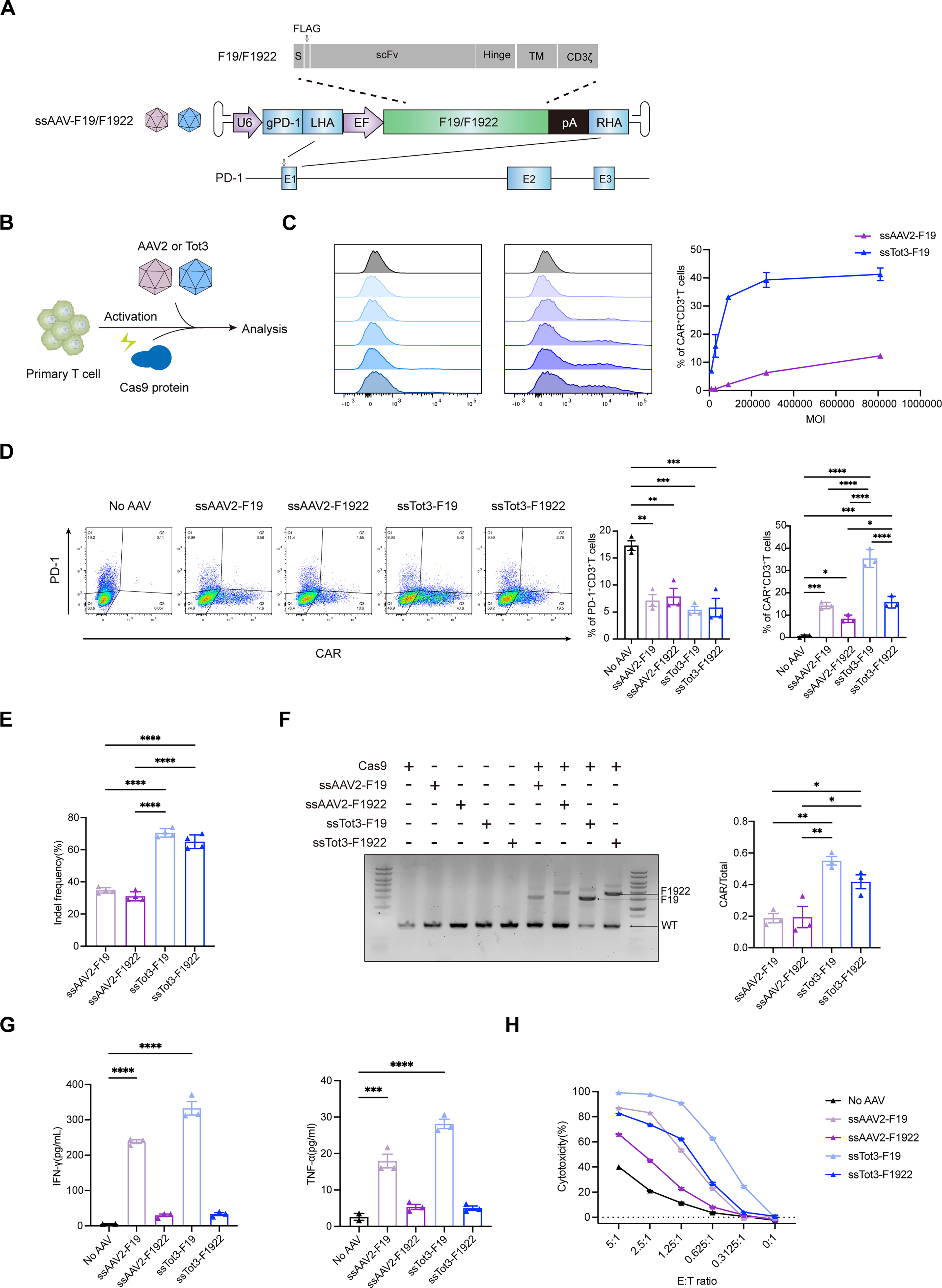
Tot3 outperforms AAV2 in CRISPR-mediated CAR integration in primary human T cells.
Considering the immune escape limit of a single antigen, the integration efficiency of a larger CAR targeting to both CD19 and CD22 antigens simultaneously was evaluated. Similar levels of PD-1 expression were observed in T cells transduced with either ssAAV-F19 or ssAAV-F1922 vectors (MOI = 5 × 10⁵) (Fig. 4D). Indel frequency analysis revealed that the Tot3-mediated knockout efficiency was 70%, significantly higher than the 35% observed in the AAV2-treated group (Fig. 4E). Moreover, the different CARs did not have a significant impact on PD-1 knockout efficiency (Fig. 4E). F19 expression driven by the same AAV serotype was consistently higher compared with F1922 (Fig. 4D, Supplementary Fig. S7A, B). PCR was performed on the genomic DNA extracted from T cells to validate the integration. The efficiency of Tot3-mediated knock-in exceeded 55% (Fig. 4F), and different CARs did not reduce the knock-in efficiency (Fig. 4F). These results indicated that CAR architecture impacts expression efficiency but not HDR-mediated integration efficiency.
To evaluate the immunological features of the CAR-T cells produced by the rAAV-CRISPR platform, rAAV-transduced T cells were cocultured with Nalm6-PD-L1. Upon antigen stimulation, T cells treated with ssAAV-F19 produced significantly higher levels of IFN-γ and TNF-α, especially Tot3 (Fig. 4G). Remarkably, T cells treated with ssTot3-F19 showed excellent antitumor activity despite a lower effector-to-target ratio, indicating that the cytotoxicity of T cells was directly related to CAR expression levels (Fig. 4H).
CD8+ CAR-T cells achieve greater tumor clearance through cytotoxic engagement with tumor cells, whereas CD4+ CAR-T cells selectively act on tumor cells from a distance by releasing IFN-γ. 42 The CD4/CD8 ratio was analyzed after the modified T cells were cocultured with tumor cells, indicating that antigen stimulation can elevate the CD4/CD8 ratios, especially for Nalm6-PDL1 cells. Meanwhile, a lower CD4/CD8 ratio was observed in the Tot3-treated group compared with AAV2. This result aligned with the performance of patients treated with CD19 CAR-T cells. 43 For clinical CD19 CAR-T products, a lower CD4/CD8 ratio was associated with a better response, highlighting the synergistic interaction between different T cell subsets (Supplementary Fig. S7C). Furthermore, we compared the differentiation status of T cells after coculturing with tumor cells based on the expression of CD45RA and CD62L. A higher proportion of naive T cells was observed in the Tot3-treated group without antigen stimulation, and fewer effector T cells appeared in the Tot3-treated group after Nalm6-PD-L1 stimulation, indicating that Tot3-treated T cells remain a less differentiated phenotype. In addition, the PD-1/PD-L1 combination reduced T cell differentiation of T cells (Supplementary Fig. S7D).
Taken together, these in vitro data indicated that Tot3 is a promising platform for effectively generating CAR-T cells with CD19 overexpression and PD-1 knockout.
Effective tumor control in a B-ALL mouse model with Tot3-derived CAR-T cells
To evaluate the antitumor activity of rAAV-treated T cells in vivo, we established a diffuse B-lineage leukemia model by introducing Nalm6-PD-L1 tumor cells into immunodeficient mice. Three days later, the tumor-bearing mice received an intravenous infusion of rAAV-treated T cells (Fig. 5A). Subsequent T cell interventions demonstrated a difference in the fate of tumors in vivo. Throughout the entire observation period, weekly imaging recorded the development of the hematoma (Fig. 5B). No AAV-treated group exhibited aggressive systemic tumor growth accompanied with rapid body weight loss. Compared with the No AAV-treated control group, the proliferation rate of leukemia cells was only slightly reduced in PD-1 knockout T cell groups that were transduced with scAAV2-sgRNA or scTot3-sgRNA (Fig. 5B, D, E). Interestingly, enhanced antitumor activity was observed when the PD-1 knockout accompanied with CAR expressed on the surface of T cells, which were transduced with ssAAV2-F19 or ssTot3-F19 (Fig. 5B, D, E). The progression of the acute proliferative phase was prolonged by 1–2 weeks, coinciding with weight stabilization. Notably, a complete response was achieved in 1/6 of the ssTot3-F19-treated group (Fig. 5B). From a longitudinal perspective, the median survival times of No AAV-, scAAV2-sgRNA-, scTot3-sgRNA-, ssAAV2-F19-, and ssTot3-F19-treated groups were 25, 26, 25, 36, and 47.5 days, respectively. Compared with the ssAAV2-F19-treated group, the survival of the ssTot3-F19-treated group was significantly prolonged (Fig. 5C).
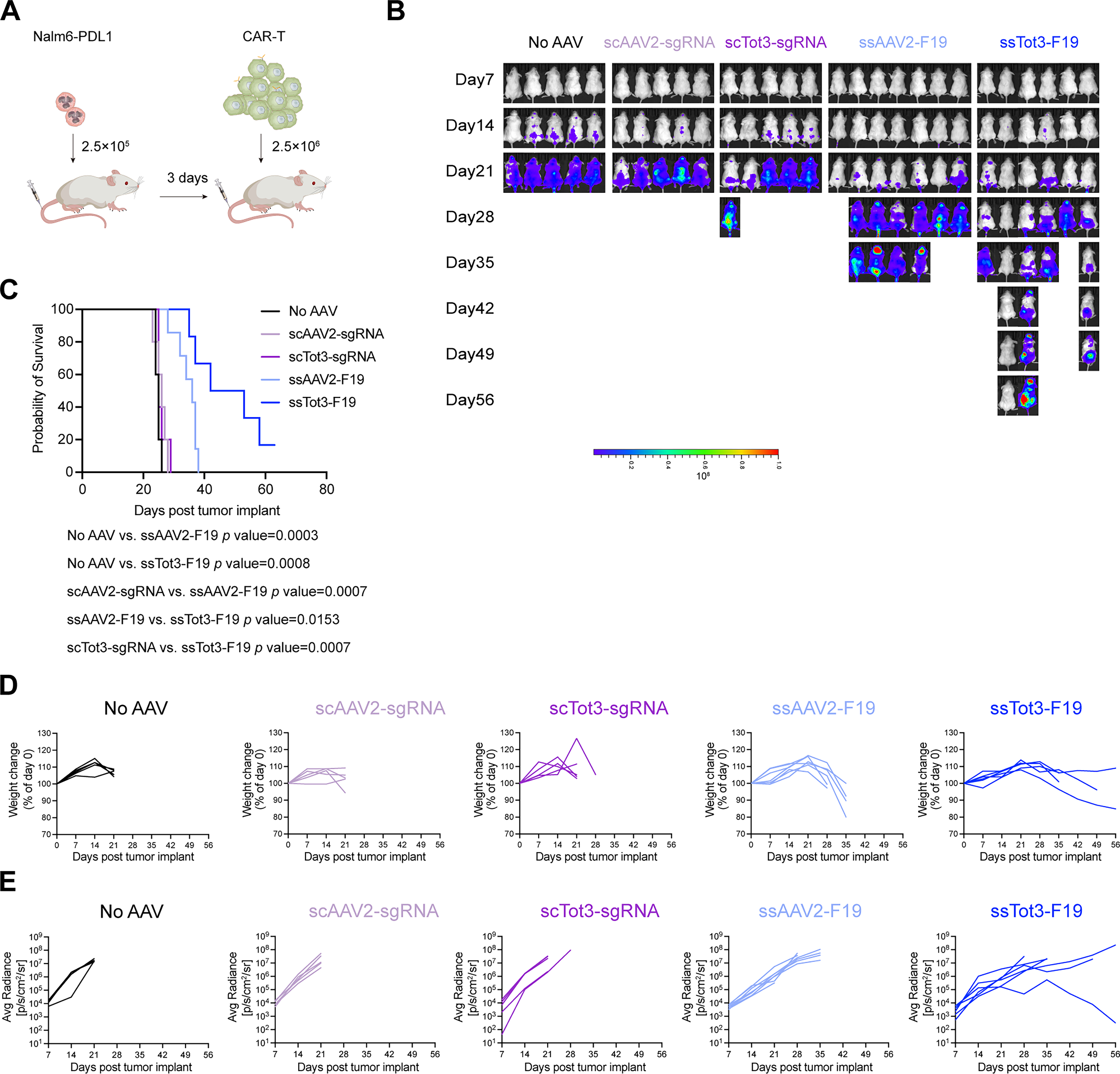
Cellular immunotherapy using T cells treated with Tot3 vector improved survival in a B-ALL model.
Together, consistent with in vitro observations, Tot3-treated T cells exhibited superior antitumor activity in vivo.
DISCUSSION
rAAV vector has become one of the most widely used viral vectors for gene replacement therapy. 30 However, due to poor transduction and expression efficiency in rapidly dividing cells, it has rarely been used to express therapeutic transgenes in primary T cells, which limits its application in cancer immunotherapy. 44 Because rAAV can serve as an effective template for gene editing, Tot3 with enhanced transduction for primary T cells was selected from a 7-mer random peptide library based on AAV2 and was used to knockout the PD-1 gene while simultaneously overexpressing CAR. The survival curve of B-ALL mice could be extended by such engineered T cells.
Previous study indicates that the transduction efficiency of LV and gamma-retroviruses (RVs) in T cells is approximately 80%. 45 In our study, based on EGFP expression, the T cell transduction efficiency mediated by Tot3 exceeds 95%. The transduction efficiency for CD4+ and CD8+ subsets was highly consistent with CD3+T cells, suggesting that neither AAV2 nor Tot3 exhibits a significant bias toward specific T cell subtypes (Supplementary Fig. S4). T cell activation commonly uses CD3/CD28 beads, which is different for residual monocytes. We hypothesize that residual monocytes are unlikely to compete effectively with CD4+/CD8+T cells for rAAV transduction. Besides, the dosing metric for AAV is the number of viral genomes, whereas for LV/RV, it is the number of particles capable of successful transduction and transgene expression. This difference results in a challenge in directly comparing AAV versus LV/RV transduction efficiency in T cells. In addition, compared with LV/RV, the greatest potential advantage of AAV as a delivery vector over an LV or RV lies in its favorable safety profile.
In a previous study, the AAV6 vector was reported as the most promising natural serotype for T lymphocyte transduction and was selected for gene editing. However, a high MOI (1 × 10⁶ vg/cell) is required to achieve transduction efficiencies of 75–80%. 19 We found that AAV2 can transduce primary T cells more efficiently than AAV6 at a lower dose of 2.7 × 10⁴ (Supplementary Fig. S1C), so we chose AAV2 as the parental serotype for selection. Tot3 selected from the AAV2 peptide library could transduce 90% of primary T cells at an MOI of 1 × 10⁵, which is 7-fold lower than AAV6 and 27-fold lower than the parental AAV2 (Fig. 2B). High MOI requirement may be attributed to the fact that activated T cells are prone to exhaustion and high metabolic stress state and induce strong antiviral immune responses. The exogenous nucleic acids trigger type I interferon response, leading T cells to silence the foreign DNA. It represents a significant factor limiting the efficiency of exogenous gene expression in T cells. Therefore, improving the capacity of T cells’ ability to express exogenous genes requires not only efficient delivery vectors but also overcoming the intrinsic defense mechanisms of T cells against exogenous genetic modification. It was suggested that AAV variants with high tropism for primary T cells could be identified from peptide libraries based on other serotypes.
For the selection, we used Jurkat cells instead of primary T cells because they were easier to obtain initially. Tot3 selected from Jurkat cells was confirmed to infect primary T cells much more efficiently than AAV6 and the parental AAV2 serotypes. Compared with AAV2, Tot3 achieved similar transduction efficiency at a 27-fold lower dose in both Jurkat cells and primary T cells (Fig. 2A, B). However, the performance of the Tot3 vectors differed between these two kinds of T cells (Fig. 2I, J). To identify novel capsids with improved transduction efficiency, direct selection of a peptide library in primary T cells is warranted.
rAAV genomic integration offers a promising approach to attain persistent transgene expression in primary cells. Research in this field is advancing along two main technical pathways. On one hand, an “all-in-one” vector system has been chosen, which is based on the hypercompact CRISPR-Cas12f1 nuclease system. 46 On the other hand, dual-AAV delivery system based on trans-splicing inteins or DNA overlapping has shown tremendous potential for precise pathogenic mutation correction. 47 In this study, we chose to knockout PD-1 and overexpress CAR simultaneously in primary T cells using rAAV vectors, which showed a significant therapeutic effect on tumor cells and the B-ALL mouse model (Fig. 5C). CAR-T cells rely on specific surface receptors toward target cells and kill tumor cells as living drugs. Cross talk between PD-1 and CAR has been confirmed.3,8,12,13
Considering the immune escape of a single antigen, CAR sequences targeting only CD19 or both CD19 and CD22 simultaneously were compared. Viral infection can differentially activate and expand CD4+ or CD8+T cells, leading to changes in the CD4/CD8 ratio. Increased rAAV infection was negatively correlated with CD4/CD8 ratio, suggesting that CD8+T cells proliferate more rapidly than CD4+T cells after viral infection. 48 Meanwhile, in vitro and in vivo functional assays revealed that CAR-T cells generated by Tot3 exhibited improved antitumor activity due to higher CAR expression. However, CD19 alone had a better therapeutic effect, suggesting negative effects on DNA fragment length in this study. The superior performance of targeting CD19 alone compared with dual targeting of CD19 and CD22 mainly shows up in higher F19 expression efficiency. We proposed that the tandem F1922 structure reduces the overall protein expression level. Further optimization efforts should be undertaken in this regard. PD-1 is sharply upregulated after the T cell activation and then declines during culture. However, this downregulation occurs more slowly in CD4+T cells than CD8+T cells, and PD-1 levels on CD8+T cells had decreased to nearly undetectable levels by day 7. No significant differences in PD-1 expression levels were observed among different treatment groups in CD8+T cells, which should be due to low baseline levels rather than differential rAAV transduction efficiency between CD4+ and CD8+T cells mediated by rAAV vectors.
High levels of Cas9 protein are crucial for efficient gene editing with AAV vectors in primary T cells. Cas9 nuclease and single-guide RNA were packaged in two rAAV vectors due to size limitations. When these two AAV vectors were transduced into Jurkat cells, gene editing and overexpression occurred simultaneously because sufficient Cas9 protein was expressed (Supplementary Fig. S5C). However, this system did not perform well in primary T cells, possibly due to low Cas9 expression. To fix this issue, the Cas9 protein was directly introduced by electroporation instead of using rAAV infection for gene editing of primary T cells. In the future, T cell-targeted lipid nanoparticle (LNP) for Cas9 mRNA delivery could be explored further.
Since five positively charged amino acids (R484, R487, K532, R585, and R588) of AAV2 are responsible for binding to the primary receptor, HSPG, disrupting the R585-R588 motif typically inhibits binding to HSPG. 35 Previous studies have shown that inserting a negatively charged 7-mer peptide into N587 disrupted the interaction between AAV2 and HSPG. This was also confirmed by the fact that, as the capsid library evolved, the affinity of heparin agarose decreased (Fig. 1D), and the proportion of acidic amino acids gradually increased (Supplementary Fig. S2A, B). In contrast, the insertion of 7 mer-NNSKLTV (Tot3) was generally associated with the restoration of HSPG-binding capacity (Fig. 2I, J).
Several biological characteristics of Tot3 were identified in this study. The thermal stability study showed that AAV2 had relatively higher stability than Tot3. Under this selection process, the lower thermal stability of Tot3 may be associated with efficient uncoating, leading to improved transgene expression. However, more attention should be given to the stability of rAAV variants selected from natural capsids in translation studies. Thus, it was proposed that Tot3 promotes transgene expression in T cells by facilitating multiple steps, including binding, internalization, and uncoating.
In summary, Tot3 selected in this study demonstrated superior transduction efficiency in human primary T cells, thereby offering a novel vector for the immunotherapy of hematological malignancies.
AUTHORS’ CONTRIBUTIONS
Conception, M.L. and B.D.; study design and execution, M.L., C.G., Z.Z., S.H., Y.L., L.Z., R.C., J.Z., L.Xiao, J.Y., Z.C., and L.Xu; and writing and editing, M.L., Y.Z., H.Y.C., C.A.V., and B.D.
Footnotes
ACKNOWLEDGMENT
The authors sincerely appreciate Li Chai, Yi Li, Xing Xu, and Haihui Yang from Science and Technology Park-Public Research Platform (Huaxi), West China Hospital, Sichuan University, for their assistance.
FUNDING INFORMATION
This work was supported by the National Key R&D Program of China (2023YFC3403300).
DATA AVAILABILITY STATEMENT
All relevant study data are available within the article or from the corresponding author upon reasonable request.
AUTHOR DISCLOSURE
B.D., Z.Z., L.Xiao, J.Y., and C.A.V. were employed by the company Sichuan Real and Best Biotech Co., Ltd. The other authors declare that they have no competing interests.