



Abstract
Aim
The aim of this review was to determine if photographic examination and subsequent image analysis provides comparable accuracy to visual inspection for the diagnosis of common dental conditions in children and adolescents.
Methods
We searched the PubMed database for studies that compared diagnostic accuracy of the two inspection techniques. Studies were screened for inclusion and were assessed for quality and risk of bias using the quality assessment of diagnostic accuracy studies tools. Findings were reported according to the preferred reporting items for systematic reviews and meta-analyses (PRISMA).
Findings
Sixteen studies met the inclusion criteria. Nine of the included studies evaluated dental caries and eight evaluated enamel defects (one study evaluated both conditions).
Conclusions
Three studies found image analysis to be superior. For the remaining six studies the diagnostic accuracy was comparable. For enamel defects, three studies found image analysis to be superior, two found visual inspection to be superior and three studies reported comparable diagnostic accuracy. Most studies have found at least comparable results between photographic and visual inspection techniques. However, the wide variation in equipment and personnel used for the collection and interpretation of photographic images made it impossible to generalise the results. It remains unclear exactly how effective store-and-forward teledentistry is for the diagnosis of common dental conditions in children.
Keywords
Introduction
The World Health Organization regards school children and adolescents as an important target group for priority actions in oral health. 1 The burden of oral disease in these age groups is significant and early diagnosis is important for the prevention of the disease’s progress. For people living in remote areas of Australia it is much more difficult to access dental services, given that the majority of dental services are located in city areas. The number of dentists available per head of population, when comparing remote communities with metropolitan areas, is about one-third. 2
Visual inspection is the most common technique to diagnose caries and enamel defects in clinical practice.3,4 Remote diagnosis using transmitted photographic images of dentition (teledentistry) may be an alternative to visual inspection, particularly for people living in remote and underserved areas.5–10 However, the photographic images used in an asynchronous mode are two-dimensional and this characteristic may affect their diagnostic accuracy.
To the best of our knowledge there has been no previous attempt to review available literature that compares the accuracy of in person visual inspection versus photographic image analysis. The aim of this review was to determine if photographic examination and subsequent image analysis provides comparable accuracy to visual inspection for the diagnosis of common dental conditions in children and adolescents.
Methods
Search strategy
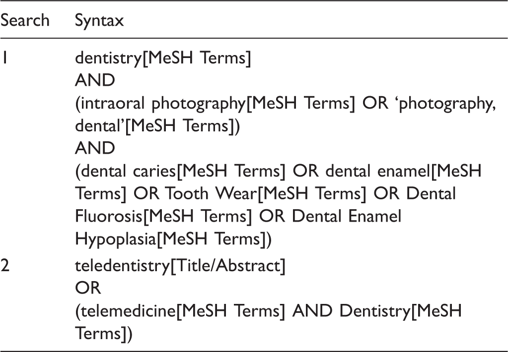
Search strategy.
Eligibility criteria
Studies were included if they compared the diagnosis from images (photographic, video or intraoral camera) to visual inspection for dental diseases common in the target population. Studies in languages other than English or with no abstract available were excluded.
Study selection
Two reviewers independently examined the titles and abstracts to determine eligibility for inclusion. Selected papers were then compared and discrepancies were resolved by consensus discussion.
Data collection and quality assessment
The full texts of studies meeting the inclusion criteria were obtained and data extracted. One reviewer extracted data from the full text. A second reviewer independently verified the extract. A form was used to record extracted data. The form included: year of publication, location, sample size, disease, index used to classify the disease, equipment used to obtain the images (digital camera, intraoral camera or video camera) and imaging protocol.
The QUADAS-2 tool tailored to this review.

QUADAS-2: quality assessment of diagnostic accuracy studies; +: added signalling question
Synthesis of results was performed narratively. Reporting of the findings of this review followed the preferred reporting items for systematic reviews and meta-analysis (PRISMA) guideline. 12
Results
We identified 16 studies that met inclusion criteria. Figure 1 represents the study selection process.
PRISMA flow-chart.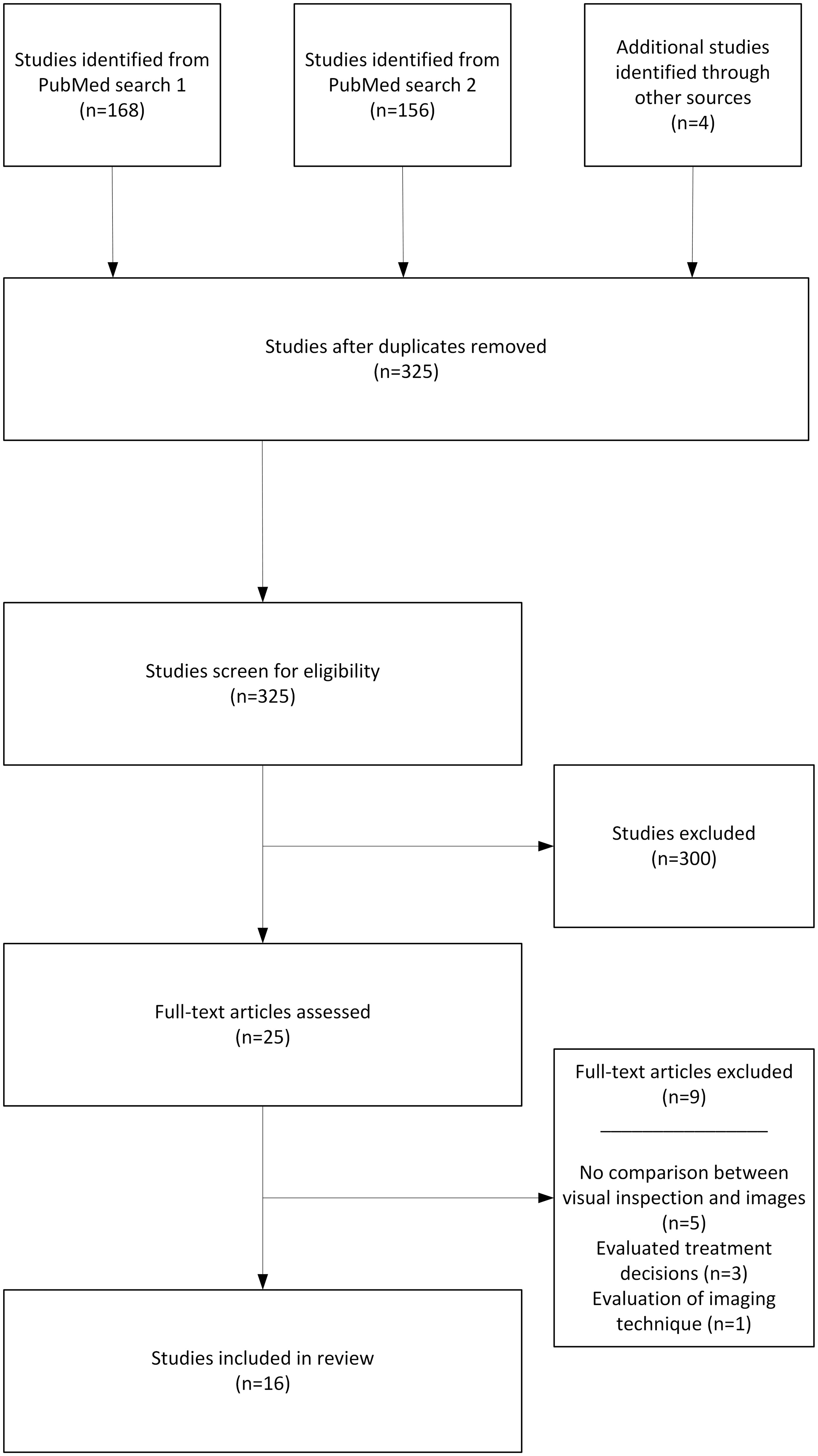
Study characteristics
Study characteristics.

DDE: developmental defects of enamel; PMH: primary molar hypomineralisation; BASCD: British Association for the study of Community Dentistry; ICDAS: International Caries Detection and Assessment System; N/A: not applicable; SLR: single-lens reflex; DMFT: decayed, missing, filled teeth (permanent); deft index: decayed, to be extracted, filled teeth (deciduous); dfs: decayed, filled surfaces; dft: decayed, filled teeth; EAPD: European Association of Paediatric Dentistry; TF: Thylstrup–Fejerskov; NR: Not reported
The type of equipment used to acquire the images showed great variation, including different intraoral cameras, photographic cameras (some film based) and video cameras (Table 3). The information about the imaging equipment used was not provided in one study. The imaging protocol for photographic cameras (frequently used with macro lens and ring flash) was largely consistent, whereas the imaging protocols for intraoral cameras were not consistent.
The professional background of the person that acquired the images was not clearly identified in most of the studies. It was possible to infer that they were either a dentist (n = 9) or a dental hygienist (n = 1). In the studies that explicitly stated the professional background of the photographer (n = 4), three were dentists and one was a trained telehealth assistant.
Comparative diagnostic accuracy
Nine of the included studies evaluated dental caries. Three studies13,14,19 found image analysis to be superior to visual inspection, one study found visual inspection superior 16 and in the remaining five studies10,17,18,20,21 the diagnostic accuracy from both inspection techniques was comparable. Comparable diagnostic accuracy of occlusal caries was found to be the most consistently reported finding.
Study results.
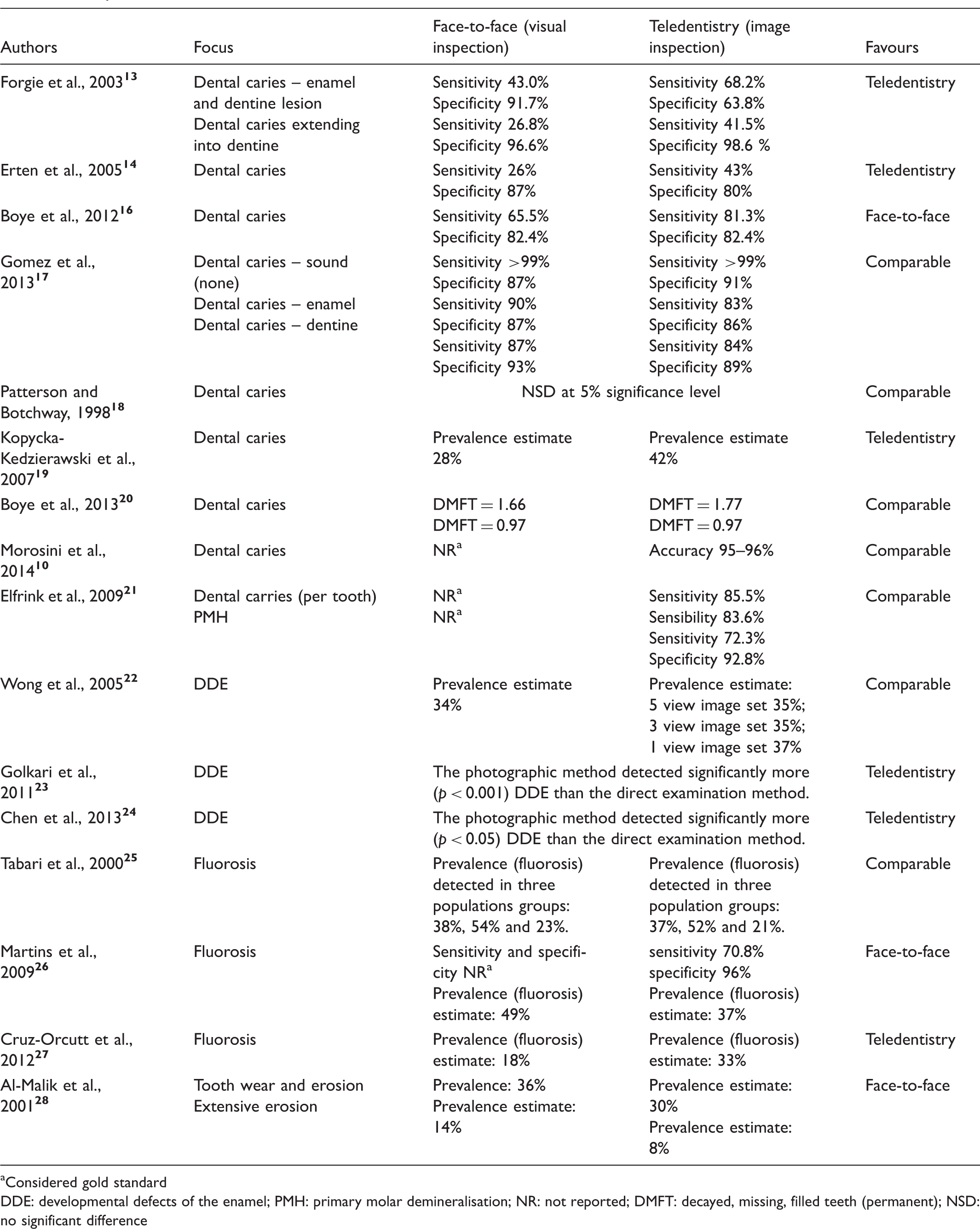
Considered gold standard
DDE: developmental defects of the enamel; PMH: primary molar demineralisation; NR: not reported; DMFT: decayed, missing, filled teeth (permanent); NSD: no significant difference
Quality and risk of bias
For the case selection domain, 19% of studies were judged to be at high risk of bias and 37% were considered unclear. This was because of lack of information about the range of disease’s severity, the prevalence of the disease or related to the choice of high-prevalence disease settings. Furthermore, all studies used convenience samples.
For the index test domain, 13% of studies were judged to be at high risk of bias and 50% were considered unclear. The main concern was the number of examiners – over 44% of studies had only one examiner. For the reference standard domain the risk of bias was judged to be high or unclear in 50% of the cases. This was because of the lack of calibration and validation by multiple examiners. For the time and flow domain, 25% of the studies were judged to be unclear. This was because there was no clear information about the washout time between visual inspection and image analysis.
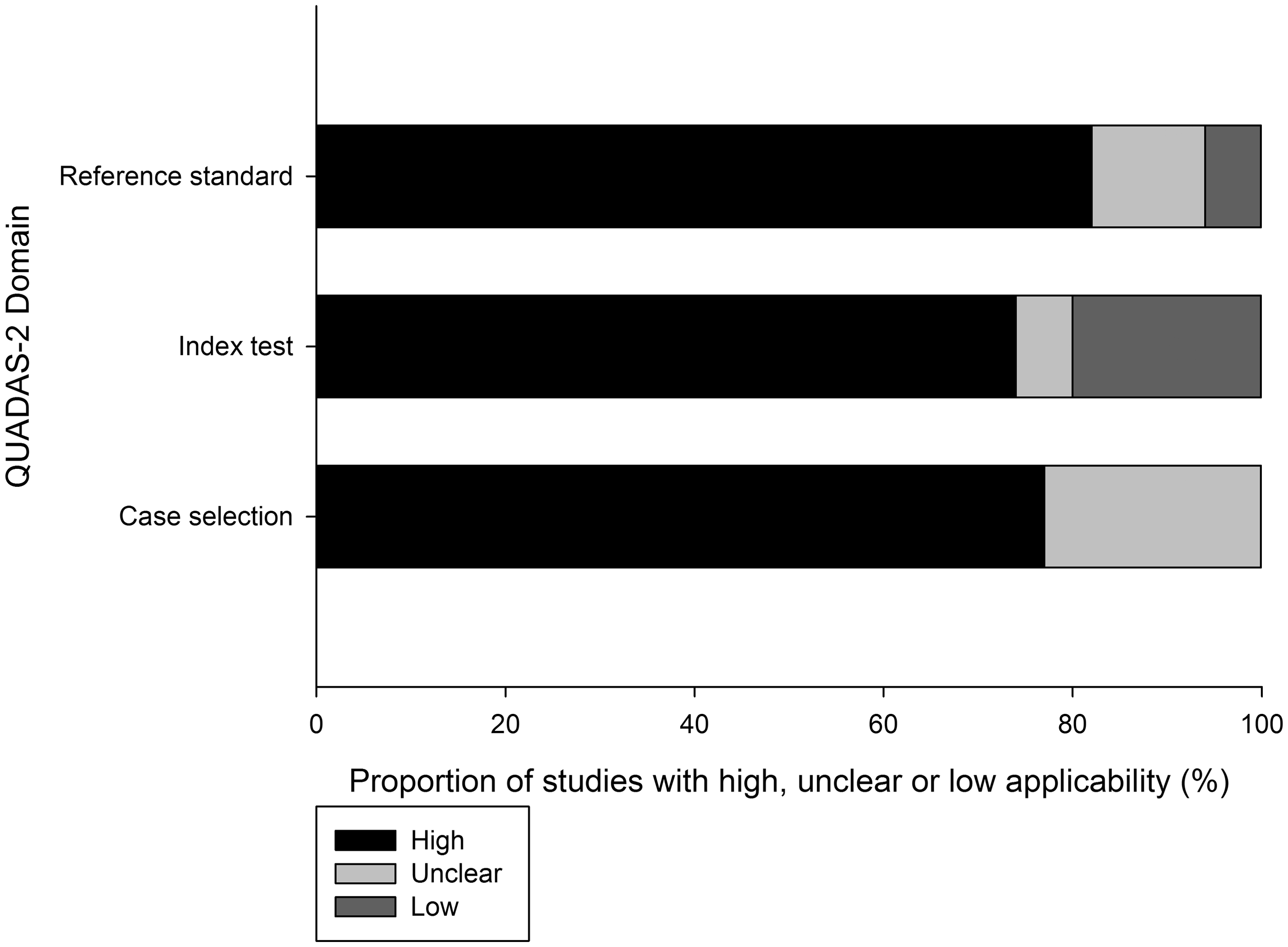
In the index test domain, 20% of studies were considered to have low applicability to the review question. Clinical applicability was also a concern for in vitro studies. The summary of the QUADAS-2 analysis is presented in Figures 2 and 3.
Summary of quality assessment of diagnostic accuracy studies (QUADAS-2) assessments for risk of bias. Summary of quality assessment of diagnostic accuracy studies (QUADAS-2) assessments for applicability.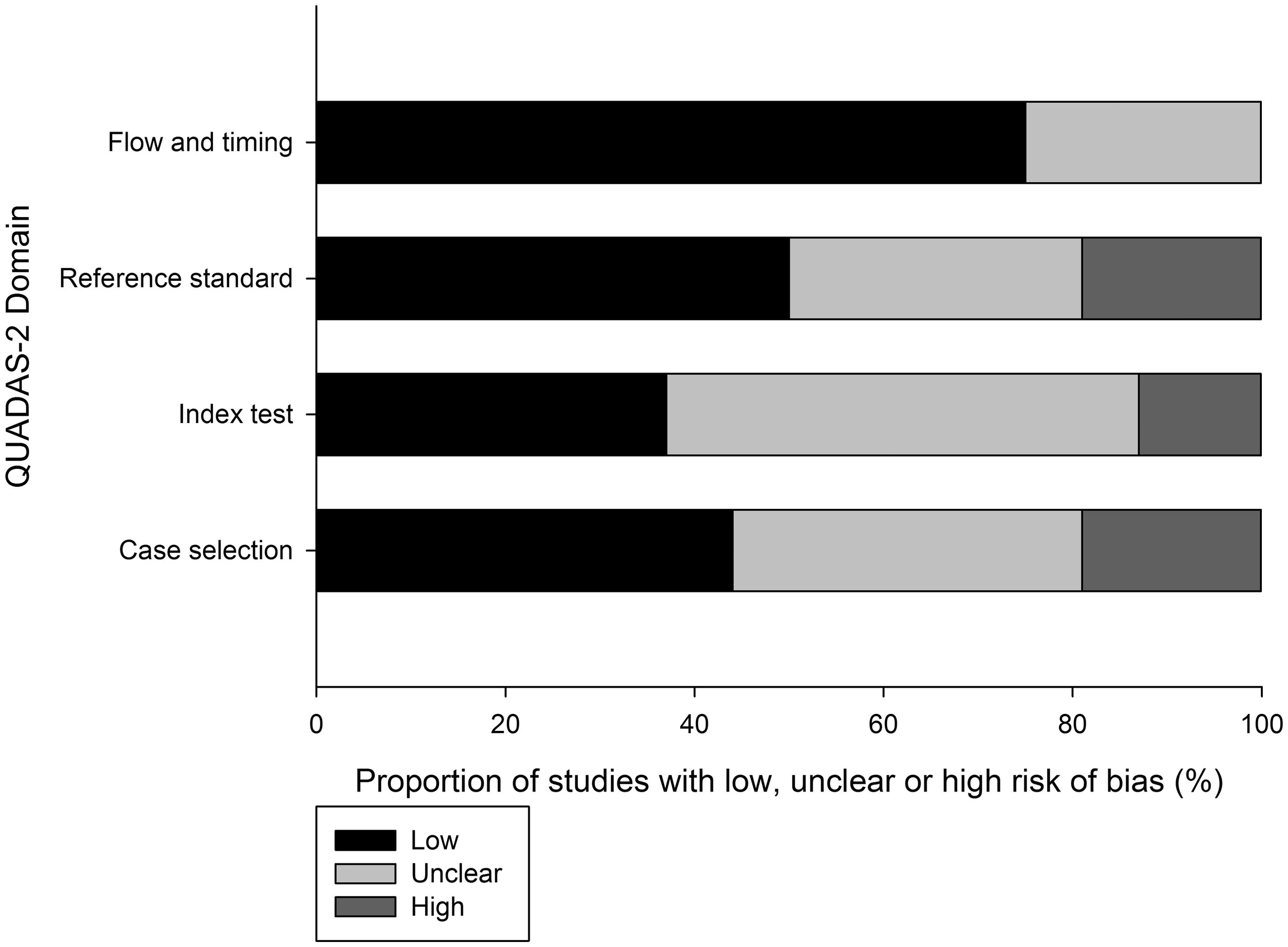
Discussion
In regards to the nine studies which compared accuracy of assessments on dental caries, three studies found image evaluation to be superior to visual inspection and the remaining six were equivalent. Two of the three studies that found image evaluation to be superior were conducted in vitro. In vitro studies are important to assess the diagnostic accuracy of dental caries because they can use the gold-standard histological confirmation of diagnosis. 29 However, in vitro studies have serious limitations in clinical applicability because the difficulties imposed by the working in oral cavity in a clinical setting (small space, poor light, presence of saliva, patient movement) are minimised.
Permanent teeth were examined in seven studies and deciduous teeth in four studies. Five of the nine studies evaluated only molars and premolars (one included canines). Almost half of them restricted the screening to the occlusal surfaces. Occlusal surfaces of posterior teeth are the most vulnerable sites for dental caries. Neither inspection technique is able to detect small interproximal lesions. Therefore, analysis of the occlusal surfaces only was considered justified. With the exception of the study that evaluated PMH, all other enamel defect studies evaluated only anterior teeth. This is justified as most consultations are for aesthetic complaints.
Visual inspection is an important tool in clinical settings for oral epidemiology and oral health surveillance. Given that photographic examination has the same limitations as visual inspection, this may indicate store-and-forward teledentistry is most applicable to the same clinical settings.
Limitations
This review has a number of limitations. The included studies were heterogeneous because different indexes were used to classify disease, personnel, equipment and methodology. The heterogeneity did not permit definitive conclusions about the performance of the two techniques. Further, many of the studies were judged to have a high or unclear risk of bias, which may further reduce confidence in the findings. We only used the PubMed database to search for studies for this review. This search strategy may have excluded articles that are only indexed by other sources.
Conclusion
There is very limited published evidence which compares the diagnostic accuracy of image analysis with in person visual inspections. Taking into account the limitations reported in this review, it seems that most studies in this area have found at least comparable results between photographic techniques and conventional methods of dental assessment for common dental conditions. However, the wide variation in equipment and personnel used for the collection and interpretation of clinical images made it impossible to generalise the results. Whilst the perceived advantages of teledentistry for patients living in remote areas is understandable in terms of access, it remains unclear exactly how effective store-and-forward teledentistry is for diagnosis of common dental conditions in children and adolescents.
Footnotes
Declaration of Conflicting Interests
The authors declared no potential conflicts of interest with respect to the research, authorship, and/or publication of this article.
Funding
The authors disclosed receipt of the following financial support for the research, authorship, and/or publication of this article: this work has been partially funded by The Brazilian National Council for Scientific and Technological Development (CNPq).