



Abstract
Cognition and brain homeostasis depends on cerebral blood flow to secure adequate oxygen and nutrient distribution to the brain tissue. Altered cerebral blood flow has previously been reported in individuals diagnosed with autism spectrum condition in comparison to non-autistics. This phenomenon might suggest cerebral blood flow as a potential biomarker for autism spectrum condition. Major technological advancement enables the non-invasive and quantitative measurement of cerebral blood flow via arterial spin labeling magnetic resonance imaging. However, most neuroimaging studies in autistic individuals exploit the indirect blood oxygen level dependent functional magnetic resonance imaging signal instead. Therefore, this review examines the use of arterial spin labeling to further investigate the neurobiology of the autism spectrum condition. Followed by a comparison of results from molecular imaging and arterial spin labeling studies and a discussion concerning the future direction and potential of arterial spin labeling in this context. We found that arterial spin labeling study results are consistent with those of molecular imaging, especially after considering the effect of age and sex. Arterial spin labeling has numerous application possibilities besides the quantification of cerebral blood flow, including assessment of functional connectivity and arterial transit time. Therefore, we encourage researchers to explore and consider the application of arterial spin labeling for future scientific studies in the quest to better understand the neurobiology of autism spectrum condition.
Lay abstract
Brain function and health depend on cerebral blood flow to secure the necessary delivery of oxygen and nutrients to the brain tissue. However, cerebral blood flow appears to be altered in autistic compared to non-autistic individuals, potentially suggesting this difference to be a cause and potential identification point of autism. Recent technological development enables precise and non-invasive measurement of cerebral blood flow via the magnetic resonance imaging method referred to as arterial spin labeling. However, most neuroimaging studies still prefer using the physiologically indirect measure derived from functional magnetic resonance imaging. Therefore, this review examines the use of arterial spin labeling to further investigate the neurobiology of autism. Furthermore, the review includes a comparison of results from molecular imaging and arterial spin labeling followed by a discussion concerning the future direction and potential of arterial spin labeling. We found that arterial spin labeling study results are consistent with those of molecular imaging, especially after considering the effect of age and sex. In addition, arterial spin labeling has numerous application possibilities besides the quantification of cerebral blood flow. Therefore, we encourage researchers to explore and consider the application of arterial spin labeling for future scientific studies in the quest to better understand the neurobiology of autism.
Keywords
Autism spectrum condition (ASC) is classified as a neurodevelopmental condition characterized by challenges in social interactions and communication as well as repetitive behavior, interests, and activity patterns (American Psychiatric Association, 2013). The recognition of ASC as a major public health concern (Simonoff et al., 2008) and the continuously increasing prevalence of ASC, the latest count affecting 2.3% of children (Maenner et al., 2021), have amplified the quantity of scientific studies investigating ASC. These studies include neuroimaging investigations, where the vast majority utilizes functional magnetic resonance imaging (fMRI). fMRI has overtaken the pole position of human brain examination and its usage is continuously increasing, contrary to molecular imaging methodologies such as positron emission tomography (PET) and single photon emission computed tomography (SPECT) by virtue of molecular imaging’s higher costs, invasiveness, and complex methodological underpinnings (Cumming, 2014). Studies utilizing fMRI have greatly enhanced the understanding of the autistic brain. Among these findings are the continuously evidenced short-range enhancement of functional connectivity (FC), a measure describing the temporal correlation in neural activity between brain structures implying neural communication between brain structures and long-range diminishment of FC compared to non-autistic (NA) individuals (Hull et al., 2017; Supekar et al., 2013). These findings essentially advocate that neighboring brain structures communicate to a higher degree while distant structures interconnect less in autistic individuals compared to NA. The fMRI-associated assessment of neural activity is based on the blood oxygen level dependent (BOLD) signal, which has been displayed to correlate with neural activity assessed via optogenetics (Vazquez et al., 2018). Albeit the knowledge expansion gained via the application of fMRI to study ASC, the BOLD signal is solitarily an indirect and qualitative measure of neural activity, based on the coupling between cerebral blood volume, cerebral blood flow (CBF) and cerebral metabolic rate of oxygen (Devor et al., 2011). Moreover, the hemodynamic response function, the BOLD signal is founded upon, has been demonstrated to be aberrant in autistic individuals, thereby potentially leading to false inference (Yan et al., 2018). Quantitative assessments of CBF were initially only obtainable via invasive methodologies such as PET and SPECT. However, arterial spin labeling (ASL) enables non-invasive and quantitative evaluation of CBF by utilizing magnetically labeled arterial blood water as an endogenous tracer (Williams et al., 1993). The quantitative nature of ASL is based upon the subtraction of control and labeled images, that differs in blood being magnetically inverted in the labeled image before the image acquisition, contrary to the control image. The difference between the labeled and control image is proportional to the tissue perfusion, thereby constituting as a measure of labeled blood from arteries delivered to the brain tissue via perfusion and enabling CBF quantification (Alsop & Detre, 1996; Williams et al., 1993). Comparisons with gold-standard 15O-H2-PET have validated CBF quantification via ASL (Puig et al., 2020) and CBF dynamics during hypercapnia challenges (Heijtel et al., 2014). Crucial methodological improvements after the development of pseudo continuous arterial spin labeling (pCASL) dramatically enhanced sensitivity and signal-to-noise ratio (SNR) of ASL (Alsop et al., 2014). Congruently, the materialization of multiple post labeling delay (PLD) sequences enabled further improved CBF quantification (Cohen et al., 2020). Importantly, the formation of the consensus paper (Alsop et al., 2014) manifested an analytical and acquisitional groundwork for appropriate ASL application. Nonetheless, fMRI is by far the most frequently utilized neuroimaging method exploring the neurovascular coupling (NVC), referring to the enhancement of nutrient transport to active neurons via amplified CBF. Reduced NVC, implying a lack of CBF response to enhanced neural activity, is associated with weakened cognitive function (Csipo et al., 2021; Tarantini et al., 2017). Importantly, brain development depends heavily on cerebrovascular maturation and integrity, as vascular beds support proliferation, differentiation, and migration of neural progenitors (Segarra et al., 2018) and ensures homeostasis and adequate oxygen and nutrient supply (Andreone et al., 2015). Therefore, cerebrovascular alterations could instigate long-lasting consequences in neurodevelopmental diseases such as ASC. Supporting this concept, postmortem examinations indicate persistent aberrant angiogenesis in ASC (Azmitia et al., 2016), exposing constant construction of new blood vessels by splitting of existing blood vessels (intussusceptive angiogenesis) rather than sprouting angiogenesis. Potentially, this continuous microvascular reorganization could trigger the local over-connectivity and long-range underconnectivity associated with ASC (Hull et al., 2017; Supekar et al., 2013), possibly counteracting complex connectivity formation involved with social behavior (Azmitia et al., 2016) in alignment with core ASC symptomatology (American Psychiatric Association, 2013). Accordingly, compromised early life angiogenesis has been demonstrated to provoke long-lasting effects on the NVC and endothelium-dependent vasodilation and induce lifelong ASC-related symptomatology (Ouellette et al., 2020). Moreover, ASC is accompanied by increased risk for and prevalence of vascular conditions (Flygare Wallén et al., 2018; Sigmon et al., 2019), overall linking ASC to vascular dysfunction. BOLD signal differences are commonly interpreted as changes in neural activity. However, the beforementioned evidence suggests that altered BOLD signal in ASC may be caused by endothelial-dependent vascular impairment and not solitarily irregular neuronal activity. Hence, further investigation of CBF in ASC is required for a better understanding of the underlying mechanisms leading to the clinical presentation of symptoms warranting an autism diagnosis. Intuitively, the advancement of ASL and its quantitative nature would encourage an amplification of ASL studies in ASC. Surprisingly, a simple PubMed search including ASL and autism only generates 12 article hits, while fMRI and autism produces close to 4000. Therefore, the following review intents to evaluate studies exploiting ASL to investigate ASC, their similarities with molecular imaging studies, and the future perspectives of ASL studies in ASC.
Method
The present review reports all studies identified based on a comprehensive literature search on PubMed, MEDLINE, PsycINFO, and Web of Sciences on 6 August 2022. Figure 1 shows an overview. Finding ASL studies examining autistic individuals was carried out utilizing the search terms: arterial spin labeling, autism spectrum disorder, autism, and cerebral blood flow. Solitarily articles using ASL and concerning autistic individuals were included in the review. In addition, ongoing studies were not included and only original articles in English were reviewed. No limitation was placed on publication date. Subsequently, references of these articles were explored to find additional relevant articles; however, none were found this way. We identified seven articles; Table 1 shows a comprehensive overview of the reviewed publications including age, sample size, ASC diagnosis, intelligence quotient (IQ), medication, region of interest and main findings.

Flow diagram illustrating the article selection process.
Overview of included studies.

AD: antidepressant; AE: antiepileptic; AH: antihypertension; AP: antipsychotics; ASC: autism spectrum condition; ADOS: Autism Diagnostic Observation Schedule; ADI-R: Autism Diagnostic Interview–Revised; ACC: anterior cingulate cortex; BD: benzodiazepine; C: caudate; CBF: cerebral blood flow; CARS: Childhood Autism Rating Scale; DSM: Diagnostic and Statistical Manual of Mental Disorders; FL: frontal lobe; GP: globus pallidus; H: hippocampus; I: insula; IFG: inferior frontal gyrus; IFO: inferior frontal operculum; IPG: inferior parietal gyrus; ITG: inferior temporal gyrus; Li: lithium; LN: lenticular nucleus; MOC: medial orbitofrontal cortex; MTG: medial temporal gyrus; MAOI: monoamine oxidase inhibitors; MS: mood stabilizer; NA: non-autistic; N.A.: not available; NS: non-stimulant ADHD medication; OFC: orbitofrontal cortex; PLD: post labeling delay; pASL: pulsed arterial spin labeling; P: putamen; PG: precentral gyrus; pCASL: pseudo continuous arterial spin labeling; RN: red nucleus; SG: subcallosal gyrus; SN: substantia nigra; ST: stimulants; STS: superior temporal sulcus; SMG: supramarginal gyrus; TL: temporal lobe; T: thalamus; TTG: transverse temporal gyrus; U: uncus; VS: ventral striatum; TR: text rev.; IQ: intelligence quotient.
Results
Hypoperfusion
Out of the seven articles incorporated in Table 1, six studies compared ASC to NA and five of these studies found decreased CBF in ASC individuals. Saitovitch et al. (2019) compared early adolescents and found significantly lower resting-state CBF in temporal regions, especially in the superior temporal sulcus (STS) using whole-brain voxel-wise analysis. Congruently, Mori et al. (2020) compared resting-state CBF in children ranging from 2 to 15 years of age, focusing on the mirror neuron system which includes motor areas, the somatosensory cortex, and the inferior parietal cortex and is involved with social learning domains. Autistic children elicited significantly lower CBF in the insula, inferior frontal gyrus, subcallosal gyrus, left superior parietal lobule, superior temporal gyrus, middle temporal gyrus, left caudate, and left lentiform. Yerys et al. (2018) investigated CBF behavior in the temporal lobe of subjects (early and middle adolescents; 12–17 years old) during a passive viewing task. Autistic individuals displayed lower CBF in the fusiform gyrus bilaterally as well as in the inferior temporal gyrus. Tang et al. (2022) examined a population ranging from 2 to 18 years of age, divided into eight age-dependent groups to investigate the age-related differences in CBF. Decreased CBF in the temporal lobe, hippocampus, and putamen was found throughout all age-segments in ASC compared to NA, with supplementary structures as the frontal lobe, thalamus, caudate, and substantia nigra displaying decreased CBF as age increased in the ASC group. Jann et al. (2015) found decreased resting-state CBF in the anterior cingulate cortex (ACC) in autistic adolescent individuals.
Hyperperfusion
Two studies found enhanced CBF in ASC compared to NA. Although Jann et al. (2015) reported hypoperfusion in the ACC, they also reported widespread hyperperfusion in frontotemporal regions including the medial orbitofrontal cortex (OFC), bilateral inferior frontal operculum, left inferior/middle temporal gyrus, and right precentral gyrus. They progressively used ASL to investigate FC, where increased local ACC FC was evident and decreased FC was found between the ACC and posterior cingulate cortex (PCC). Peterson et al. (2019) found hyperperfusion in the caudate, lenticular nucleus, ventral striatum, OFC, and amygdala in adult autistic individuals. In addition, examination of white matter (WM) demonstrated enhanced CBF of the centrum semiovale, corona radiata, and internal capsule (Peterson et al., 2019).
CBF correlation with symptomatology
Saitovitch et al. (2019) demonstrated a negative correlation between Autism Diagnostic Interview–Revised (ADI-R) score and CBF in the STS, indicating a linkage between STS hypoperfusion and advancing ASC symptomatology. Congruent results were evidenced by Saitovitch et al. (2016), who solitarily investigated autistic individuals, demonstrating a significant positive correlation between number of fixations during an eye-tracking task and CBF in the STS. The fixations of interest involved looking at characters eyes, a well-established surrogate for sociability in ASC (American Psychiatric Association, 2013). Essentially, both studies advocate that STS hypoperfusion affects social skills negatively in ASC (Saitovitch et al., 2016, 2019). Similarly, Yerys et al. (2018) discovered a significant positive correlation between left fusiform gyrus perfusion and Benton facial recognition score, while correlation with ASC symptomatology was insignificant. Again, these findings point to a relationship between hypoperfusion in ASC and social difficulty. Mori et al. (2020) investigated the correlation between IQ and CBF without displaying any significant association. Contrastingly, Peterson et al. (2019) found CBF and Autism Diagnostic Observation Schedule (ADOS) scores to positively correlate, especially the ADOS social affect domain score and the Social Responsiveness Scale (SRS) subscale score regarding social awareness, which correlated positively with temporal regions, hypothalamus, basal ganglia, amygdala, as well as CBF of WM tracts, implying a connection between enhanced CBF and degree of the social challenges. In analogous lines, Jann et al. (2015) displayed a positive correlation between perfusion and SRS score in the middle temporal gyrus, inferior temporal gyrus, and frontal operculum, and between ADOS severity score and OFC perfusion. In addition, SRS scores correlated negatively with CBF in the ACC but positively with FC of the ACC. Indicating that higher social difficulties in ASC were correlated with decreased CBF and high local FC in the ACC. While, two studies advocated for a positive correlation between CBF and ASC degree (Jann et al., 2015; Peterson et al., 2019), three support the opposite (Saitovitch et al., 2016, 2019; Yerys et al., 2018). These discrepancies between the studies are possibly age-related, as Jann et al. (2015) and Peterson et al. (2019) examined autistic adolescent and adults, while the studies demonstrating an association between hypoperfusion and ASC degree were performed in children (Mori et al., 2020; Saitovitch et al., 2016, 2019) except for Yerys et al. (2018) who also investigated adolescents. However, these tendencies could also be caused by differences in ASL acquisition and analysis. The age-related differences, analytical, and acquisitional limitations will be further assessed in the “Discussion” section.
Discussion
ASL study tendencies and resemblance with SPECT/PET
Of the six studies comparing ASC to NA, four studies found only hypoperfusion in ASC (Mori et al., 2020; Saitovitch et al., 2019; Tang et al., 2022; Yerys et al., 2018), one study solitarily hyperperfusion (Peterson et al., 2019), and another study demonstrated both (Jann et al., 2015). However, the existing number of ASL studies is scarce, consequently at risk of being unrepresentative or misinterpreted. Therefore, comparison with molecular imaging is appropriate. The majority of human PET studies investigating CBF in ASC are heavily underpowered, for an overview see (Bjørklund et al., 2018; Zürcher et al., 2015). However, a smaller number of well-powered PET studies in autistic children demonstrated hypoperfusion in the temporal lobes (Boddaert et al., 2002; Zilbovicius et al., 2000) which was most pronounced in the STS (Boddaert et al., 2002; Duchesnay et al., 2011). Conversely, a well-powered PET study in ASC adults demonstrated widespread hyperperfusion in the temporal cortex, putamen, caudate, substantia nigra, hippocampus, PCC, and visual cortex (Pagani et al., 2012). In line with this, inadequate power is similarly the Achilles heel of most SPECT studies. However, compatible results are present among the highest-powered studies, pointing to hypoperfusion in temporal, prefrontal, occipital, thalami, and basal ganglia in autistic children (Ohnishi et al., 2000; Starkstein et al., 2000). SPECT studies exclusively including ASC adults are scarce; however, one highly powered study including more than 140 participants demonstrated hypoperfusion in the basal ganglia and hyperperfusion in the cerebellum (Mcdonald et al., 2020). In summary, the majority of molecular imaging studies performed in autistic children demonstrated significant hypoperfusion, displaying a compliance between ASL and molecular imaging results (Bjørklund et al., 2018). In contrast, the quantity of studies performed in ASC adults is limited, and the findings regarding CBF are more discrepant than in children.
Methodological limitations and its influence on results
Albeit the clear similarity between ASL and molecular imaging results, several included studies could have optimized their neuroimaging acquisition and analysis (see overview in Table 2). For example, Yerys et al. (2018) utilized a two-dimensional (2D) pCASL instead of three-dimensional (3D) sequence and Peterson et al. (2019) used the inferior pulsed arterial spin labeling (pASL) sequence, which has lower SNR than pCASL (Wong et al., 1998). It is recommended to use background suppression to enhance SNR (Alsop et al., 2014); however, only Jann et al. (2015) mention the use of it. Furthermore, Saitovitch et al. (2019) acquired data from a 1.5 Tesla scanner, meaning lower SNR than if acquired with the recommended 3 Tesla (Alsop et al., 2014). In addition, Peterson et al. (2019) examined a wide age range with a rather short PLD (1300 ms). This means that the PLD is probably shorter than the arterial transit time (ATT) especially among the older segment of the population, which can produce both overestimation and underestimation of CBF (Hu et al., 2020), thereby introducing insecurity in the results. In addition, no study mentions the use of partial volume correction. Since gray matter (GM) perfusion is 2–4.5 times higher than in WM, CBF quantification is heavily influenced by partial volume effects and voxel tissue composition (Asllani et al., 2008). Therefore, differences in brain morphometry and anatomy, which ASC is associated with (Prigge et al., 2021), could have influenced the reported results. In addition, medicament use varies significantly within and between studies, which heavily influence the quantification of CBF (Martins et al., 2020).
Overview of imaging parameters.
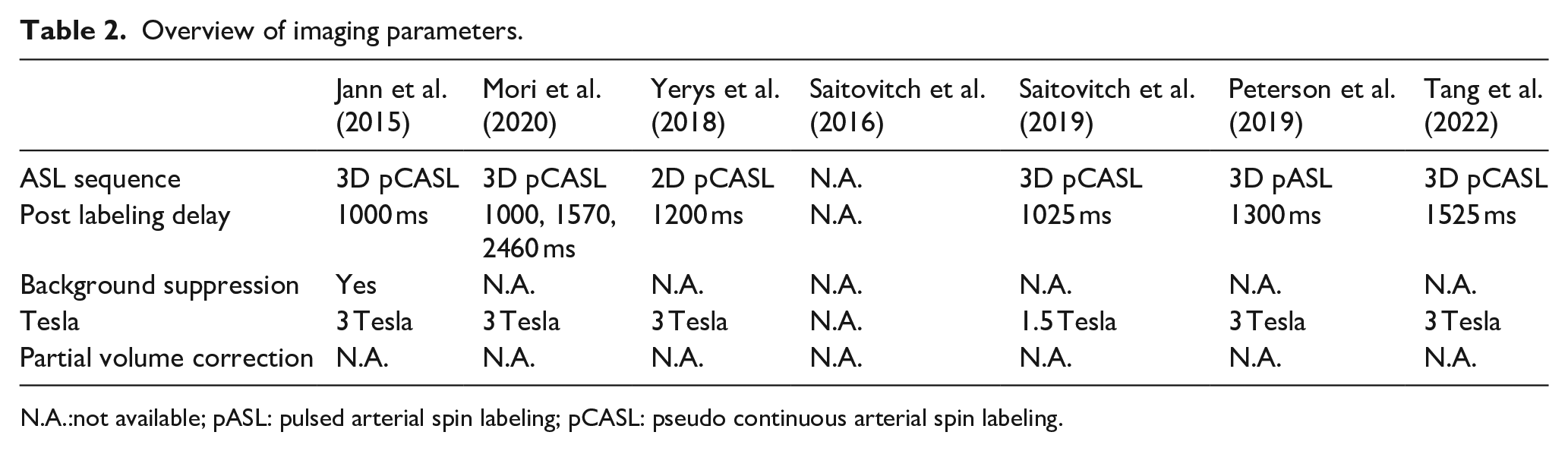
N.A.:not available; pASL: pulsed arterial spin labeling; pCASL: pseudo continuous arterial spin labeling.
Interconnection of ASL results with the remaining neuroimaging literature
The two studies performed by Saitovitch et al. (2016, 2019) demonstrated STS hypoperfusion in ASC resembling findings from molecular imaging (Ohnishi et al., 2000; Starkstein et al., 2000). The correlation between STS perfusion and ASC symptomatology is consistent with the entrenched role of STS in the social brain (Blakemore, 2008; Lahnakoski et al., 2012) and evidence linking social difficulty in ASC to aberrant STS activity during both emotion recognition tasks and resting-state (Alaerts et al., 2014). In addition, a novel fMRI study examining the neural responds to motherese, referring to an older person speaking to an infant, demonstrated that ASC children with lower degree of social abilities and eye-tracking attention to motherese displayed a lower neural response in the STS. Essentially, significantly weakened reaction to motherese in ASC was related to compromised development of temporal areas commonly associated with maternal affective speech (Xiao et al., 2022). The STS is interconnected with the fusiform gyrus (Lahnakoski et al., 2012), where Yerys et al. (2018) displayed hypoperfusion in ASC which correlated with reduced facial recognition ability, thereby supporting results from existing fMRI studies (Di Martino et al., 2009). In line with this, a recent study employing a face and car recognition fMRI paradigm, displayed an ASC age-independent decrease in FC between the fusiform gyrus, frontal and primary visual cortices during facial recognition, and underconnectivity with the OFC during both car and face recognition in ASC (Lynn et al., 2018). The OFC is another region associated with the social brain (Blakemore, 2008), in which Jann et al. (2015) found hypoperfusion in ASC and association with symptom levels. Atypical OFC neural activity is associated with ASC (S. A. Green et al., 2015) and OFC damage with instigation of ASC-related symptomatology (Sabbagh, 2004). The OFC is connected with several sensory association cortices as the ACC and amygdala, manifesting the OFC as an essential node for sensory, emotional processing, learning, and decision-making (Barbas & Zikopoulos, 2006). Importantly, this links ASC with OFC abnormality, due to common symptomatology including sensory and emotional processing disruption (American Psychiatric Association, 2013). A recent postmortem examination discovered a significant decrease in the density of OFC excitatory neurons in ASC, potentially causing diminished excitatory inputs and outputs followed by enhanced local inhibition in the OFC, thereby supporting the findings of hypoperfusion by Jann et al. (2015) and insinuating dysfunctional long-range communication in ASC (Liu et al., 2020). The OFC is closely interconnected with the ACC, where Jann et al. (2015) also evidenced hypoperfusion, which correlated with ASC symptoms, corresponding with earlier molecular imaging CBF findings (Ohnishi et al., 2000). In addition, the finding of enhanced local ACC FC in this study resembles earlier fMRI studies that correspondingly discovered positive correlations between FC and ASC symptom degree (Agam et al., 2010). The findings of enhanced FC within the ACC and diminished FC between the ACC and PCC (Jann et al., 2015) support the short-range hyperconnectivity and long-range hypoconnectivity hypothesis of ASC (Supekar et al., 2013). The ACC is also involved in social and emotional processing, with recent evidence linking ACC abnormalities to ASC core symptoms such as repetitive behavior and symptoms regarding social engagement (Zhang et al., 2020). Conversely, Peterson et al. (2019) presented hyperperfusion in social brain regions as the OFC and amygdala in autistic adults, potentially implying an age-related change in metabolism. Interestingly, Peterson et al. (2019) demonstrated hyperperfusion in the frontal WM in ASC, which correlated positively with social difficulty in ASC. Hyperperfusion overlapped with reduced N-acetylaspartate (NAA) metabolites, involved in axonal signaling, which indicates reduced density and function of axonal mitochondria (Rae, 2014). The overlap of reduced NAA and hyperperfusion in WM could suggest that glial cells incompletely or unproductively attempt to compensate reduced axonal metabolism in ASC. The results harmonize with a recent fluor-deoxy-glucose (FDG) PET study, demonstrating increased metabolic rate in WM of the prefrontal regions and internal capsule in ASC (Mitelman, Buchsbaum, et al., 2018). However, the results from Peterson et al. (2019) should be interpreted very carefully, due to the low SNR caused using pASL, not using background suppression, combined with the high influence of partial volume effects when investigating CBF in small regions as WM tracts and the WM volumetric differences associated with ASC (Prigge et al., 2021). In summary, the results from ASL studies agree with findings from other neuroimaging methodologies, where temporal regions appear closely linked to ASC symptomatology (Mori et al., 2020; Saitovitch et al., 2016, 2019). In addition, novel insights concerning metabolic WM alterations in ASC are intriguing but demand reliable inspection henceforth.
Future research questions in autism research
Cerebral blood flow in an age-related perspective
ASC is associated with an unconventional developmental trajectory. A longitudinal study in an autistic population displayed enlarged GM volume in early childhood, which normalized and resembled volumes found in NA by late childhood, before progressing toward increased ventricles indicating atrophy in early adulthood and reduced corpus callosum WM volume in adulthood (Prigge et al., 2021). Furthermore, fMRI studies have demonstrated enhanced FC with increasing age in ASC, contrary to the trajectory in NA (Ma et al., 2021; Padmanabhan et al., 2013), these results suggest an immaturity of neural networks and impaired pruning in ASC (Supekar et al., 2009). In a recent study by Sharma et al. (2018), the metabolic developmental trajectory in ASC was assessed via FDG PET. By dividing subjects into age-segments, <5 years, 5–10 years, and 10–15 years, a clear negative correlation between age and glucose metabolism was displayed. Interestingly, when comparing ASC with NA, the <5 years ASC group displayed hypermetabolism in a multitude of structures, including structures from all brain lobes, yet the hippocampus was the only structure demonstrating hypometabolism compared to NA. However, the age groups of 5–10-year and 10–15-year olds both demonstrated hypometabolism in all included GM structures compared to NA (Sharma et al., 2018). Essentially, ASC displayed a completely opposite metabolic trajectory compared to NA, as the ASC group diminished glucose metabolism while the NA group intensified glucose metabolism with increasing age during childhood and adolescence. In contrast to this, studies in autistic adults have displayed hypermetabolism (Mitelman, Bralet, et al., 2018; Rumsey, 1985) signifying a potential metabolic trajectory shift in ASC. This tendency was also demonstrated by Tang et al. (2022), where the participants were divided into eight age-dependent groups: 2, 3, 4, 5, 6–8, 8–10, 10–14, and 14–18-year-old. Comparisons between the 2-year-old ASC and NA group displayed hypoperfusion in the temporal lobe, hippocampus, and putamen in the ASC group. These results were consistent throughout all groups; however, the quantity of structures showing hypoperfusion in the ASC groups increased with increasing age (Tang et al., 2022). However, sedation was used especially in the young groups, which is believed to influence CBF quantification (Ogawa et al., 2015). Nonetheless, equivalent trajectories were demonstrated in other ASL studies, where ASC children elicited hypoperfusion (Mori et al., 2020; Saitovitch et al., 2019; Yerys et al., 2018), whereas the only study involving autistic adults displayed hyperperfusion (Peterson et al., 2019), thereby adding to the scarce and discrepant literature involving CBF in autistic adults, which displays both hyperperfusion (Pagani et al., 2012) and hypoperfusion (Mcdonald et al., 2020) compared to NA. However, additional studies are needed to verify the results on regional specificity considering the discrepancies found in different studies. Based on the findings regarding glucose metabolism, it appears that these regions are focused around the limbic, temporal, and deep nuclei (Mitelman, Bralet, et al., 2018; Rumsey, 1985). However, regional developmental variability is an important aspect that one should consider, as brain regions mature in a posterior to anterior trajectory, meaning posterior regions mature prior to anterior brain structures. In addition, the interpretation of results are enigmatic as both hyper- and hypo-perfusion can indicate abnormal neurodevelopment (Taki et al., 2011), signifying that results should be carefully interpreted. Taken all the previous findings together, additional studies exploring the longitudinal trajectory of CBF in ASC and additional studies involving adults would be of great importance for a better understanding of the neurobiology of ASC.
Cerebral blood flow in a sex-related perspective
The research involving ASC documented a marked male-sex-bias (Mo et al., 2021). This is also evident in this review, since merely half the included ASL articles encompassed female participants (Jann et al., 2015; Peterson et al., 2019; Saitovitch et al., 2019; Tang et al., 2022). Surprisingly, only one study investigated sex-related differences. Peterson et al. (2019) displayed that the tendency of enhanced CBF in the amygdala, ACC, and ventral striatum in NA females compared to NA males was reversed in ASC.In this study, ASC females displayed lower CBF than autistic men, the men also demonstrated enhanced CBF compared to NA females and males (Peterson et al., 2019). Essentially, autistic men contradicted the known phenomenon of higher CBF in females compared to males (Kaczkurkin et al., 2019), thereby suggesting this as a possible feature of ASC. To the best of our knowledge, similar investigations have not been carried out using molecular imaging. Nevertheless, since the prevalence of ASC is 4.2 times greater among male individuals (Maenner et al., 2021), the higher numbers of male participants included in the studies are somewhat understandable. Lately, increased attention has been directed toward the investigations of sex-related discrepancies. A recent study demonstrated enhanced FC between the substantia nigra and regions involved with somatosensory processing in young autistic males which correlated with ASC-related genetic probability (Hernandez et al., 2020). However, neither enhanced FC nor association with genetic probability manifested among autistic females. Subsequently, the investigators performed the same sex discrepancy examination in NA individuals, where solitarily NA males demonstrated cumulative ASC-related genetic probability to correlate positively with FC (Hernandez et al., 2020). The substantia nigra and somatosensory-related regions are associated with altered sensory processing and repetitive behavior, which are core ASC symptomatology (American Psychiatric Association, 2013). Interestingly, FC in these regions is positively associated with repetitive behavior and sensory processing symptoms, which has been reported to be significantly less prevalent in female autistic individuals (American Psychiatric Association, 2013; McFayden et al., 2020). These findings suggest that there may be a protective mechanism at play for females, or that these sex-related discrepancies are caused by the amplified quantity of genes linked to inflammatory and neuroimmune processes and diminished gene manifestation associated with neural and synaptic function in males (Voineagu et al., 2011; Werling et al., 2016). In contrast to this, female autistic individuals appear to be particularly affected by WM alterations, such as amplified diminishment of WM integrity (Lei et al., 2019; Nordahl et al., 2015). Nevertheless, the emerging emphasis on sex differences in ASC is necessitated as the enhanced camouflaging ability of ASC females and the associated mental health risks (Hull et al., 2019) obligate further exploration of ASC-related sex differences, including CBF examinations, where sex-related discrepancies are well documented in NA populations (Kaczkurkin et al., 2019). Therefore, CBF potentially constitutes as a biomarker for ASC, perhaps especially for autistic females, which would be tremendously advantageous as considerable difficulties are coupled with particularly the diagnosis of ASC in females (Hull et al., 2019).
Future possibilities of arterial spin labeling
Functional connectivity
ASL encloses diverse analytical possibilities. Besides CBF quantification, the typically fMRI-related measure of FC can be evaluated via ASL (Viviani et al., 2011), exemplified via its utilization by Jann et al. (2015). Comparisons of ASL and fMRI derived FC analysis show high similarity (Jann et al., 2016). Part of the analytical procedure of resting-state fMRI involves the detection of resting-state functional networks, commonly via independent components analysis (ICA). ASL displays the same detection ability (Dai et al., 2016; Jann et al., 2013), and furthermore, ASL has been displayed to derive fewer artifactual networks than BOLD fMRI (Dai et al., 2016). This is supposedly caused by lower vulnerability of ASL to motion-related artifacts because of background suppression (Ye et al., 2000) and less vulnerability to air–tissue susceptibility artifacts commonly demonstrated in the OFC and medial/temporal cortex (Dai et al., 2016). Moreover, ASL is less affected by low-frequency confounds such as scanner drifts and non-neural noise than BOLD fMRI (Dai et al., 2016). In addition, the other common resting-state analytical method referred to as seed-based FC is accessible using ASL (Jann et al., 2013), which oppositely to the network-specific valuation of ICA focuses on individual brain structures connectivity. Essentially, it appears that ASL can investigate FC in an equivalent manner to BOLD fMRI. Importantly, increasing attention on the optimal ASL preprocessing pipelines (Jann et al., 2016) helps the reliability and validity of future ASL FC studies. Condensed, previous work has established a foundation for FC assessment via ASL MRI, permitting acquisition of an ASL sequence to conduct the same investigations as an fMRI BOLD sequence, on top of quantification of CBF.
Arterial transit time
As previously mentioned, the PLD refers to the time epoch allowing labeled blood to flow from the labeling area to the brain, before the labeled image is acquired (Cohen et al., 2020). The manifestation of multiple PLD sequences enables investigation of the ATT. Conversely to PLD the ATT is the actual transport time from the large arteries to the specific brain area (Petersen et al., 2010), and therefore varies throughout the brain, where superiorly located regions exhibit lengthier ATT compared to inferior regions due to longer distances (Gonzalez-At et al., 2000). The discrepancy in ATT throughout the brain and between ATT and PLD is considered the most pronounced limitation of ASL, potentially generating both underestimation and overestimation of CBF (van Osch et al., 2018). Therefore, assessment and inclusion of ATT in kinetic modeling empowers improved CBF quantification (Cohen et al., 2020). Of the included ASL studies, only Mori et al. (2020) exploited multiple PLD ASL, yet without examining ATT comparisons between ASC and NA. Nonetheless, of the included ASL studies, the utilization of multiple PLD sequences would have improved the reliability of CBF quantification in the included ASL studies, especially in investigations including a diverse age-range like Tang et al. (2022) and Peterson et al. (2019), where single PLD sequences may generate falsely significant differences, as the ATT changes with age (Dai et al., 2016). Diminished ATT has previously been linked to inferior cognition (MacIntosh et al., 2015), thereby not just constituting ATT as a methodological and analytical optimization tool (Cohen et al., 2020) but additionally as an interesting physiological measure to investigate.
Pharmacological challenges
To this date, there is no approved pharmacological treatment targeting the core symptoms of ASC. However, medication is often utilized to treat symptoms such as irritability, aggression, and other challenging clinical presentations that accompany ASC (Posey, 2009). However, the neuropeptide oxytocin (OXT) is forthcoming as a potential treatment prospect (Green & Hollander, 2010), due to its fundamental regulative role in complex social cognition and behavior (Meyer-Lindenberg et al., 2011). A prior meta-analysis displayed a significant association between variations in oxytocin receptor (OXTR) gene expression and ASC (LoParo & Waldman, 2015), harmonizing with the results of OXTR knock-out animal studies exhibiting social difficulty (Takayanagi et al., 2005). In contrast, there are marked effects of OXT administration on human behavioral and neuroimaging studies (Baumgartner et al., 2008; Grace et al., 2018; Quintana et al., 2016; Riem et al., 2011). In line with the general ASC neuroimaging literature, fMRI studies have vastly investigated OXT administration. However, the subtractive methodology of ASL instigates cancelation of signal drifts making ASL preferable to the application of BOLD signal when assessing pharmacological longitudinal changes (Wang et al., 2011), due to ASLs high reproducibility and quantitative nature. Two studies have investigated intranasal OXT administrations effect on CBF in NA (Martins et al., 2020; Paloyelis et al., 2014), demonstrating a decrease in perfusion of the amygdala (Martins et al., 2020) and tendencies of decreased whole-brain CBF after OXT administration compared to placebo (Paloyelis et al., 2014). These CBF results are congruent with fMRI outcomes, as diminished FC is evident effects of OXT administration in NAs (Grace et al., 2018; Quintana et al., 2016; Riem et al., 2011). Yet, as OXT occasionally instigates contrarily directed activity alterations in NA compared to ASC (Domes et al., 2013), even demonstrating altering effects in autistic females and males (Hernandez et al., 2020), a comprehensive investigation of OXTs influence on CBF in autistic populations is warranted.
Neurotransmitter systems
Recent evidence verified fluctuations in CBF to reflect metabolic burdens from underlying neurotransmitter systems (Dukart et al., 2018). In a novel study, participants were exposed to drugs with established dopaminergic, serotonergic, glutamatergic, and GABAergic mechanisms or placebo. Interestingly, a consistent spatial relationship between CBF alterations and receptor density was demonstrated (Dukart et al., 2018). Moreover, associations concerning drug affinity of the respective receptors and CBF were established (Dukart et al., 2018). Successive studies confirmed these relationships (Bojesen et al., 2018; Selvaggi et al., 2019) ascertaining a direct linkage between CBF and pharmacodynamics, and thereby the potential of ASL MRI in translational medicine. Considering the variety of neurotransmitter-driven theories in ASC such as the excitatory/inhibitory imbalance hypothesis involving glutamate and GABA (Coghlan et al., 2012), the dopamine (Paval & Miclutia, 2021), and serotonin hypothesis (Harrington et al., 2013), ASL CBF constitutes as a prime candidate for non-invasive investigation of the beforementioned hypotheses.
Conclusion
The included articles demonstrate a clear tendency of hypoperfusion in autistic children (Mori et al., 2020; Saitovitch et al., 2019; Tang et al., 2022; Yerys et al., 2018), while a propensity of hyperperfusion was evident in the only study involving autistic adults (Peterson et al., 2019). These CBF discrepancies are age-dependent and regionally specific, primarily located in limbic, temporal, deep nuclei, and frontal areas. The results are comparable to those of molecular imaging (Bjørklund et al., 2018). However, the evidence is primarily from when molecular imaging was less sensitive and heavily influenced by analytical biases as partial volume effects, severely manipulating CBF quantification (Law et al., 2000). To enable reliable CBF quantification via ASL in the future, appropriate methodological considerations are necessitated, including the acquisition of multiple PLD sequences and the use of partial volume correction and background suppression. Furthermore, additional attention should be directed toward age and sex-related differences. Throughout the review, the advantages of ASL compared to BOLD fMRI have been discussed. Not to discredit or neglect BOLD fMRIs crucial importance for neuroimaging research, but to inspire researchers to utilize ASL, diversify ASC neuroimaging research and most importantly expand our knowledge regarding the autistic brain. ASL enables quantification of the physiological measurement of perfusion (Gonzalez-At et al., 2000), contrary to BOLD signals’ enigmatic relative units without explicit relationship to neurophysiology. The quantitative nature and development during the last decade emphasize the potential of ASL as an instrument for ASC biomarker identification, ASC subtype segmentation, and utilization in pharmacological challenges, and thereby hopefully contributing to improved quality of life and earlier diagnosis in the constantly growing autistic population.
Footnotes
Declaration of conflicting interests
The author(s) declared no potential conflicts of interest with respect to the research, authorship, and/or publication of this article.
Funding
The author(s) received no financial support for the research, authorship, and/or publication of this article.
