



Abstract
Background
White matter hyperintensities (WMH) are prominent neuroimaging markers of cerebral small vessel disease (CSVD) linked to cognitive decline. Nevertheless, the pathophysiological mechanisms underlying WMH remain unclear.
Objective
This study aimed to assess the structural decoupling index (SDI) as a novel metric for quantifying the brain's hierarchical organization associated with WMH in cognitively normal older adults
Methods
We analyzed data from 112 cognitively normal individuals with varying WMH burdens (43 high WMH burden and 69 low WMH burden). Neuroimaging data were used to calculate SDI, and gene enrichment analysis was conducted to explore related molecular pathways.
Results
An increased spatial gradient of SDI from the sensory-motor cortex to the associative cortex was observed. Compared to the low WMH burden group, the high WMH group exhibited elevated SDI in the right superior frontal gyrus, bilateral orbital gyrus, bilateral precentral gyrus, bilateral cingulate gyrus, bilateral thalamus, and bilateral striatum. In the high WMH burden group, SDI in the left thalamus and right cingulate gyrus negatively correlated with memory, while SDI in the right orbital gyrus and left precentral gyrus positively correlated with processing speed. Gene enrichment analysis highlighted associations with pathways involved in neural system function, potassium ion transmembrane transport, synaptic signaling, neuron projection development, and cell secretion regulation.
Conclusions
The findings suggest SDI alterations as a potential mechanistic pathway in WMH, which is associated with significant molecular pathways and cognitive impairments. This study provides a theoretical framework for understanding the pathophysiology of WMH progression and subsequent cognitive deficits.
Keywords
Introduction
White matter hyperintensities (WMH) are common in older adults and represent a salient radiological hallmark of cerebral small vessel disease (CSVD), discernible as regions exhibiting heightened signal intensity in T2-weighted or fluid-attenuated inversion recovery (FLAIR) images. 1 Previous investigations have illuminated a putative nexus between WMH formation and underlying pathophysiological cascades, including chronic hypoperfusion, 2 compromised cerebrovascular reactivity, 3 and disruptions in blood-brain barrier integrity. 4 These pathological conditions are evident across various cohorts, including individuals with Alzheimer's disease (AD), vascular cognitive impairment, and cognitively normal older adults. Convergent evidence5,6 suggests a strong association between WMH and cognitive decline. However, a significant proportion of individuals with WMH do not exhibit clinically detectable cognitive impairment. Furthermore, prior studies7,8 have indicated that in populations with cognitive impairment, neurodegenerative and vascular pathologies often coexist, mutually contributing to the progression of WMH and cognitive decline. Therefore, focusing on cognitively normal older adults with WMH may offer a more robust foundation for investigating the etiology of WMH and provide a novel perspective for early detection and mechanistic exploration of cognitive impairment related to WMH.
Convergent evidence suggests that both aging and cognitive impairment are associated with the dysfunction of the brain's hierarchical organization from unimodal to cross-modal regions through disconnection mechanisms.9,10 Hence, it is imperative to elucidate the alterations in hierarchical structure connectivity (SC) and functional connectivity (FC) for exploring the underlying mechanisms of WMH progression and consequent cognitive decline in the aging population. Specifically, the configuration of FC within the brain may be intricately shaped and constrained by underlying SC. 11 Meanwhile, FC patterns themselves may serve as reflections of the underlying structural architecture.12,13 Hence, the structure-function decoupling provides a promising perspective to depict the brain's hierarchical organization. To date, several studies investigating the relationship between brain structure and function mainly relied on direct correlation 14 or subtraction 15 methods, potentially overlooking the intricate nuances inherent in their relationship. In response to these challenges, Preti and De Ville have introduced a novel metric known as the structural decoupling index (SDI). 16 This metric offers a refined approach to quantify the degree of dependency between brain structure and function for each specific region. In essence, functional activity within various brain regions, as detected through functional magnetic resonance imaging (fMRI) signals, can be delineated within the graph domain, reflecting different connectome harmonics spanning from low to high frequency. By employing the SDI, researchers can systematically evaluate the degree to which functional activity aligns with or deviates from structural constraints.
Neuroimaging genetic methodologies are essential for linking brain connectomics with transcriptomics, 17 offering a robust framework for exploring the molecular mechanisms underlying various disorders, such as AD,18,19 and major depressive disorder. 20 Previous research 10 has demonstrated that the SDI can uncover relevant gene pathways while quantifying hierarchical brain structures. Therefore, conducting genetic analyses informed by SDI alteration patterns in a cohort of cognitively normal older adults with distinct WMH burden allows us to gain critical insights into the molecular pathways involved in the development of WMH.
The primary objective of this study is to elucidate the potential role of SDI alterations as a mechanistic pathway underlying WMH. To this end, we commenced by computing individual SDIs within a cohort comprising 112 cognitively normal older individuals exhibiting varying degrees of WMH burden, achieved through the decoupling of SC and FC. Subsequently, intergroup disparities in SDI were quantified, alongside an assessment of their association with cognitive performance and WMH volume. Finally, leveraging gene enrichment analysis, we endeavored to delineate the underlying molecular mechanisms governing changes in SDI observed in the context of WMH.
Methods
Subjects
The participants were all recruited from the Neurology Department's outpatient service of Tongji Hospital, Wuhan, China. The study received approval from the Ethics Committee of Tongji Hospital, Tongji Medical College, Huazhong University of Science and Technology. Each participant provided written informed consent before their inclusion in the study. This study encompassed a cohort of 112 individuals with normal cognitive function, characterized by comprehensive demographic and psychological profiles, alongside neuroimaging data comprising fMRI, diffusion tensor imaging (DTI), and 3D T1-weighted images. The study delineated stringent inclusion criteria for participant selection, stipulating: (1) an age range between 50 and 80 years, with right-handedness; (2) educational attainment exceeding 5 years; (3) varying degrees of WMH from mild to severe; and (4) the absence of cognitive impairment, as defined by a score of 0 across all six Clinical Dementia Rating (CDR) domains, a Mini-Mental State Examination (MMSE) score of ≥24 for those with primary education or ≥27 for those with junior school education or higher, and no objective evidence of impairment in any of the three cognitive domains. Conversely, exclusion criteria were meticulously delineated to ensure the integrity and homogeneity of the participant cohort, encompassing: (1) cerebral hemorrhage or infarcts exceeding a diameter of 15 mm; (2) incapacity to undergo comprehensive neuropsychological assessments or MRI scans; (3) a documented history of epilepsy or neurodegenerative conditions such as Parkinson's disease or AD; (4) exclusion of conditions mimicking WMH, such as multiple sclerosis or leukodystrophy; (5) severe large vessel diseases, exemplified by carotid artery stenosis exceeding 50%; and (6) systemic diseases including cancer or connective tissue disorders. These stringent criteria were employed to ensure the homogeneity of the study cohort and minimize confounding variables that could potentially obfuscate the interpretation of study findings.
Utilizing the WMH Fazekas scoring system as a criterion, 21 we stratified the included individuals into two distinct groups. Specifically, 43 individuals exhibited high WMH burden, as determined by periventricular WMH Fazekas scores of 3, or deep WMH Fazekas scores ranging from 2 to 3 on FLAIR sequence images,22,23 while the remaining 69 subjects were categorized as low WMH burden group.
Neuropsychological tests
Each participant underwent a comprehensive battery of neuropsychological assessments and clinical interviews, encompassing the following instruments: (1) CDR; (2) MMSE; (3) the Symbol Digit Modalities Test (SDMT); (4) the Trail Making Test (TMT); (5) the Digit Span Test (DST); and (6) the Auditory Verbal Learning Test (AVLT). The neuropsychological tests were subsequently aggregated into three distinct cognitive domains: processing speed (TMT-A and SDMT), executive function (DST backward and TMT-B), and memory (AVLT immediate recall, short delay recall, long delay recall, and long delay recognition). To facilitate comparisons across cognitive domains, the raw scores from neuropsychological tests were converted to z-scores using the means and standard deviations derived from the entire cohort of subjects in this study. Furthermore, we calculated the compound z-score of each domain by averaging z-scores for the corresponding tests to present the cognitive performance. 24
MRI acquisition
MRI scans were conducted utilizing a 3.0-Tesla MRI scanner (Discovery MR750, GE Healthcare, Milwaukee, WI, USA) equipped with a 32-channel head array coil. The parameters of MRI Sequences are presented in Supplemental Tables 1–3. Throughout the imaging session, participants were provided with explicit instructions to maintain ocular closure, minimize bodily movements, and sustain a state of relaxation.
MRI preprocessing
The fMRI data were preprocessed using SPM12 (Statistical Parametric Mapping, Institute of Neurology, London, UK). The initial 10 volumes were discarded, followed by slice timing correction to address acquisition delays. The remaining images were realigned to the first volume to correct for head motion, with subsequent analyses limited to participants exhibiting no more than 3° of angular rotation or 3 mm of translation. The images were then normalized to MNI space and resampled to 3 × 3 × 3 mm voxels. Linear regression was performed to account for nuisance variables, including motion parameters and signals from white matter and cerebrospinal fluid. Finally, a temporal band-pass filter (0.01–0.08 Hz) was applied to remove low-frequency drifts and high-frequency noise.
The DTI data preprocessing was performed using MRtrix3 (http://www.mrtrix.org/) and involved several key steps. First, DTI images were converted to MRtrix format. For each scan, diffusion images were denoised (dwidenoise in MRtrix3), Gibbs ringing correction (mrdegibbs in MRtrix3), head motion correction, eddy current and susceptibility-induced distortion correction (dwibiascorrect and dwifslpreproc in MRtrix3), followed by bias field correction to address intensity non-uniformities (dwibiascorrect in MRtrix3). A brain mask was generated to isolate brain tissue. Response functions for white matter, gray matter, and cerebrospinal fluid were estimated using the Dhollander method, enabling multi-shell, multi-tissue constrained spherical deconvolution (MSMT-CSD) for fiber orientation distribution (FOD) estimation. T1-weighted anatomical images were coregistered to the diffusion data. For fiber tracking, the iFOD2 algorithm was employed with parameters including a cutoff threshold of 0.06, an angular cutoff of 45 degrees, and a maximum streamline length of 250 mm. This approach generated a comprehensive set of streamlines, which were subsequently sifted to retain a specified number of tracks for connectivity analysis, culminating in the creation of a brain structure network connection matrix (fiber number network, FN, sized N × N).
Subsequently, Pearson's correlation coefficients were computed, establishing associations between the mean time series of distinct brain regions delineated within the Brainnetome Atlas. 25 This facilitated the derivation of FC matrices for each subject. The SC network was delineated based on fiber connections between regions within the Brainnetome Atlas, as defined by the FN metric. The data analysis pipeline is presented in Figure 1.

Data analysis pipeline. (A) Structural decoupling index (SDI) construction. (B) Analysis of the SDI group differences between the WMH low burden and high burden groups. (C) An examination of the correlation between cognitive function and SDI. (D) Gene enrichment analysis. fMRI: functional magnetic resonance imaging; DTI: diffusion tensor imaging; PLS: partial least squares; WMH, white matter hyperintensities.
WMH volumes were quantified using FLAIR images through the application of the lesion prediction algorithm (LPA), integrated within the LST toolbox version 3.0.0 (accessible at http://www.statistical-modelling.de/lst.html), designed for use with SPM. The LPA employs a binary classifier trained on manually segmented data to automatically detect and segment hyperintensities in FLAIR images. Prior to segmentation, the FLAIR images were co-registered to the corresponding T1-weighted images, and the resulting lesion maps were normalized to MNI space. The total WMH volume for each participant was calculated by summing the segmented lesion voxels, and volumes were adjusted for intracranial volume to account for individual brain size differences. The segmented WMH template has been carefully reviewed and revised by two experienced neuroimaging experts. Furthermore, we performed a log transformation of WMH volume prior to conducting the subsequent statistical analyses.
Function-structure coupling index
Building upon prior investigations, 16 we implemented a graph signal processing framework to elucidate the relationship between FC and SC. Initially, we utilized eigen decomposition of the Laplacian matrix derived from the structural connectome to project individual functional data onto corresponding structural harmonics (FN). Subsequently, employing graph signal filtering, we partitioned the active signal into two components: one representing coupling and the other decoupling signals with the underlying structural architecture. The SDI was then introduced as a metric quantifying the ratio between the norms of coupling and decoupling signals across temporal data points. Through this methodological approach, individual SDIs were computed for each FN matrix. Brain regions exhibiting an SDI > 1 denote reduced coupling between SC and FC, indicative of activity signals diverging from underlying structural pathways. Conversely, regions with an SDI < 1 demonstrate the opposite trend, signifying a closer alignment between functional activity and structural connectivity.
SDI group differences and correlation with cognition
To quantify the aberrant characteristics of the structural-functional connectome in individuals with varying degrees of WMH, we computed the disparity in individual SDIs between low WMH burden and high WMH burden cohorts using a two-sided t-test, with significance thresholds set at p < 0.05 after False Discovery Rate (FDR) correction. Additionally, to further elucidate the clinical implications of structural and functional connectome alterations, we conducted an exploratory correlation analysis to investigate the relationship between WMH, SDI, and cognitive performance within both low WMH burden and high WMH burden groups, with statistical significance similarly defined at p < 0.05 (uncorrected). To mitigate the confounding effects of demographic variables such as age, sex, education, and mean framewise displacement (mFD), we employed linear regression techniques in the above statistical analysis.
Sensitivity analysis
Considering that hypertension 26 and diabetes 27 are well-known risk factors for cognitive decline and WMH, we added them as covariates in the analysis of SDI differences between groups, in addition to the four previously mentioned covariates. Furthermore, we correlated the T-map plots from the sensitivity analyses with the original results to assess the robustness of our findings.
Gene enrichment analysis
The gene enrichment analysis was conducted utilizing the group difference map derived from the SDI. Initially, gene expression data was acquired from the Allen Human Brain Atlas (http://human.brain-map.org/) and spatially mapped onto the Brainnetome Atlas utilizing the Abagen toolbox (https://abagen.readthedocs.io/en/stable/), resulting in a gene expression matrix of dimensions 242 × 15633. Subsequently, the extensive pool of 15,633 genes was prioritized based on their respective weight values through partial least squares (PLS) analysis. This process facilitated the identification of genes most strongly associated with the observed group differences in SDI. Specifically, the top 500 genes were selected based on the sorting of gene weights by absolute value, as determined through PLS analysis. Finally, gene enrichment analysis was performed on this subset of genes utilizing the Metascape platform (https://metascape.org/gp/index.html#/main/step1).
Results
Participant characteristics
A summary of the demographic, cognitive, and neuroimaging characteristics is given in Table 1. Briefly, there was a significant age gap (p = 0.006) between the two groups. The high WMH burden group had higher WMH volumes (p < 0.001) and Fazekas scores (p < 0.001) than the low WMH burden group. Despite both groups being cognitively normal, the high WMH burden group still showed some decline in execution function (p = 0.004) compared to the low burden group, while there were no significant differences in other cognitive domains.
Demographic and clinical features of subjects.
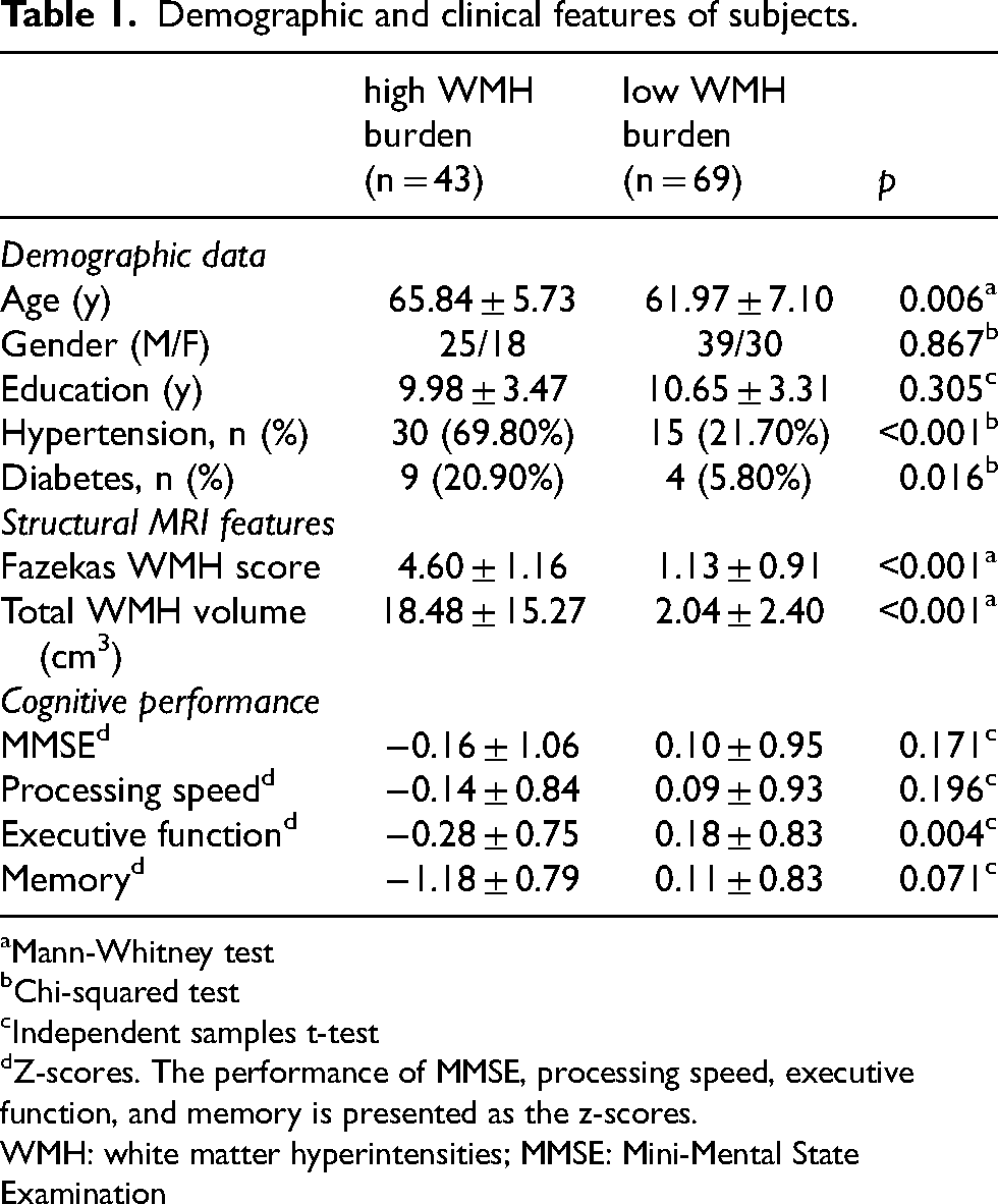
Mann-Whitney test
Chi-squared test
Independent samples t-test
Z-scores. The performance of MMSE, processing speed, executive function, and memory is presented as the z-scores.
WMH: white matter hyperintensities; MMSE: Mini-Mental State Examination
SDI alteration patterns in WMH high burden and low burden patients
As depicted in Figure 2A, the distribution patterns of SDI in both groups exhibit similarities: SDI values were higher in the associative cortex (especially the thalamus, parahippocampal gyrus, and amygdala), and lower in the sensor and motor cortices, particularly in the postcentral gyrus, revealing a spatial gradient of SDI extending from higher-order functional areas to sensorimotor regions. Regionally, there were notable differences in SDI between the two groups that were widely dispersed among multiple brain areas. When compared to the low WMH burden group, patients with high WMH burden had a distinctly elevated pattern, primarily including the right superior frontal gyrus (SFG), bilateral orbital gyrus, bilateral precentral gyrus, bilateral cingulate gyrus, bilateral thalamus, and bilateral striatum (p < 0.05, FDR-corrected, Figure 2B and Table 2).

Structural decoupling index (SDI) distribution and difference maps. (A) The low WMH burden and high WMH burden groups’ respective SDI pattern and tables detailing the top five highest and lowest SDI scores for each group. (B) Significant statistical distinctions between patients with low WMH burden and high WMH burden in terms of SDI. In individuals with high WMH burden compared to low WMH burden, the warmer and colder hues represent greater and lower SDI measurements, respectively. Amyg: amygdala; Cun: cuneus; Pcun: precuneus; PhG: parahippocampal gyrus; PoG: postcentral gyrus; ROI: region of interest; SDI: structural decoupling index; SPL: superior parietal lobule; Tha: thalamus; WMH, white matter hyperintensities. Detailed information of the brain regions is shown at http://atlas.brainnetome.org/.
All regions showed higher SDI in patients with high WMH burden.

Detailed information of the brain regions is shown at http://atlas.brainnetome.org/.
SDI: structural decoupling index; WMH: white matter hyperintensities
Sensitivity analysis
The T-map plots from the sensitivity analyses for group differences exhibited a strong correlation with the original results (r = 0.93, p < 0.001, Supplemental Figure 1).
Relationships between WMH, SDI, and cognitive performance
Within the low WMH burden group, a notable positive correlation was observed between the SDI in the bilateral cingulate gyrus and WMH volume (p < 0.05, uncorrected). However, no significant correlations were identified between SDIs within brain regions and measures of processing speed, executive function, or memory. Conversely, within the high WMH burden group, significant positive correlations between SDIs and WMH volume were detected in the right SFG, bilateral orbital gyrus, and bilateral cingulate gyrus (p < 0.05, uncorrected). Additionally, SDIs in the left thalamus and right cingulate gyrus exhibited significant negative correlations with memory, while SDIs in the right orbital gyrus and left precentral gyrus displayed significant positive correlations with processing speed (p < 0.05, uncorrected). No significant correlations were observed between WMH volume and cognitive performance in either group. Nevertheless, none of the correlation analyses remained statistically significant after applying multiple comparison corrections (Details are shown in Table 3).
Relationships between WMH, SDI, and cognitive performance.

WMH, white matter hyperintensities; SDI: structural decoupling index; EF: executive function; PSL: processing speed; CG: cingulate gyrus; SFG: superior frontal gyrus; OrG: orbital gyrus; Tha: thalamus; PrG: precentral gyrus. WMH volume has been adjusted for intracranial volume and log-transformed. Detailed information of the brain regions is shown at http://atlas.brainnetome.org/.
Gene enrichment pathways linked to changes in SDI
The results of this investigation showed that the PLS1 corresponded with the T-map of SDI in high WMH burden versus low burden (r = −0.50, p < 0.001) and explained 25.07 percent of the variability in gene expression. Furthermore, our gene enrichment analysis demonstrated the significant enrichment of multiple Gene Ontology (GO) biological processes, such as potassium ion transmembrane transport (GO:0071805, p = 2.63e-10, FDR-corrected), synaptic signaling (GO:0099536, p = 2.63e-10, FDR-corrected), neuron projection development (GO:0031175, p = 2.57e-08, FDR-corrected), and regulation of secretion by cell (GO:1903530, p = 3.63e-06, FDR-corrected). Moreover, a Reactome pathway study revealed a substantial association between SDI and the neural system (R-HSA-112316, p = 4.47e-12, FDR-corrected) (Figure 3 and Table 4). Comparable findings were also obtained from the top 1000 gene enrichment analysis (Supplemental Figure 2).

Gene enrichment analysis. (A) The outcomes of the Reactome pathway and Gene Ontology (GO) term. The amount of input genes determines the node's size. (B) The relationship between the structural decoupling index (SDI) T-map in high white matter hyperintensities (WMH) burden versus low WMH burden and the partial least squares (PLS1) score.
The detailed information of the top 20 results for pathway and process enrichment analysis.
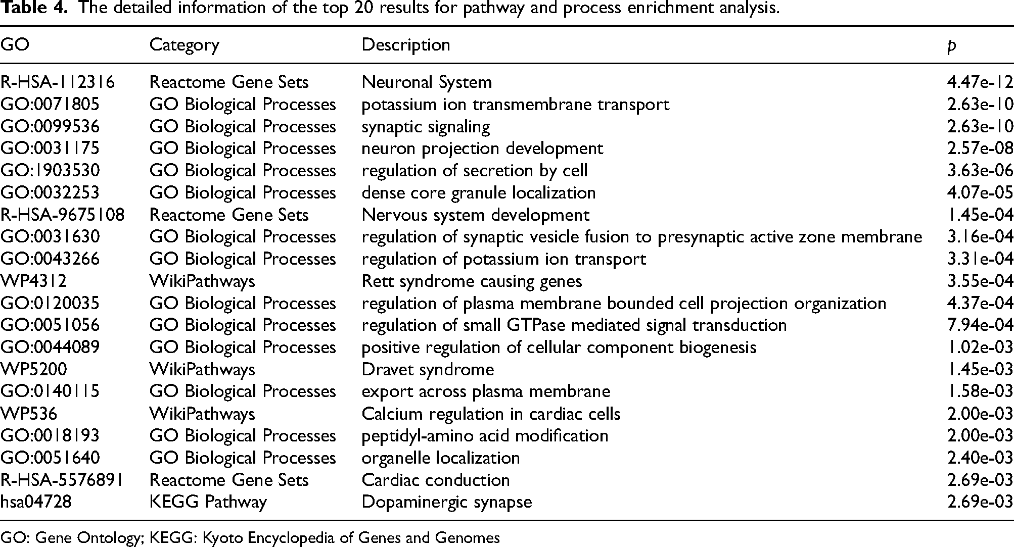
GO: Gene Ontology; KEGG: Kyoto Encyclopedia of Genes and Genomes
Discussion
This study systematically investigated the SDI alterations in cognitively normal older individuals with distinct WMH loads (FDR-corrected) and demonstrated that the SDI can serve as a novel metric for characterizing cerebral hierarchical structural-functional alterations associated with WMH. Specifically, SDI in the bilateral cingulate gyrus exhibited positive correlations with WMH volume in subjects with both high and low WMH burden (uncorrected). Moreover, within the high WMH burden group, SDI in the right cingulate gyrus showed a negative correlation with memory function, and SDI in the right orbital gyrus displayed a positive correlation with processing speed (uncorrected), whereas no SDI-cognition associations were observed in the low WMH burden group. Through gene enrichment analysis, the molecular underpinnings of SDI were elucidated. These findings indicated that variations in gene expression linked to WMH and cognitive abilities may be manifested in SDI, offering a theoretical foundation for understanding the hierarchical organization changes of the brain underlying WMH.
Previous research has indicated that the SDI may be an effective tool for characterizing the structure-function relationship, underscoring the brain's hierarchical organization. This organization extends from robustly coherent “unimodal” sensory cortices to loosely affiliated “transmodal” association cortices.9,10,28–30 In this study, the SDI distribution patterns within both high and low WMH burden groups exhibited a similar structural constraint on function across brain regions, consistent with the hierarchical structure of the brain. 10 Specifically, associative cortices (higher SDI) engage in polysynaptic indirect connections in addition to direct interactions via white matter fibers. 31 In contrast, direct white matter fibers predominantly mediate interactions between sensory-motor cortices (lower SDI). 32 Consequently, a higher SDI is the result of the concurrent functional activation of areas that are not physically related. Thus, our study reaffirms that the SDI is a valuable neuroimaging index capable of reflecting the hierarchical structure of the brain.
In our study, the high WMH burden group exhibited significantly higher SDI values in the right SFG, bilateral orbital gyrus, bilateral precentral gyrus, bilateral cingulate gyrus, bilateral thalamus, and bilateral striatum compared to the low burden group. The sensitivity analyses conducted thereafter yielded consistent result patterns, indicating that hypertension and diabetes may be unlikely to offer a plausible explanation for the SDI differences between subjects with high and low WMH burden. Notably, within the high WMH burden group, the SDIs of the left thalamus and right cingulate gyrus showed significant negative correlations with memory function. The thalamus, serving as the brain's relay center, is crucial for information transmission and integration, 33 while the cingulate gyrus is pivotal for emotional processing, cognitive control, and memory. 34 The elevated SDI in these key regions of cognitive networks suggests that disruption of direct structural connectivity in WMH may lead to hierarchical re-organization of the brain and the potential decline of higher cognitive functions, especially memory. The orbital gyrus, essential for emotion regulation 35 and decision-making, 36 and the precentral gyrus, a primary motor cortex region, 37 also showed significant SDI alterations. Interestingly, SDI in the right orbital gyrus and left precentral gyrus exhibited a positive correlation with processing speed. This conflict could be attributed to a compensatory mechanism: in high WMH burden populations, due to the destruction of direct fiber connections, elevated SDI in these regions corresponds to more indirect synaptic connections and increased neuronal information exchange, thereby facilitating faster processing speed. As neurofibrillary disruption advances, the loss of compensatory indirect synaptic connections could subsequently decelerate information processing speed. It is important to note that participants in this study were cognitively normal older adults, indicating that observed cognitive declines are indicative of a pre-clinical stage. Our hypothesis posits that changes in SDI may quantify the characteristic alterations of the brain's hierarchical organization and play a role in the pre-clinical phase of cognitive decline associated with WMH, offering potential insights for early detection and mechanistic investigations into WMH-related cognitive impairment.
Our investigation into the genetic determinants of SDI alterations revealed significant gene pathways including neural system, potassium ion transmembrane transport, synaptic signaling, neuron projection development, and regulation of secretion by cell. The transmembrane transport of potassium ions is crucial for maintaining cell membrane potential, neural signaling, regulation of osmotic pressure, cardiac function, and participation in metabolism and cell growth.38–40 Previous research has shown that potassium channels play a significant role in the pathophysiological processes of various neurodegenerative diseases. 41 For instance, the potassium channel Kv1.3 is highly expressed in the microglia of AD patients, 42 and the expression of Tandem pore domain halothane-inhibited K + channel 1 induces activation of glial cells in AD and Parkinson's disease. 43 Additionally, disruptions in potassium channel Kir4.1 and the calcium-dependent potassium channel MaxiK in the endfeet of astrocytes are closely associated with cognitive deficits following cerebrovascular disease. 44 Our study identified a significant association between potassium ion transmembrane transport and WMH-associated structural changes in brain function. Given that WMH is a specific marker of CSVD, these findings reinforce the critical role of potassium ion transmembrane transport in the pathophysiological processes of cerebrovascular disease. Polysynaptic indirect connections represent neural pathways involving intermediate neurons or circuits and multiple synapses for information transmission.45–47 Disruption of direct fiber connections in individuals with high WMH burden leads to slowed information processing and transmission, necessitating compensation through indirect synaptic connections, consequently increasing SDI values. The plausible causal relationship between SDI changes and alterations in synaptic connections was substantiated by the enrichment of gene pathways, including synaptic signaling and neuron projection development. Identification of these critical pathways is paramount for understanding and managing WMH.
Several limitations of this study should be acknowledged. First, to differentiate between sporadic and hereditary WMH, future research should explore whether the coupling of brain connectivity across various subgroups forms a continuum. Second, given that all study subjects were cognitively normal and the sample size was relatively small, the results of the exploratory correlation analysis were not corrected. Future research with larger sample sizes and appropriate correction methods is necessary to further validate these preliminary findings and establish more definitive conclusions. Thirdly, although we specifically selected a pre-clinical population to better explore the underlying mechanisms of SDI in relation to WMH burden, the potential influence of undetected AD pathology cannot be entirely excluded due to the absence of the amyloid beta biomarker testing. Future studies incorporating the amyloid beta testing will be necessary to provide a more comprehensive understanding of the observed relationships. Fourthly, Given the lack of field map files in the original fMRI dataset and no state-of-the-art method could be widely applied for precise distortion correction in the fMRI dataset without field map, the distortion correction was not applied in the preprocessing for fMRI data, which may have introduced susceptibility-induced field inhomogeneities and impacted data accuracy. To further verify the robustness of the fMRI main results, we experimentally employed a new tool called SynBOLD-DisCo, 48 which utilizes the topup-like algorithm to reduce susceptibility-induced off-resonance fields without the need for a field map. Specifically, we calculated the FC across brain regions using both the original data and the topup-corrected data, yielding a mean correlation coefficient of r = 0.64, with correlation p-values in all regions demonstrating significant results (all p-values < 1 × 10⁻¹⁰). Although SynBOLD-DisCo has yet to undergo extensive validation and may not be entirely reliable for fMRI, 48 the exploratory verification indicates that susceptibility-induced inhomogeneities likely exert a minimal influence on our results, thereby reinforcing the validity of our findings. Finally, to deepen our understanding of the molecular mechanisms underlying SDI, additional gene expression studies specifically focusing on WMH are warranted.
The present study identified that alterations in the SDI across hierarchical brain regions may serve as a potential pathophysiological mechanism underlying WMH and subsequent cognitive decline. Furthermore, the gene enrichment pathways associated with these alterations were also elucidated. These findings may provide a robust framework for understanding the pathophysiological mechanisms involved in WMH progression and the early prediction of associated cognitive deficits.
Supplemental Material
sj-pdf-1-alz-10.1177_13872877241309098 - Supplemental material for Structure-function coupling alterations in cognitively normal individuals with white matter hyperintensities
Supplemental material, sj-pdf-1-alz-10.1177_13872877241309098 for Structure-function coupling alterations in cognitively normal individuals with white matter hyperintensities by Junyong Du, Shabei Xu and Wenhao Zhu in Journal of Alzheimer's Disease
Footnotes
Acknowledgments
The authors have no acknowledgments to report.

Author contributions
Junyong Du (Conceptualization; Data curation; Formal analysis; Investigation; Methodology; Resources; Software; Validation; Visualization; Writing – original draft); Shabei Xu (Data curation; Supervision; Writing – review & editing); Wenhao Zhu (Funding acquisition; Investigation; Methodology; Project administration; Supervision; Visualization; Writing – review & editing).
Funding
This work was supported by the Natural Science Foundation of Hubei Province (No. 2021CFB382), and the Medical Advanced Research Fund of Bethune Charitable Fundation (No. 2023-YJ-152-J-004 and 2023-YJ-152-J-022).
Declaration of conflicting interests
The authors declared no potential conflicts of interest with respect to the research, authorship, and/or publication of this article.
Data availability
The data supporting the findings of this study are available on request from the corresponding author.
Supplemental material
Supplemental material for this article is available online.
References
Supplementary Material
Please find the following supplemental material available below.
For Open Access articles published under a Creative Commons License, all supplemental material carries the same license as the article it is associated with.
For non-Open Access articles published, all supplemental material carries a non-exclusive license, and permission requests for re-use of supplemental material or any part of supplemental material shall be sent directly to the copyright owner as specified in the copyright notice associated with the article.
