



Abstract
Background
Cortico-basal degeneration (CBD) is a neurodegenerative disease typically responsible for cortico-basal syndrome (CBS) or progressive limb apraxia. Half of CBD patients, however, present atypical symptoms, making the diagnosis difficult.
Objective
We reported the case of a woman in her late sixties (BM208), an unusual case of autopsy-proven CBD, showing early signs of Benson's syndrome or posterior cortical atrophy. In addition, we compared cognitive performance and atrophy in different brain regions of BM208 with other neurodegenerative diseases patients to highlight clinical signs that could have guided the diagnosis earlier.
Methods
We retrospectively compared BM208 to patients with typical amnestic Alzheimer's disease (AD) (n = 18, Mini-Mental State Exam (MMSE) scores between 18 and 24), Benson's syndrome due to AD (n = 3), CBS/progressive supranuclear palsy (PSP) syndrome (n = 5), and Lewy body dementia (LBD) patients (n = 3) and a control group (n = 24). All these participants underwent an MMSE, a complete neuropsychological examination and 3DT1 MRI.
Results
Although BM208 was more severely cognitively impaired overall, her cognitive performance was more similar to Benson's syndrome patients’ cognitive profile compared to CBS patients or any other degenerative pathology (typical AD/LBD). Consistently, although BM208 was more atrophic than all other groups, she showed cortical atrophy that matched a Benson's syndrome pattern more than typical AD or CBS. However, the analysis of subcortical atrophy revealed atrophy of the basal ganglia corresponding CBS cases. Furthermore, visual analyses on sagittal T1 images showed atrophy of the midbrain, characteristic of CBS/PSP syndrome.
Conclusions
These results highlight the additive value of fine-grained MRI subcortical quantification to diagnose non-AD rare neurodegenerative disorders.
Keywords
Introduction
Cortico-basal degeneration (CBD) is a neurodegenerative disease characterized by the accumulation of hyperphosphorylated tau (pTau) protein in cortical and subcortical neurons and gray matter astrocytes,1,2 therefore making it a primary tauopathy, more specifically a 4R-tauopathy. 3 Cortico-basal syndrome (CBS) is the typical clinical manifestation of CBD.4,5 It is characterized by lateralized motor (dystonia, myoclonus, akinetic-rigid syndrome, postural instability) and cognitive dysfunction (apraxia, aphasia, or alien limb).6–8 However, CBS is observed in only half of the patients with underlying CBD. 9 In 50% of the cases, CBD is manifested by atypical symptoms that initially suggest other neurodegenerative diseases such as progressive supranuclear palsy (PSP),10,11 primary progressive aphasia,12–14 frontotemporal lobe degeneration,8,15 or, more rarely, Alzheimer's disease (AD).8,16,17 The variety of symptoms that CBD can produce can make the in vivo diagnosis challenging.
On the other hand, posterior cortical atrophy (PCA) is a rare degenerative syndrome, also called Benson's syndrome, characterized by progressive decline in visuospatial, visuoperceptual, and praxis abilities. 18 Most of the time, PCA is due to AD pathology and is called the ‘visual variant’ of AD.19–21 It is caused by the accumulation of amyloid and neurofibrillary tangles (NFTs) in the occipital and parietal lobes containing primary and associative visual cortices. 19 Although AD frequently causes PCA, there are rare cases due to other neurodegenerative diseases as Lewy body dementia (LBD), Parkinson's disease, Creutzfeldt-Jakob disease, and also CBD.22–24
In this clinicopathologic report, we describe the case of a woman in her late sixties (included in our study as BM208), an unusual case of autopsy-proven CBD, showing Benson's syndrome as her first symptoms. We compared neuropsychological and imaging data of BM208 with patients suffering from various diagnostic including typical amnestic AD, Benson's syndrome due to AD, CBS, PSP syndrome, and LBD as well as to a control group to highlight signs that could have suggested in vivo a diagnosis of CBD.
Case report
Initial visit
BM208 was a woman in her late sixties, right-handed and retired from teaching. She was presented to our neurology department one year after symptoms’ onset. She complained about visual deficits (e.g., could not see objects in front of her) and balance issues. She was no longer able to count and wanted to say swear words although she was able to refrain. She had a history of hypertension and type II diabetes, which were treated and stabilized. She had an unremarkable neurological history, but her mother died from amyotrophic lateral sclerosis at 70 years old.
The neurological evaluation was limited as the patient had difficulties performing simple instructions. She had no motor deficits or ataxia. Her cranial nerves were normal, as well as her spontaneous oculomotricity. At the first clinical evaluation, she had no extrapyramidal signs, myoclonus or asymmetry. The neurologist, however, reported ideational apraxia (e.g., she did not know how to enter her bank code or how to open or close a door or window) as well as ideomotor apraxia (e.g., she was no longer able to do her hair), and a grasping reflex, suggesting a frontal syndrome. At that time, she scored 21/30 on the Mini-Mental State Examination (MMSE): she lost one point in temporal orientation, five in calculation, one in memory, one in language and one for praxis.
Biological results
A blood analysis demonstrated that the patient's complete blood count, glucose and cholesterol levels and hepatic, thyroid, and renal functions were normal.
A lumbar puncture revealed normal amyloid-β (Aβ42) protein (565 pg/ml; norm of laboratory (NL) > 469), normal pTau (54 pg/ml; NL < 61) and slightly increased total tau protein levels (482 pg/ml; NL < 274). Aβ40 and therefore Aβ42/40 ratio were not quantified.
Neuropsychological evaluation
A neuropsychological examination was performed two months later. At that time, the MMSE had already decreased to 16/30 (6/10 in orientation, 2/3 in memory and 2/3 in recall, 0/5 in calculation, 6/8 in language and 0/1 in praxis) (Table 1). The patient was nosognosic.
Raw and standardized scores of the neuropsychological assessment of BM208.

The standards presented are those established and used in clinics. No norms are available for MMSE and Clock test.
The patient, who was particularly anxious on the day of the evaluation, was accompanied by a friend. During the anamnesis, she had difficulties answering factual questions (age, education, professional background). In terms of episodic memory, she confused events and asked repetitive questions. Regarding language, her friend described an occasional anomia and difficulties understanding when several sentences followed one another. She had various praxic and neurovisual complaints including difficulty brushing her hair and occasionally wearing her clothes inside out. She complained about difficulties in reading or writing and signed only with her initials. Additionally, she struggled with neurovisual tasks such as differentiating between her shoes, locating personal belongings, telling the time, plugging in an outlet, or finding her way around her. She was also unable to type her bank code, although she could say it from memory; she could no longer cook or open or close a door. It was difficult to determine whether these latter complaints stemmed from praxis or neurovisual difficulties. Finally, the patient's friend explained that the troubles evolved quickly, and that the patient was much more anxious than before and suffered from panic attacks.
The evaluation revealed impaired spontaneous information retrieval, deficit in lexical evocation under semantic and phonological induction, as well as an alteration of oral naming capacities and visuo-constructive apraxia. BM208 was only able to mark the number ‘12’ on the circle during the clock test (on command) 25 and explained that she could not read the time on her watch. During the CERAD visuospatial test, 26 the patient was only able to copy the circle (Figure 1). She only drew a line for the diamond and explained that she could not copy the tangled rectangles. She was then offered to draw on the line that she could see but she could not do it over all the segments. Spontaneous verbal expression during the examination was fluent, devoid of paraphasia or dysarthria but was characterized by a slight anomia. Executive functioning could not be assessed: BM208 was unable to complete the Trail Making Test 27 because she could not see the numbers in part A. Based on the patient's complaints, we could also suspect the presence of dressing apraxia. The details of BM208 scores in this neuropsychological examination can be found in Table 1.
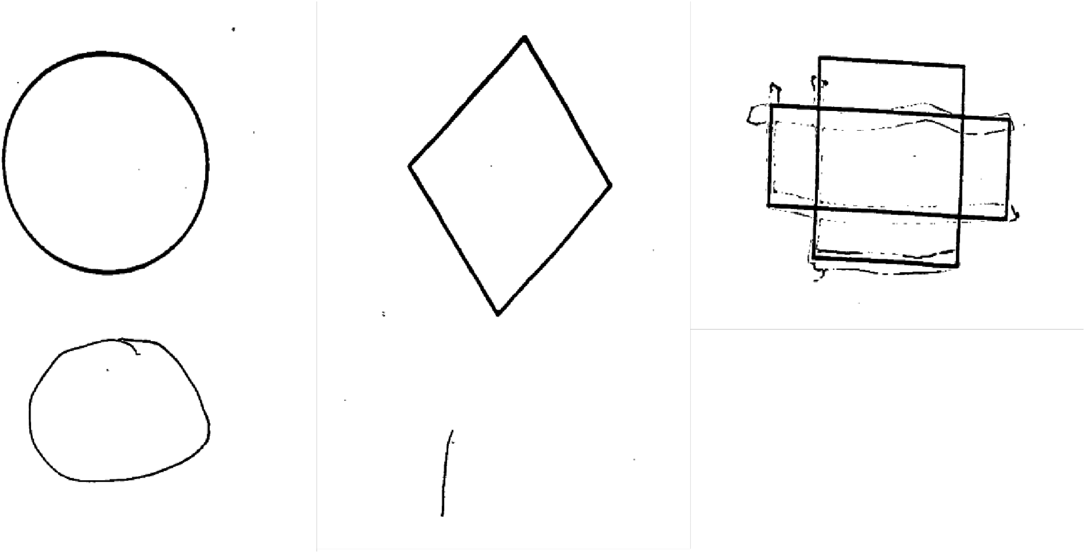
CERAD figure copy task. BM208 performance in the CERAD figure copy task.
Combined with the neurological examination demonstrating both ideational and ideomotor apraxia, the neuropsychologist raised the hypothesis of Benson's syndrome due to AD which was possible as detailed by Dubois, Vilain and colleagues but a second amyloid measure was necessary to confirm or infirm the diagnosis. 28
Imaging
Magnetic resonance imaging (MRI)
A first clinical brain MRI was done outside our hospital at the symptoms’ onset and showed leukoaraiosis and no hippocampal atrophy. However, the specific scores and detailed data from this scan were not available. A second scan was performed in our hospital one year later and revealed supraphysiological atrophy with a Scheltens scale of 2 (Figure 2) 29 and a Fazekas score of 2. 30 Perivascular spaces were also enlarged and atrophy of the body of corpus callosum was observed (Figure 2).

Evolution of MRI scans.
Amyloid-positron emission tomography (PET)
As part of her inclusion in a research study, the patient underwent a [18F]-Flutametamol-PET (VizamylTM, GE Healthcare), which is commonly used to quantify amyloid-β protein deposition in the brain and was necessary in confirm or infirm the AD diagnosis. 28 This examination was negative (Centiloid = -19), thereby confirming the absence of amyloid pathology and formally excluding the hypothesis of atypical AD.28,31 At that time, neuropsychological assessment and imaging examination raised the hypothesis of Benson's syndrome due to a non-AD tauopathy. The neurologist prescribed a fluorodeoxyglucose (FDG)-PET scan to determine whether any metabolic asymmetry could be demonstrated. However, the patient refused to undergo that scan.
Follow-up
One year after her first visit (two years after symptoms’ onset), the MMSE was rated at 11/30 and revealed temporal orientation and episodic memory decline. A release of archaic reflexes with echolalia was observed as well as attitude tremor. There were no signs of parkinsonism, asymmetry, dystonia or myoclonus. As the patient's oculomotricity was limited, an ophthalmological evaluation was requested but turned out to be unreadable because the patient did not understand the instructions.
Six months later, BM208 showed a serious deterioration: she had decreased oculomotricity in all directions but primarily superior, walked with small steps, had retropulsion and an inability to execute requested movements as well as a discrete cogwheel. At the time, apraxia of walking was clearly identifiable, as well as signs of parkinsonism. Motor impairment was bilateral, with no significant asymmetry. Her family reported movement and gesture difficulties. MMSE was evaluated at 11/30. At that time, a DAT-scan was prescribed to investigate progressive supranuclear palsy or α-synucleopathy with an unusual onset. However, the patient never underwent the scan because of her health condition.
BM208 joined a research study conducted at Saint-Luc University Hospital, Brussels and approved by the local Ethical Committee (Eudra-CT number: 2011-001756-12) and whose methodology was already described by Ivanoiu and colleagues. 32 As part of this research program, she performed three volumetric two, three and 3.5 years after her first symptoms (Figure 2). They only reveal that global atrophy had progressed. Retrospectively, once the neuropathological diagnosis was known, we were able to observe that the globus pallidus (GP) showed a slight bilateral hypointensity that evolved across time. 33 Midbrain atrophy could also be observed on the 3D-T1 research sequences that are not routinely done and analyzed by a neuroradiologist. Retrospectively, a hummingbird sign was observed in sagittal view, i.e., a disproportionate atrophy of the rostral aspect of the midbrain, which appears elongated in the midsagittal plane. 34 A “Mickey Mouse” sign was also observed in the axial view referring to rounded midbrain peduncles and suggesting 4R-tauopathy as PSP or CBD (Figure 2). 34 However, we observed a normal midbrain/pons ratio. The last MRI (3.5 years after symptoms’ onset) showed general severe bilateral atrophy as described before but more prominent in the motor and sensory cortices. When combined and examined longitudinally, MRIs scans showed that leukoaraiosis was progressing: the Fazekas scale rating gradually progressed from 2 to 3 in two years (Figure 2).
BM208 underwent a last neurological evaluation 3.5 years after the symptom's onset. The rigidity and parkinsonian signs continued to progress, making walking, and standing impossible. The patient no longer used her left hand. The asymmetry of this symptom raised the hypothesis of CBD. The MMSE score was 5/30 at that time.
The patient died at 70 years old, almost four years after symptoms onset, from aspiration pneumonia.
Neuropathology
A brain autopsy was performed to establish the origin of her symptoms, as the in vivo evaluation had not allowed making a definite diagnosis.
The fresh brain weighed 950 g. Macroscopically, the left hemisphere showed severe atrophy in the temporal cortex, mostly in the mesio-temporal lobe. The circle of Willis showed moderate atherosclerosis with three out of four vessels affected.
Severe neuronal loss accompanied by gliosis were observed in the entorhinal cortex and hippocampus, particularly in the subiculum and CA1-2 subfields. Mild to moderate neuronal loss and gliosis were observed in the parietal lobe (mainly in the postcentral region) as well as in the amygdala and midbrain.
Cerebral small vessel disease was found in a high number of regions representing moderate severity.
Mild amyloid pathology corresponding to amyloid-β phase 2 with very few amyloid deposits in the occipital cortex was observed. 35 CERAD score was rated as 1, meaning that the AD neuropathologic changes were low.36,37
Neurofibrillary pTau tangles were found mainly in the transentorhinal region and entorhinal cortex, characteristic of a Braak NFT stage II.38,39 Notably, abundant ballooned neurons showing pTau immunoreactivity (pTau S202/T205, clone AT8, 1:1000, ThermoFisher) were observed in the frontal and insular cortices and claustrum, as well as temporal and occipital cortices, caudate nucleus, putamen and GP. Astrocytic plaques were found in the frontal, occipital and insular cortices while coiled bodies were observed in the frontal white matter and dentate gyrus of the hippocampus (Figure 3, Table 2). 3- and 4-repeat tau isoform immunostainings (3R and 4R tau, respectively) were also performed in the frontal cortex and hippocampus (3R, clone 8E6/C11, 1:500, Merck Millipore; 4R, clone 1E1/A6, 1:1000, Merck Millipore). As expected, fibrillary tau lesions were observed in frontal cortex with 4R but not 3R tau (Figure 4(a) and (b)). Of note, the immunoreactivity observed with 3R tau in the frontal cortex corresponds to endogenous, but not pathological tau (Figure 4(a)). In the hippocampus, both isoforms were observed in tau lesions, consistent with AD-related tau pathology, typical of Braak NFT stage II (Figure 4(e) and (f)). An AD case was immunostained in parallel for 3R and 4R tau as a positive control (Figure 4(c), (d), (g) and (h)). The presence of all these pTau lesions led to the final post-mortem diagnosis of CBD. 2
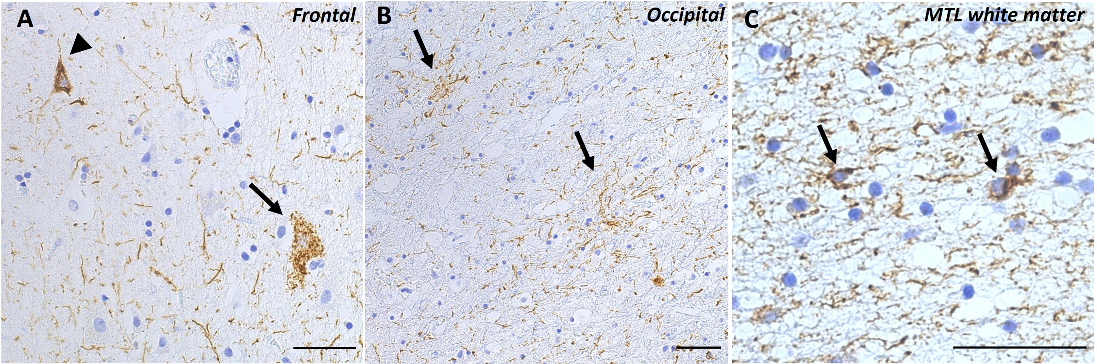
Neuropathological images.
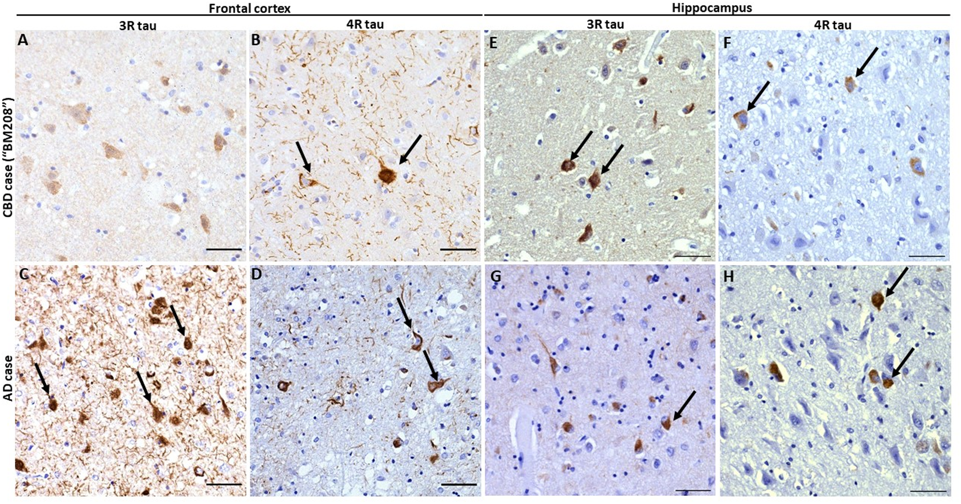
3- and 4-repeat tau isoform immunostainings. Immunohistochemistry of the frontal cortex and hippocampus (cornu ammonis 2 subregion) with anti 3-repeat tau (clone 8E6/C11, 1:500, Merck Millipore) and 4-repeat tau isoforms (clone 1E1/A6, 1:1000, Merck Millipore). The BM208 case described here shows fibrillar 4R but not 3R immunoreactivity in tangles (arrows) and neuropil in the frontal cortex (a-b). In the hippocampus (e-f), BM208 shows both 3R and 4R tau lesions, typical of AD-related Braak NFT stage II pathology (arrows). A symptomatic AD case (age at death = ∼ 70 years, Braak NFT stage VI, Aβ phase 5) was stained in parallel in both regions as a positive control, and displays strong fibrillary immunoreactivity for both 3R and 4R tau (c-d and g-h arrows) in both regions. Scale bars = 50 μm.
Neuropathological assessment of pTau-positive lesions.
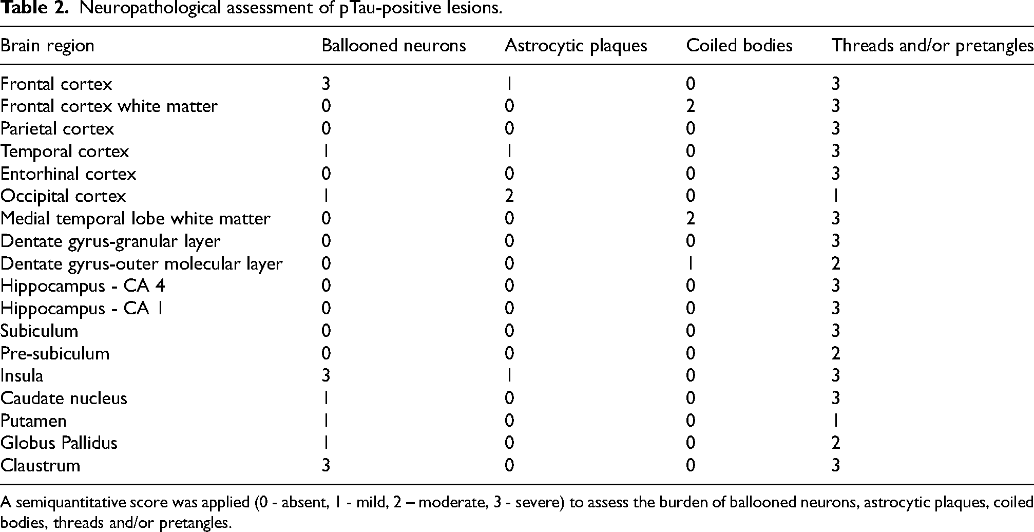
A semiquantitative score was applied (0 - absent, 1 - mild, 2 – moderate, 3 - severe) to assess the burden of ballooned neurons, astrocytic plaques, coiled bodies, threads and/or pretangles.
TDP-43 and α-synuclein pathologies were not observed.
Methods
Participants
To identify any factors that might have supported the diagnosis of CBD, we retrospectively compared BM208 to patients with typical amnestic AD (n = 18), Benson's syndrome due to AD (n = 3), CBS (n = 3), PSP syndrome (n = 2), and LBD (n = 9) as well as to a control group (cognitively normal Aβ negative participants, n = 24) from the UCL-2011-001756-12 32 and UCL-2018-0034/73-9440,41 studies. These participants were diagnosed clinically15,42,43 by a neurologist, but no autopsy has been carried out to confirm these diagnoses as these patients were included in in vivo studies. Given the small number of CBS and PSP syndrome patients, we grouped these two conditions together. We only selected typical AD patients who had an MMSE score ranged from 18 to 24/30 to include patients with a relatively similar level of dementia from BM208. However, we were unable to impose this same criterion on other pathologies, given that these neurodegenerative diseases are less frequent than AD. All the participants had an MMSE and a 3DT1 research MRI.
Ethical approval for these studies was granted by the Ethics Committee of UCLouvain (Eudra-CT number: 2011-001756-12, 2018-0034/73-94 and UCL-2020-355). Informed consent was obtained from all individual participants in accordance with the principles of the Declaration of Helsinki.
Cognitive assessment
All participants underwent a complete neuropsychological testing evaluating four cognitive domains: (1) verbal episodic memory using the Free and Cued Selective Reminding Test (FCSRT), 44 French version, (2) language using the Lexis Naming Test, the Category Fluency Test for animals, and the Letter Fluency Test for the letter ‘P’, 45 (3) executive functions using the Trail Making Test (TMT) part A and B 27 and Luria's Graphic Sequences,46,47 and (4) visuospatial functions using the Clock Drawing Test 25 and the Praxis part of the CERAD battery. 26 However, due to the extent of her difficulties, BM208 did not perform FCSRT, TMT and Luria's Graphical Sequences. Regarding memory, she performed the Dubois 5-word test. 48 Moreover, the executive z-score 32 was not available for BM208 and will therefore not be compared. An independent sample of 32 clinically normal individuals who remained cognitively stable over an eight-year period was used to calculate cognitive z-scores. 32 Z-scores were computed for each cognitive domain based on three measures and averaged to create a global cognitive z-score. A detailed explanation of this method has already been published. 32
A description of our sample can be found in Table 3.
Demographics of our sample.

Education level 1 = elementary school, Education level 2 = completed high school, Education level 3 = at least one year of post high school education
Brain MRI
Thirty-five of the 43 anatomical MRI scans, including BM208's (February 2018), were recorded using protocol described by Ivanoiu and colleagues in 2015, 32 while the 8 remaining scans were recorded using protocol described by Malotaux and colleagues in 2023. 40
Subcortical segmentation and cortical parcellation of MRI data were performed using FreeSurfer v7.2. 49 Meta-ROIs for each lobe were computed based on the Desikan-Killiany atlas. 50 The deep structures (basal ganglia and hippocampal volumes) were extracted unchanged from the Destrieux atlas. 51 All structures volumes were adjusted for total intracranial volume.
Statistics
To compare BM208's performances with other patients, we calculated z-scores for each group on the different sections of the MMSE (May 2017, total score = 21/30) as well as on neuropsychological examination and on MRI volumes, adjusting MRI volumes for intracranial volume.
It should be remembered that that BM208 only performed the Dubois 5-word 52 test which is an easier test than the FCSRT. Therefore, in order to enable a comparison of her performance against other groups, we have assigned her the worst available free recall score from our databases and the average total recall score.
Results
Cognitive sub-scores
On the MMSE, BM208 had better temporal orientation and memory encoding than all other groups (Figure 5(a)). She had more pronounced calculation and visuoconstructive difficulties than typical AD, CBS/PSP syndrome, and LBD patients. These clinical deficits matched those observed in Benson's syndrome due to AD, although, she had better memory recall compared to this group. In the Dubois 5-word test, her learning and memory scores were in the normal range, but her immediate and delayed free recalls were pathologic (Table 1), suggesting retrieval difficulties in verbal episodic memory.
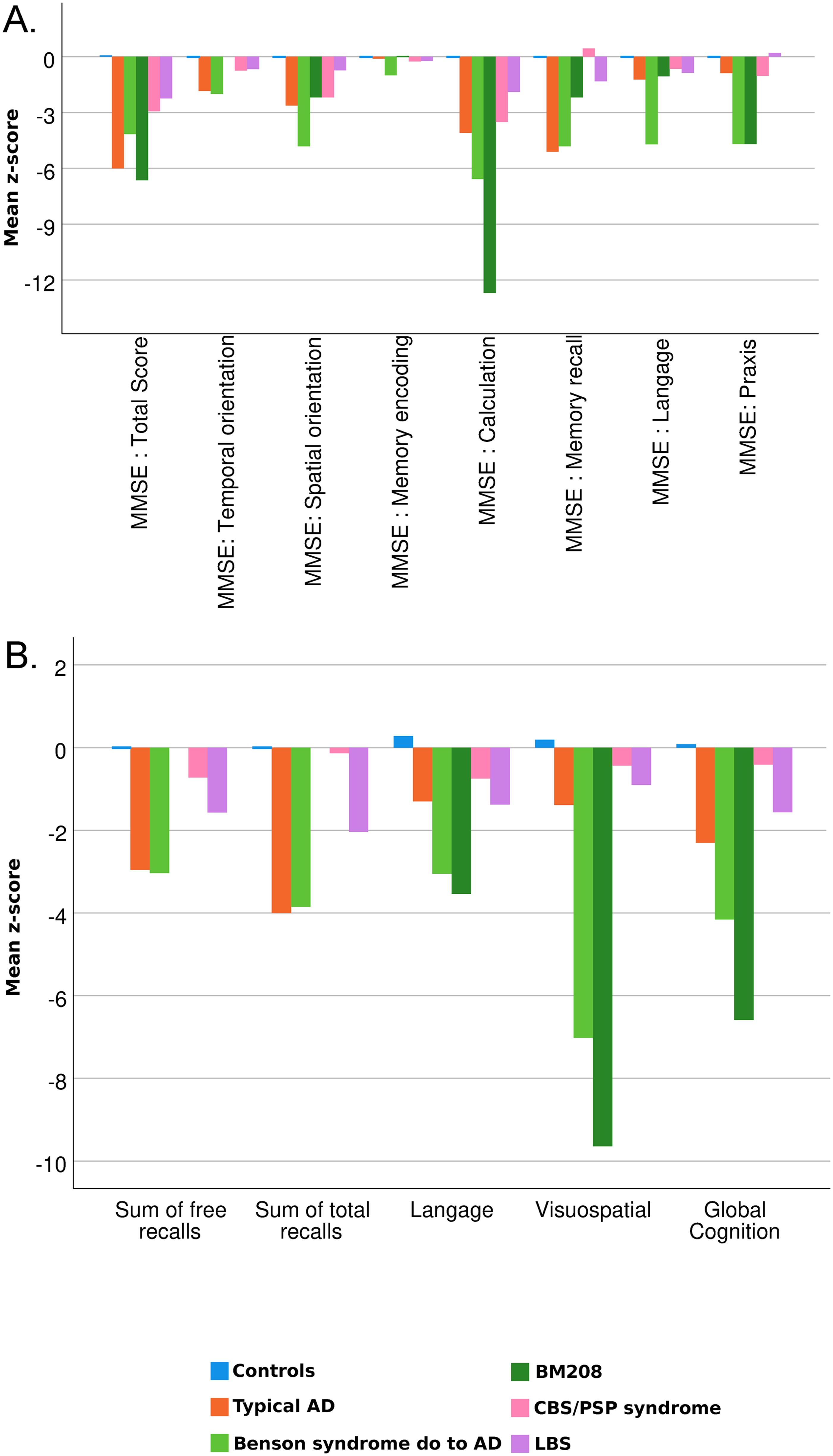
Z-score representation of MMSE subscores and neuropsychological assessment of different groups of patients and our CBD patient (BM208) compared to a control group.
We then compared BM208's cognitive z-scores to other patients. She showed greater episodic memory difficulties in free recall than those observed in typical AD and Benson's syndrome patients. On the other hand, unlike AD patients, she had no difficulties with total recall (including both free and cued recall). Her language difficulties were relatively comparable to those observed in Benson's syndrome patients, but greater than those observed in the other groups. BM208 also had important visuospatial difficulties similar to those observed in Benson's syndrome. Globally, her neuropsychological examination was more in line with the deficits observed in Benson's syndrome patients than those observed in CBS or any other group (Typical AD/LBD; Figure 5(b)).
MRI volumes
In terms of imaging, volumetric MRI showed global atrophy (compared to typical AD, Benson's syndrome, CBS/PSP syndrome, LBD). She particularly showed bilateral parietal and occipital atrophy that was also observed in Benson's syndrome, although frontal atrophy was more important in BM208 than in Benson's syndrome. She also showed atrophy of all basal ganglia structures including bilateral accumbens and caudate nuclei, putamen and thalami. Left caudate nucleus and putamen were also slightly reduced in CBS/PSP syndrome patients. She finally had bilateral hippocampal atrophy that was more pronounced than in typical AD patients (Figure 6).

Representation of the cerebral regions volumes z-score deviation of different groups of patients and our CBD patient (BM208) compared to a control group.
Discussion
Clinical case description and differential diagnosis
We described the case of a woman in her late sixties (BM208) who presented with autopsy-proven CBD for whom no in vivo diagnosis was made other than non-AD proteinopathy. The hypothesis of CBD was only suggested at the end of her life, five months before death, due to the late development of unilateral motor symptoms. The symptomatology began with visual-constructive impairment. Memory, language, and executive functions then progressively deteriorated over two years. Only rare cases of CBD patients with inaugural cognitive predominant syndrome instead of the typical asymmetric movement syndrome have previously been described. 53 These cases presented with a frontal-behavioral-spatial syndrome, a clinical phenotype characterized by early prominent executive, behavioral and visual disorders, 15 while the usual asymmetric presentation was initially absent and appeared later. BM208 showed this atypical progression, initially presenting with visual and behavioral difficulties, followed by a progressive parkinsonian syndrome and gait apraxia two years later, while asymmetrical signs only became evident at the end of her life.
The patient was initially referred to us with suspected cognitive impairment of vascular origin. The neuropsychological assessment then raised the possibility of Benson's syndrome. However, the absence of amyloid pathology ruled out the hypothesis of Benson's syndrome due to AD. The hypothesis of prion disease, such as Creutzfeldt-Jakob disease (including the Heindenhain characterized by posterior cortical symptoms 54 ), was also excluded because cerebrospinal fluid (CSF) total tau protein concentration was only slightly elevated, CSF 14-3-3 protein concentration was in the normal range and we did not observe any typical MRI abnormalities. Gerstmann syndrome was also excluded as, despite her impaired numeracy, BM208 was able to write, and did not complain about left-right disorientation or digital agnosia (although these were not formally tested). The presence of this syndrome could have helped the neurologist to show parietal lobe involvement which can either suggest PCA or CBD. The clinical diagnosis remained a non-AD proteinopathy for a long time, as total tau levels were slightly increased in the CSF. Finally, as motor symptoms evolved asymmetrically, the hypothesis of CBD was raised five months before the patient's death. Neuropathological analysis showed the presence of astrocytic plaques, ballooned neurons and coiled bodies, characteristic of CBD.1,2,55 The final diagnosis was thus confirmed as CBD. However, low burdens of AD neuropathological changes were also observed. 56 The probability that AD pathology explained her symptomatology was low, according to NIA-AA criteria. 57 Considering the extent of the CBD lesions compared with AD pathology, specifically in the occipital cortex, and considering the predominant visuospatial apraxia among the first clinical symptoms, we attributed her clinical syndrome to CBD.
When BM208 presented her first symptoms suggesting PCA, the hypothesis of AD was immediately ruled out due to the absence of amyloid-β positivity in CSF, and [18F]-Flutemetamol-PET a few months later. 28 In their recent multicenter study, Chapleau and colleagues showed that even in the absence of positive amyloid biomarkers in CSF, 94% of PCA patients had a positive Aβ-PET, suggesting atypical AD in most of the cases. 24 The normality of amyloid-β biomarkers in a case such as the one presented should be considered a red flag, prompting the search for another etiological cause. However, no biomarker could demonstrate CBD except the slight increase in CSF total tau that suggested non-AD proteinopathy as other proteinopathies inducing neuronal death can indeed increase CSF tau concentration.
Uncommon presentation of CBD as PCA
In a previous study, it was shown that out of 21 cases of in vivo diagnosis of PCA, more than half (13/21) were due to AD and only two to CBD while other cases were due to Parkinson's disease (1), LBD (3), and prion-associated diseases (2). 23 In his report, Renner et al. found no evidence of PCA due to other tauopathies. 23 Similarly, three other studies presented cases of PCA due to CBD21,58,59 and one case of PCA due to FTD-17, 60 another tauopathy. Finally, a recent international study showed that out of 145 patients with PCA syndrome who underwent autopsy, ten were non-AD cases, including four with LBD, two with CBD, two with brain infarcts, and one with Pick's disease. 24 However, our literature review did not identify PCA due to any other tauopathies. Besides the fact that the rare cases of PCA due to non-AD tauopathy were almost all due to CBD, we looked for elements that would have helped the neurologist to suspect CBD earlier.
Neuropsychological comparison and cognitive manifestations
In addition to literature review, we compared BM208 with patients suffering from other neurodegenerative diseases and showed that her cognitive performance was more similar to the one of patients with Benson's syndrome due to AD than the CBS patients’ one. More specifically, BM208 had greater visuo-constructive and calculation difficulties. The only inconsistency with a neuropsychological profile of Benson's syndrome/AD was her memory difficulties, which were most evident in free recall and normalized using the cue. This memory performance profile is more similar to the one observed in CBS/PSP syndrome. Moreover, several of these complaints suggested the presence of neurovisual difficulties as well as different types of apraxia (dressing, writing, ideomotor and ideational), which is a typical symptom of CBS. Indeed, it has been shown that 50% of patients suffering from CBD presented with limb apraxia, while no typical AD patient presented this disorder. 53 Constantinides and colleagues 61 even mentioned that 84% of CBD patients are affected by this symptom. More specifically, ideomotor apraxia is the most frequent kind of apraxia in CBD and is due to dysfunction of the supplementary motor area. 62 Among ten CBD patients, these authors reported seven patients with ideomotor apraxia, three of whom also suffered from ideational apraxia. In another study, among four CBD patients, all of them only suffered from ideomotor apraxia. 63 CBD patients with more severe apraxia (presenting both ideomotor and ideational apraxia) were more cognitively impaired in other cognitive functions than patients who only presented ideomotor apraxia, which correspond to our observation with BM208. This additional difficulty may result from the additional parietal or diffuse cortical damage. 62
Imaging findings and distinctive features supporting CBD diagnosis
Regarding imaging, BM208's brain was more atrophic in all regions than all other patients. In addition to severe global atrophy, BM208 demonstrated specific parietal and occipital lobe atrophy that was also observed in Benson's syndrome due to AD patients and that can be associated with the Benson's syndrome-like symptoms that the patient exhibited. More interestingly, she showed basal ganglia atrophy that was only observed in PSP syndrome/CBS patients. Visual analysis of the research sequences (repeated 3D-T1) also showed midbrain atrophy that evolved over time. This type of imaging sequence is not routinely acquired or analyzed by neuroradiologists in a clinical setting. However, the findings it reveals could have guided the neurologist toward an earlier consideration of a 3R/4R tauopathy. In addition to cortical and subcortical atrophy, we observed T2-FLAIR putamino-pallidal hypointensity which is not only observed in CBD 33 but also in PSP, 64 Parkinson's disease, 65 multiple sclerosis, 66 and multiple system atrophy. 67 This T2 hypointensity would be linked to increased iron deposition64,68 that is commonly observed in tauopathy and could be associated with tau deposition. 69 Hence, the observed T2-FLAIR putamino-pallidal hypointensity would indicate a high tau deposition in the putamen and globus pallidus, thus suggesting CBD. 33 Combined with the atrophy of the basal ganglia and the midbrain, this could have guided clinicians to a CBD hypothesis earlier.3,8
However, some clinical and MRI features suggested PSP, a clinical diagnosis that was considered before death. PSP is another 4R tauopathy that is often misdiagnosed with CBD given the overlap in clinical symptoms and imaging characteristics between both pathologies. PSP is generally characterized by supranuclear ophthalmoplegia, dystonia, rigidity of the neck and upper trunk, pseudobulbar palsy, and dementia. 70 On the other hand, CBS patients present progressive asymmetric rigidity, apraxia, tremor, and cortical and extrapyramidal symptoms. 4 While PSP and CBS are the clinical presentations most typically associated with PSP and CBD respectively, there is well recognized clinical heterogeneity in these pathologies. 71 Indeed, PSP may be one of the clinical phenotypes seen in CBD, just as CBS is one of the clinical phenotypes that may be seen in PSP. 72 In both conditions, patients may also present with frontal symptoms or primary progressive aphasia. 73 Not only the clinical motor symptoms associated with basal ganglia dysfunction (e.g., akinetic rigidity syndrome), but also midbrain and basal ganglia atrophy and T2-FLAIR putamino-pallidal hypointensity are often found in PSP, making in vivo differential diagnosis difficult. In the present report, BM208 showed late asymmetric motor signs, since at the end of her life she no longer used her left arm, thus tipping the diagnostic hypothesis towards CBD. A recent large CBS autopsy study (n = 113) showed that clinical features differed by pathology: apraxia of speech in CBD, ocular motor impairment and postural instability in PSP, and myoclonus, episodic memory loss and posterior cortical signs in CBS due to AD. 74
Intriguingly, BM208 also showed severe bilateral hippocampal atrophy that was presumably due to the pTau load in the hippocampus associated with CBD. Hippocampal atrophy is not common in typical CBD 75 although a low density of pTau deposits can be observed in the hippocampus, mainly in CA1-2. 76 Despite this significant hippocampal atrophy, BM208's encoding capacity was relatively preserved on tests requiring encoding very short word (MMSE, 5 words of Dubois), while retrieval was altered. One hypothesis to explain this intriguing relative preservation of memory capacity could be that memory impairment would not be solely linked to hippocampal atrophy, but also to the type of neurons and the affected cortical network as observed in semantic dementia.77,78
In addition, the 2019 MRIs showed atrophy of the pre- and postcentral gyri, which is more specific to CBD.3,8 Moreover, both visual and volumetric analysis reported midbrain atrophy, a specific feature of PSP, another 4R-tauopathy.34,64 Unlike MMSE score and sub-scores, which only allow to exclude a typical AD pathology, these fine-grained MRI quantifications could have guided the neurologist towards a possible diagnosis of CBD if they had been performed antemortem. However, such analyses are not part of the clinical routine MRI work-up, that only evidenced a generalized atrophy. These fine differences were observed using Freesurfer, 49 a segmentation software that quantified the volumes; we then compared these results with data from cognitively healthy controls, allowing us retrospectively to detect unusual areas of atrophy. Cases like BM208 advocate for the use of fined-grained MRI quantification in clinical practice when the diagnosis is difficult to make clinically.
Conclusion
In conclusion, this autopsy-proven case report confirmed that CBD is a possible cause of PCA syndrome. Furthermore, the comparative approach highlighted the additive value of acquiring 3D-T1 MRI sequence as well as in vivo fine-grained MRI quantification methods in the diagnosis of a PCA syndrome due to a non-AD tauopathy. CBD is characterized by an atrophy of the basal ganglia, midbrain and the precentral gyrus that is not typically observed in other neurodegenerative disorders. The search for this structural signature may help orienting the in vivo diagnosis in such atypical cases.
Footnotes
ORCID iDs

Author contributions
Yasmine Salman (Conceptualization; Data curation; Formal analysis; Investigation; Methodology; Visualization; Writing – original draft; Data Collection); Lara Huyghe (Conceptualization; Data curation; Formal analysis; Investigation; Methodology; Visualization; Writing – original draft; Data collection); Lisa Quenon (Methodology; Supervision; Validation; Writing – review & editing); Olivia Ghysens (Writing – review & editing; Data collection); Vincent Malotaux (Writing – review & editing; Data collection); Sandra O Tomé (Methodology; Resources; Visualization; Writing – review & editing); Dietmar Rudolf Thal (Formal analysis; Investigation; Supervision; Validation; Writing – review & editing; Data collection); Bernard J Hanseeuw (Conceptualization; Investigation; Methodology; Supervision; Validation; Visualization; Writing – review & editing).
Funding
The authors disclosed receipt of the following financial support for the research, authorship, and/or publication of this article: YS was funded by the Belgian Fund for Scientific Research (FNRS), grant number FRIA40014635 LH was funded by the FNRS, grant number ASP40016560. BH was funded by the FNRS, grant number CCL40010417 and the FRFS-WELBIO, grant number 40010035. We also thank the Fondation Louvain and Saint-Luc Foundation who provided in-kind contributions.
Declaration of conflicting interests
DRT & SOT received consultant honorary from Muna Therapeutics (Belgium). DRT collaborated with Novartis Pharma AG (Switzerland), and GE-Healthcare (UK). DRT is a member of Acta Neuropathologica editorial board.
The remaining authors declared no potential conflicts of interest with respect to the research, authorship, and/or publication of this article.
Data availability
Anonymized data are available on justified request to Bernard.Hanseeuw@uclouvain.be.