



Abstract
Background
There is growing evidence that cardiorespiratory fitness (CRF) mitigates the likelihood of dementia caused by Alzheimer's disease and may underlie the cognitive benefits observed from aerobic exercise. Previous evidence further demonstrates neurodegeneration is the biological substrate for cognitive deterioration and younger brain age may protect the brain from the deleterious effects of neurodegeneration. However, little is known about the relationships between CRF, brain age, and neurodegeneration in older adults with amnestic mild cognitive impairment (aMCI).
Objective
The aim of this cross-sectional study was to examine associations between CRF, brain age, and neurodegeneration among individuals with aMCI, using baseline data from the Aerobic exercise and Cognitive Training (ACT) trial, which examined the cognitive effects and underlying mechanisms of a 6-month ACT in older adults with aMCI.
Methods
CRF was measured with peak oxygen uptake (VO2peak), from a symptom-limited peak cycle-ergometer test. Brain age and hippocampal volume were obtained from structural magnetic resonance imaging. Brain age was estimated using brainageR. Descriptive statistics and bivariate correlations were assessed. Linear regression models were used to analyze the relationships between CRF, brain age, and hippocampal volume, while adjusting for covariates. All analyses were conducted using R (version 4.3.2).
Results
The sample (N = 141) averaged 73.66 ± 5.78 years of age, 16.91 ± 2.89 years of education, 27.46 ± 5.15 in BMI, and 23.49 ± 2.16 on Montreal Cognitive Assessment, with 53% male and 92.2% White. The mean brain age was 72.37 ± 7.89 years with 3157.31 ± 449.35 mm3 hippocampal volume. No association was found between CRF, brain age, and hippocampal volume.
Conclusions
Future studies need to explore other brain indicators related to CRF.
Trial Registry
ClinicalTrials.gov, https://clinicaltrials.gov/study/NCT03313895, NCT03313895, October 18, 2017
Introduction
Cardiorespiratory fitness (CRF), the heart and lungs’ ability to supply oxygen to muscles during physical activity, is a crucial element of both physical health and overall fitness. 1 CRF declines 3%–6% during the third decade of life, and this decline accelerates each decade to about 20% per decade after age 70. 2 Low CRF is a significant predictor of mortality and morbidity. Individuals with inadequate CRF are more likely to have chronic diseases such as Alzheimer's disease (AD) and related dementias. 3 However, the evidence regarding the association between CRF and cognition is mixed. A review showed that changes in CRF over time may not directly impact cognitive changes in older adults without known cognitive impairment. 4 Conversely, other research has indicated that high CRF, measured with peak oxygen consumption (VO2peak) is correlated with better performance in global cognition, memory, executive function, and motor skills among older adults. 5 Little is known about whether an association exists between CRF and cognition in individuals at risk for AD-related dementias.6,7
Existing studies on CRF and cognition often involve healthy older adults without cognitive impairment.4,8 A few studies examining CRF and brain integrity in individuals with early-stage AD show a strong association.9,10 VO2peak was higher among older adults with intact cognition than those with mild cognitive impairment (MCI) or mild dementia (1.7 ± 0.6 ml/kg/min versus 1.4 ± 0.4 ml/kg/min based on graded treadmill test). 10 Cross-sectional studies examining the relationships between CRF levels and hippocampal volume among adults with intact cognition have shown mixed findings. Some studies of older adults suggest a negative relationship between CRF and brain atrophy,10–12 and between CRF and hippocampal volume,13–15 while others have found no relationship. 10 Moreover, longitudinal studies show no significant association between CRF and hippocampal volume, indicating that CRF may not affect the hippocampus in the same way it affects other regions of gray matter.6,16
A possible factor explaining the mixed associations between CRF and cognition is brain age, a biomarker of cognitive well-being discovered in the past ten years. 17 Brain age is conceptually defined as an estimate of an individual's cognitive age derived from structural magnetic resonance imaging (MRI).18–20 Machine learning has been used to determine brain age and identify patterns that distinguish younger brains from older ones. 21 An older brain age may contribute to poor cognitive function and reduced brain health. 22
Little is known about the relationship between CRF and brain age, with some studies suggesting a negative correlation between CRF and brain age.23,24 For example, compared to individuals who have been aerobically fit in the past ten years, those who have maintained a high level of CRF throughout adulthood are predicted to have a younger brain age. 23 Another study showed that brain age decreases by 0.58 years for each additional flights of stairs climbed. 24 Individuals with higher CRF levels often exhibit a lower brain age compared to their sedentary counterparts, suggesting a protective effect of fitness on cognitive aging. 24 Currently, how CRF and brain age independently and interactively contribute to cognition and AD remains unknown.
Emerging research has highlighted the role of physical activity as a moderator in the relationship between CRF and brain age.25,26 Studies indicate that higher levels of physical activity can enhance cognitive function and brain health, potentially mitigating the effects of aging on brain structure and function.25,26 Physical activity appears to amplify the neuroprotective benefits associated with higher CRF, promoting neurogenesis and improving vascular health, both of which are critical for maintaining optimal brain function. 27 However, some studies reveal a complex interaction between CRF and physical activity, with varying outcomes depending on age and baseline fitness levels. 28 Thus, understanding how physical activity influences the relationship between CRF and brain age is essential for developing targeted interventions aimed at enhancing cognitive health across the lifespan.
This study examined the relationships between CRF, brain age, and neurodegeneration in older adults with amnestic MCI and how physical activity moderated these relationships. We hypothesized that (1) higher CRF was associated with younger brain age and greater hippocampal volume; (2) a younger brain age was associated with greater hippocampal volume; and (3) physical activity moderated the relationship between CRF, brain age and hippocampal volume.
Methods
Design
This study used baseline data from the Aerobic Exercise and Cognitive Training (ACT) Trial, which tested the efficacy and mechanisms of a six-month ACT intervention in older adults with amnestic MCI. The ACT Trial was a Stage II, single-blinded, 3-site RCT. Trial randomized 146 participants equally to one of four arms: ACT, moderate-intensity cycling, speed of processing cognitive training, or stretching/mental leisure activity control for six months with 12-month follow-up. The Institutional Review Board of each site approved the study.
Setting
Screening and all data collection occurred in a private room at the clinical research facility at each site. CRF was assessed in exercise testing laboratories. MRI was conducted at the facility on campus or in a local hospital.
Sample
A variety of recruitment strategies were mobilized to proactively recruit participants, including clinic, community, and word-of-mouth referrals; recruitment material distributions; exhibits at local events, such as health fairs and festivals; media outreach, such as Facebook and email blasts; press releases and newspaper advertisement; and Alzheimer's Prevention Registry. Potential participants initiated contact with us and underwent a phone screen, in-person interview, medical clearance, symptom-limited, peak cycle-ergometry cardiopulmonary exercise, and MRI. Details of the trial protocol were previously published. 29
The inclusion criteria for the ACT Trial were Montreal Cognitive Assessment (MoCA) between 18 and 26 after education adjustment, less than one standard deviation below age- and education-corrected population norms on Rey's Auditory Verbal Learning Test, less than 30 on Activity of Daily Living–Prevention Instrument, no dementia, community-dwelling, 65 years old or older, English-speaking, adequate visual acuity, exercise and MRI safety verified by providers, stable on cognitive drugs, and capacity to consent. Potential participants were excluded for the following reasons: Geriatric Depression Scale score of more than five with contextual evidence for major depression; resting heart rate at or below 50 or at or above 100 beats/min; neurological, psychiatric, or substance disorders in the past five years; American College of Sports Medicine contraindications to exercise; new and unevaluated signs and symptoms; abnormal exercise test; abnormal MRI; and enrollment in another intervention study.
Among the 146 participants enrolled in the ACT Trial, data on brain age (n = 4) and CRF (n = 1) were missing. As a result, 141 were included in this study.
Variables and measures
The independent variable, CRF, was measured with VO2peak. The dependent variables, brain age and hippocampal volume, were measured with MRI. Potential covariates were sex; race; marital status; age; education; body mass index (BMI); global cognition; number of comorbidities; total brain volume (TBV), representing the overall size of the brain and encompassing all its structures, including gray matter, white matter, and cerebrospinal fluid; and activities of daily living (ADL).
Independent variable
CRF was measured with VO2peak, obtained from the symptom-limited cardiopulmonary exercise test, and performed on a cycle ergometer. VO2peak was defined as the average oxygen consumption in the final 30 s prior to test termination. A participant began cycling on a recumbent stationary cycle at a comfortable speed. The cycling intensity was increased at one metabolic equivalent (=3.5 ml oxygen/kg body weight/minute) every 3 min until the participant achieved volitional fatigue or satisfied the test termination criteria per American College of Sports Medicine. 30 Peak hemodynamic responses, such as heart rate and rhythm, were continuously monitored via electrocardiogram. The Borg Rating of Perceived Exertion (RPE) was administered, and blood pressure was assessed during the last minute of each stage and at peak exercise. Oxygen consumption was measured by indirect calorimetry. Breath by breath analyses were averaged using 30 s means.
Moderator variable
Physical activity was evaluated through self-reporting and categorized into four levels: sedentary, light, moderate, and vigorous. Participants were deemed sedentary if they reported no exercise. Individuals engaging in light activity reported activities for less than three days at moderate intensity, such as slow walking (2.0 mph) or playing the piano. Participants were classified as moderately active if they reported activities at moderate intensity for three or more days, or participating in vigorous activities for fewer than two days, for example, walking at 3 mph or doing calisthenics. Finally, participants were categorized as engaging in vigorous exercise if they reported participating in intense activities, such as brisk walking (over 3 mph) or climbing hills, for two or more days.
Dependent variables
Brain age was calculated using brainageR to generate a brain-predicted age value from each participant's raw T1-weighted MRI scan. 31 The software performs segmentation and normalization on raw T1-weighted MRI scans, then applies a Gaussian Processes regression, implemented in R (kernlab package), 32 to predict an age value with the pre-trained brainageR model.21,33,34
Automated volumetric segmentation was performed with Freesurfer v7.3.2 using the hippocampal and amygdala segmentation module. 35 The whole hippocampal volume was extracted from Freesurfer stats output.
Covariates
Sex was categorized as male or female. Age was recorded in years. Race was classified into two groups: White and Others (i.e., Asian and African American). Educational attainment was measured by the number of years of education completed. Marital status was classified as either married or not married. The number of comorbidities was measured as the total number of chronic conditions (e.g., cardiovascular disease, diabetes, pulmonary conditions). Global cognition was assessed using the MoCA. 36 TBV was measured as brain segmentation volume without ventricles, CSF, or choroid plexus, and generated with Freesurfer v7.4.1. Finally, BMI was measured as weight (kg)/height (m2), and ADL was measured using the Activities of Daily Living-Prevention Instrument (ADL-PI, self-reported). 37
Analyses
Statistical analyses were conducted using RStudio (R version 4.3.2). Descriptive statistics were analyzed with chi-square, ANOVA, and/or Kruskal-Wallis tests based on variable type (continuous or categorical). Correlations were measured utilizing bivariate correlation. Linear regression assumptions were confirmed. Multiple linear regression models were used for analyses, adjusting for covariates. Moderation was tested using a moderated linear regression model.
Results
Univariate analysis
This study included 141 participants (Table 1). Briefly, most participants were male (n = 75, 53%) and non-Hispanic White (n = 130, 92.2%). The mean age of the participants was 73.66 ± 5.78 years, with an average education of 16.91 ± 2.89 years. Most participants were married (n = 99, 70%). The mean brain age was 72.37 ± 7.89 years, with a mean hippocampal volume of 3157.31 ± 449.35 mm³ and a mean TBV of 1,105,297.88 ± 133,233.56 mm³. Physical activity was categorized as sedentary (6 [4.25%]), light (57 [40.43%]), moderate (50 [35.46%]), and vigorous (28 [19.86%]). Due to the small number of participants in the sedentary group, we included these 6 participants in the light activity group for the moderation analysis.
Descriptive statistics.

BMI: body mass index; MoCA: Montreal Cognitive Assessment; ADL: Activities of Daily Living.
Mean (SD); n / N (%).
Number of comorbidities (e.g., cardiovascular disease, diabetes, pulmonary conditions).
Bivariate analysis
Bivariate correlations (see Table 2) were estimated to assess the relationships between the study variables. The results indicated that men had older brain age (r = −0.2, p < 0.05) and larger hippocampal volumes (r = −0.21, p < 0.05) compared to women. Age (r = 0.56, p < 0.001) and comorbidities (r = 0.17, p < 0.05) were correlated with brain age. Brain age was negatively related to hippocampal volume (r = −0.33, p < 0.001) and total brain volume (TBV) (r = −0.22, p = 0.007). Additionally, CRF (r = 0.22, p = 0.008), BMI (r = 0.18, p = 0.03), MoCA scores (r = 0.18, p = 0.03), and TBV (r = 0.57, p < 0.001) were correlated with hippocampal volume.
Correlations among study variables.
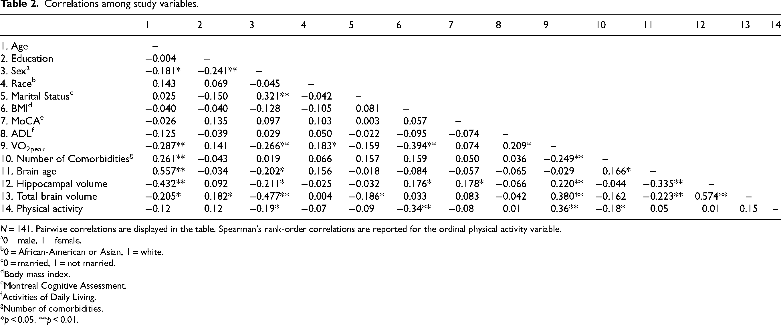
N = 141. Pairwise correlations are displayed in the table. Spearman's rank-order correlations are reported for the ordinal physical activity variable.
0 = male, 1 = female.
0 = African-American or Asian, 1 = white.
0 = married, 1 = not married.
Body mass index.
Montreal Cognitive Assessment.
Activities of Daily Living.
Number of comorbidities.
*p < 0.05. **p < 0.01.
Relationships of VO2PEAK to brain age
In the unadjusted model, VO2peak and brain age were not significantly related (β = −0.04, SE = 0.13, p = 0.73) (See Figure 1). The relationship again was not significant after adjusting for covariates in the model (β = 0.27, SE = 0.15, p = 0.07) (See Table 3).
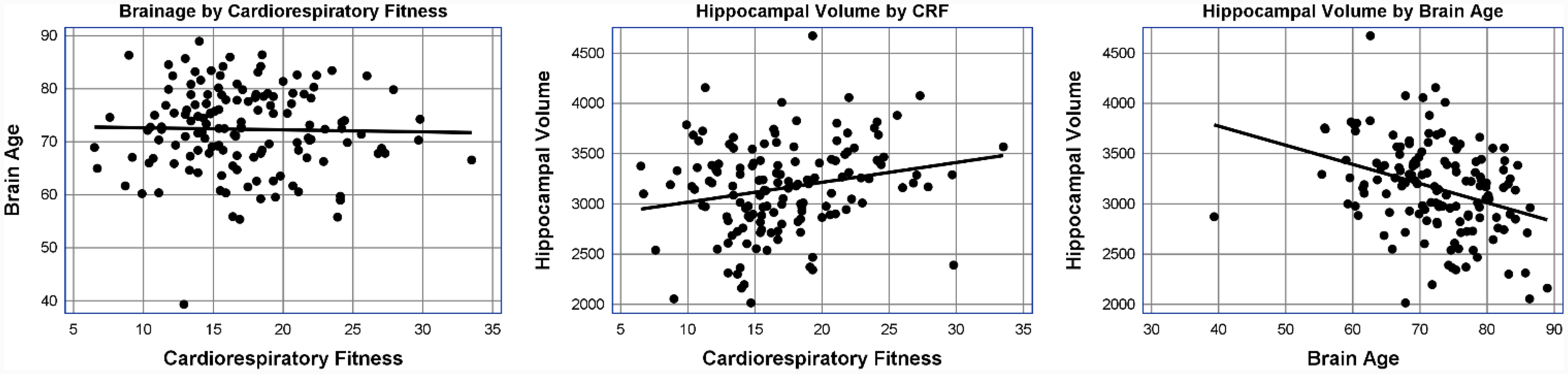
Relationships between cardiorespiratory fitness, brain age, and hippocampal volume.
Associations of cardiorespiratory fitness with brain age and neurodegeneration and associations of brain age with hippocampal volume (neurodegeneration).

N = 141. Estimate is a raw score regression coefficient. CI = is the lower and upper limit of a 95% confidence interval.
0 = male, 1 = female.
0 = African American or Asian, 1 = White.
0 = married, 1 = not married.
Body mass index.
Montreal Cognitive Assessment (MoCA).
Activities of Daily Living.
Number of comorbidities (e.g., cardiovascular disease, diabetes, pulmonary conditions).
*p < 0.05. **p < 0.01.
Relationships of VO2PEAK to hippocampal volume
With no covariates in the model, VO2peak is significantly related to hippocampal volume (β = 20.01 SE = 7.51, p = 0.008) (See Figure 1). Hippocampal volume is higher by 20.01 mm3 for every 1 ml/kg/min increase in VO2peak. However, with covariates in the model, VO2peak is not significantly related to hippocampal volume (β = −1.42, SE = 7.95, p = 0.85) (See Table 3).
Relationships of brain age to hippocampal volume
In the unadjusted model, brain age is negatively related to hippocampal volume (β = −19.06, SE = 4.55, p < 0.001) (See Figure 1). The results indicate that when the brain age is higher by one year, the hippocampal volume decreases by 19.06 mm3. After adjusting for covariates, brain age is not related to hippocampal volume (β = −3.77, SE = 4.52, p = 0.40) (See Table 3).
Relationships of VO2PEAK to brain age and hippocampal volume as a function of physical activity
Physical activity did not moderate the relationships between VO2peak and the dependent variables. Specifically, for brain age, the contribution of the VO2peak × physical activity interaction was minimal, with ΔR² = 0.003, F(2, 125) = 0.353, p = 0.703. Similarly, for hippocampal volume, the interaction accounted for a negligible amount of variation, with ΔR² = 0.009, F(2, 125) = 1.090, p = 0.339. Table 4 illustrates that the relationships between VO2peak and the dependent variables did not significantly differ (each p > 0.30) between the moderate and low physical activity groups, nor between the high and low physical activity groups.
Moderator analysis results for brain age and hippocampal volume.

N = 141. Estimate is a raw score regression coefficient. CI = is the lower and upper limit of a 95% confidence interval.
Mean-centered.
Discussion
We examined the associations of CRF with brain age and hippocampal volume and whether physical activity moderates these associations among older adults with amnestic MCI. The analyses showed a non-significant association between CRF and brain age. CRF was positively associated with hippocampal volume, but not after controlling for covariates. Moreover, physical activity did not moderate the associations between CRF, brain age, and hippocampal volume.
Previous studies have found a link between CRF, cognitive health, and brain structure, including reduced cognitive decline,12,38,39 enhanced brain connectivity, 40 and improved brain neuroplasticity in older adults with or without cognitive impairment. 41 However, little is known regarding the association between CRF and brain age. This study addressed a significant gap in the literature by examining the association between CRF and brain age among community-dwelling older adults with amnestic MCI.
While other studies have linked CRF and brain age in healthy older adults,23,24 this study did not show a correlation between CRF and brain age among older adults with amnestic MCI. These discrepant findings may be attributable to differences in participant populations. 24 Variations in CRF assessment methods, study designs (i.e., longitudinal versus cross-sectional), sample sizes, and the precision of brain-age metrics, may also contribute to these differing results. This study had a smaller sample size compared to studies in healthy older adults.23,24 This study utilized the brainageR software to generate a brain-predicted age from T1-weighted MRI scans, primarily focusing on structural MRI for age prediction. In contrast, other studies adopted a multimodal approach, incorporating a variety of brain metrics derived from multiple imaging modalities, including structural MRI, diffusion tensor imaging (DTI) and functional MRI. 23
Additionally, other studies used different measures to assess CRF, such as the number of stairs climbed 24 or calculated a composite score derived from several health-related measurements, including resting pulse, blood pressure, maximum grip strength, BMI, and waist circumference. While these measures can show overall physical health, they do not directly measure CRF. 23
The relationship between CRF and brain age is influenced by a variety of factors, including the severity of MCI and demographic characteristics of the sample, such as age, living situation, marital status, and educational level. Previous studies have often overlooked the psychosocial and environmental variables affecting the relationships among CRF, brain age, and hippocampal volume. Our findings underscore the complexity of investigating CRF's impact on brain health and emphasizes the necessity of considering important covariates when interpreting research findings.
Previous studies examining CRF and hippocampal volume have shown mixed findings. Some studies reported higher CRF levels are linked to larger hippocampal volume, 22 whereas other studies found no statistically significant associations. 6 In our study, CRF was related to hippocampal volume. However, this relationship was no longer significant after controlling for variables such as sex, race, marital status, age, education, BMI, global cognition score, number of comorbidities, TBV, and ADL. The variability in findings across studies and the influence of confounding variables highlight the need for further research.
Future research should investigate how these confounding variables interact with CRF, affecting neurodegeneration throughout the AD spectrum. Additionally, it is crucial to consider these confounding variables when developing physical activity interventions aimed at improving neurocognitive outcomes. Tailoring interventions to account for age, sex, BMI, and other factors could enhance their effectiveness in mitigating neurodegeneration.42,43
In addition, the moderating effect of physical activity merits further investigations. While physical activity is essential for overall health, it is a multi-dimensional concept including mode, frequency, intensity, and duration. A meta-analysis highlighted that consistent physical activity positively impacts cognitive function, but it may not necessarily amplify the benefits of CRF. 44 Lastly, brain health can be influenced by various lifestyle factors, including diet, stress, and sleep, alongside physical fitness. 45 This highlights the complexity of brain age and the need for a multifaceted approach in future studies. Longitudinal research is warranted to delve deeper into how CRF influences brain health over time, particularly considering that cognitive decline is a gradual process influenced by various lifestyle factors. 46
This study has several strengths, such as a carefully characterized and geographically diverse sample with amnestic MCI and innovative and gold-standard assessments of CRF, brain age, and hippocampal volume. However, the cross-sectional nature of this study prevents us from making causal inferences regarding the associations of CRF with brain age and neurodegeneration. Furthermore, this study relied on self-reported measures of physical activity, which is prone to recall bias. In contrast, objective measures, such as accelerometers, offer a more accurate assessment of physical activity levels by capturing actual movement data. Moreover, this study relied on a single imaging modality, T1-weighted MRI, and it may not capture the full complexity of brain age. We focused only on older adults aged 65 years and older with MCI; thus, the findings would not be generalizable to healthy older adults without cognitive impairment or older adults with AD dementia. Last, participants in this study were predominantly White and were more educated than the general US population.
Conclusion
This study's findings indicate that CRF is not significantly associated with brain age or neurodegeneration in older adults with amnestic MCI. Future studies, particularly longitudinal studies, are essential to verify these results and determine whether higher CRF could potentially lead to a younger brain age in individuals with amnestic MCI or AD dementia. Additionally, further research should investigate other brain metrics associated with CRF to provide a more comprehensive understanding of its effects on brain health.
Footnotes
Acknowledgments
The CTSI and Center for Magnetic Resonance Resources at the University of Minnesota were supported by the National Institutes of Health National Center for Advancing Translational Sciences of the National Institutes of Health Award Number UL1TR000114 and the National Institute of Biomedical Imaging and Bioengineering Award Number P41 EB1058941, respectively. The content is solely the responsibility of the authors and does not necessarily represent the official views of the National Institutes of Health. We sincerely appreciate the contributions of study participants and study staff.

Ethical considerations
All procedures involving human subjects in this study were conducted in accordance with the ethical standards of the Committee on Human Experimentation at our institution and were also in compliance with the principles outlined in the Helsinki Declaration of 1975, as revised in 2013.
Consent to participate
Informed consent was acquired from all individual participants included in the study. Participants received an explanation of the study's objectives, methodologies, potential risks, and advantages. They were also informed about the right to withdraw from study at any moment without repercussion. Written consent was secured prior to engagement in the research.
Consent for publication
The authors confirm that written informed consent was obtained from all participants for the publication of their data.
Author contributions
Grace Derboghossian (Formal analysis; Writing – original draft; Writing – review & editing); Keenan Pituch (Formal analysis; Writing – review & editing); Mia Anthony (Investigation; Writing – original draft; Writing – review & editing); Dereck Salisbury (Investigation; Writing – review & editing); Feng Vankee Lin (Conceptualization; Funding acquisition; Investigation; Writing – review & editing); Fang Yu (Conceptualization; Formal analysis; Funding acquisition; Investigation; Writing – original draft; Writing – review & editing).
Funding
The author(s) disclosed receipt of the following financial support for the research, authorship, and/or publication of this article: The National Institute on Aging of the National Institutes of Health funded the ACT trial under Award Number R01AG055469-01A1. It was registered on ClinicalTrials.gov on 10/18/2017 (Identifier: NCT03313895).
Declaration of conflicting interests
The author(s) declared no potential conflicts of interest with respect to the research, authorship, and/or publication of this article.
Data availability
The datasets generated and analyzed for the current study are available from the corresponding author upon reasonable request.