



Abstract
Background
Alzheimer's disease (AD) is the most prevalent neurodegenerative disorder characterized by cognitive deficit and pathological accumulation of amyloid-β (Aβ) and tau proteins. The rodent models have contributed greatly to unravel AD pathogenesis, but these AD models have been shown a modest clinical translational effectiveness.
Objective
Therefore, developing mass-producible primate AD models is promising for more effective drug development.
Methods
Here, we constructed the AD monkey models by simultaneously infusing AAV-Tau and Aβ into different brain regions.
Results
The induced monkeys showed a durable cognitive impairment lasting for at least 10 months after the modeling. Simultaneously, the increased levels of total tau and hyperphosphorylated tau (pTau) at several AD-associated sites, and neurofilament light chains (NfL) with altered Aβ level were detected at different time points in cerebrospinal fluid and/or plasma by using MSD kits. The increased brain accumulation of Aβ and tau proteins was also detected by positron emission tomography/magnetic resonance imaging and immunohistochemical staining. The model monkeys also had significant glial activation; an indicator of inflammation commonly seen in the brains of AD patients.
Conclusions
Together, this study provides mass-producible monkey models showing durable AD-like hallmark pathologies (Aβ, tau, NfL, i.e., ATN) and cognitive deficits. As monkeys are genetically and metabolically the closest to humans, these models will offer more effective drug discovery and development for AD.
Keywords
Introduction
Alzheimer's disease (AD) is the most common neurodegenerative disorder, and the prevalence of AD is rapidly increasing with a global population aging. The main clinical feature of AD patients is the cognitive decline with pathological accumulation of amyloid-β (Aβ) and tau proteins, the most recognized hallmarks of AD brain pathologies.1,2
The extracellular accumulation of Aβ forming senile plaques is one of the recognized hallmark pathologies in the AD brains. Regarding the driven molecular mechanisms underlying AD pathologies, the amyloid cascade hypothesis still dominates the field. 2 It is generally believed that the Aβ pathology occurs before tau, whereas tau pathology comes with more solid link to cognition/behavior issues. In familial AD (FAD), Aβ accumulation is a hallmark of early AD development and/or a triggering event. Based on the N-terminal cleavage of the amyloid-β protein precursor (APP), the produced Aβ peptides show different lengths, commonly from 39∼43 amino acid residuals. Among them, Aβ1–42 in its soluble form, particularly Aβ oligomers, is believed to be the most toxic form in disease progression. 3 The roles of neuroinflammation and abnormal activation of glial cells in mediating Aβ toxicity have received great attention. 4 Additionally, an impaired Aβ clearance is an important mechanism leading to Aβ accumulation in the brain. 5 The drug development against Aβ has been the main direction in the AD field for over two decades. The strategies include active immunity (by injecting Aβ peptide to induce antibody), passive immunity (by directly injecting antibodies against Aβ), use of β- or γ-secretase inhibitors, and blood replacement. Recently, several Aβ targeting antibodies have shown promising effects in clinical application.6–8 However, ongoing large-scale clinical trials are needed to confirm their clinical efficacy and side effects of the antibodies.
Accumulation of hyperphosphorylated tau forming neurofibrillary tangles is another hallmark of the AD brain. As a major microtubule-associated protein, the normal function of tau is to promote microtubule assembly and maintain stability of the microtubules. Recently, tau proteins have been found to play a crucial role in multiple functions beyond microtubules or cytoskeletons. For instance, tau proteins can actively regulate cell viability and phosphorylation of tau endows resistant of the cells to apoptosis induced by various apoptotic inducers.9–11 In the AD brains, tau protein is abnormally hyperphosphorylated. The hyperphosphorylated tau dissociates from microtubules which causes microtubule disassembly and axonal transport deficits, and eventually leads to synapse damages and cognitive impairment. 9 Tau phosphorylation mutually promotes its other types posttranslational modifications, such as acetylation, sumoylation, etc., 12 which makes tau more vulnerable to aggregation. The aggregated tau protein induces multiple cellular dysfunctions, such as disrupting synaptic transmission and neural circuits directly leading to memory deficits,13–16 inhibiting autophagy that aggravates tau accumulation,17,18 damaging mitochondrial dynamics, 19 and so on.
In addition to Aβ and tau, an increased level of neurofilament light chain (NfL) has also been proposed to be a marker of neurodegeneration. As a neuronal cytoskeleton protein, NfL is involved in maintaining cell shape and the regulation of axonal and dendritic trafficking. The axon dysfunction and degeneration are important steps in the pathogenesis of neurodegenerative diseases, which occur often before the detectable misfolded protein deposits. During these processes, the NfL can be released into the extracellular space and then into the cerebrospinal fluid (CSF) and plasma, from where it serves as a biomarker of neurodegeneration. Currently, the ATN (i.e., Aβ, tau, and NfL) has been applied for the clinical diagnosis of AD. 20 Studies also suggest that glial activation and chronical inflammation are involved in AD pathogenesis. It is proposed that these pathological markers are closely related to the severity of the disease in AD patients. 21
Studies based on rodents have made great progress in revealing AD pathogenesis, however, the related clinical drug development has been disappointing. Compared with the rodents, non-human primates (NHP) are genetically and phylogenetically closer to the human being, and the NHPs have more complex brain network and the functions than the rodents. 22 For example, APP, Aβ42 or Aβ40 peptide in Macaca fascicularis, has a sequence homology of up to 100% with humans, and the longest form of tau protein in macaques has a 98% homology with humans. 23
The currently existing NHP models of AD include spontaneous and artificially induced. The spontaneous NHP model has a long generation cycle, high cost and big heterogeneity, which severely limits the application.24–27 The existing artificially induced AD monkey models have been produced by using transgenic technique, 28 or overexpressing human tau, or injecting Aβ oligomers (AβOs) into the brain.28–31 As tau and Aβ are synergistically but not independently promoting AD progression, simultaneous tau and Aβ treatment has been recomended. 32 In addition, transgenic techniques for making monkey model show great limitations in mass production for the single birth reproduction monkeys.
In the present study, we investigated whether mass-producible AD monkey models could be made by combined infusion of Aβ fibers (AβF) or AβOs and AAV-Tau into specific brain regions. We found that AβF or AβOs plus AAV-Tau infusion to bilateral prefrontal cortex (PFC), or to the PFC plus hippocampus, or to the hippocampus alone could induce a durable cognitive deficit with whole set of clinically recognized AD-like pathologies, including an increased brain Aβ and tau accumulation, an increased CSF and plasma total tau and the phosphorylated tau, an increased CSF and plasma NfL level, and an increased or reduced CSF Aβ42 or Aβ42/Aβ40 ratio depending on different brain region infusion. Simultaneously, the model monkeys also show glial activation. These novel AD-like monkey models can be applied not only for in-depth pathogenesis studies, but also for effective drug development.
Methods
Reagents and antibodies
The anesthetics, analgesics, and sedatives used in this study included isoflurane (Ruipu Biological Pharmaceutical, Tianjin China), atropine sulfate (Shanghai whole Yu biotechnology animal pharmaceutical, China), Sutai 50 (Vic LTD, France), ceftriaxone sodium (Shanghai Shangyao Xinya Pharmaceutical, China), lidocaine hydrochloride (Huamu animal health products of Jilin Province, China), and propofol (Guangdong Gabor Pharmaceutical, China).
The primary antibodies used in this study were as follows: AT8 (Invitrogen, MN1020), tau181 (Invitrogen, 701530), tau404 (Invitrogen, 44-758G), tau396 (Invitrogen, 35-5300), HT7 (Invitrogen, MN1000), tau5 (Abcam, ab80579), tau217 (Abcam, ab291080), β-actin (Proteintech, 66009-1-AP), ThioS (Merck, T1892), NfL (Proteintech, 12998-1-AP), NfH (Proteintech, 21471-1-AP), GFAP (Abcam, ab7260), Iba-1 (Abcam, ab178876). The secondary antibodies used for immunohistochemistry: anti-mouse (Jackson 168226); anti-rabbit (Jackson 169882). The secondary antibodies used for western blotting: anti-mouse (abclonal AS003), anti-rabbit (abclonal, AS014). DAPI and thioflavin-S were obtained from Roche (10236276001) and Sigma (T1892-25G).
The kits in this study: DAB color development kit (Vector, ZL0229), human amyloid beta 42 ELISA kit (Thermo Fisher, KHB3441), human amyloid beta 40 ELISA kit (Thermo Fisher, KHB3481), S-PLEX Human Tau (p217) kit (MSD, K151APFS), S-PLEX NHP Tau (p181) kit (MSD, K156AGMS), S-PLEX NHP Tau (total) kit (MSD, K156APSS), S-PLEX NHP GFAP kit (MSD, K156AMPS), R-PLEX Human Neurofilament L Assay Kit (MSD, K1517XR).
Preparation of AAV/Aβ fiber/AβOs
All AAVs used in this study were produced by OBiO Technology (Shanghai, AAV-P301L-EGFP, H20370), and AAVs were aliquoted and stored at −80 °C before use.
Human Aβ1−42 peptide was commercially synthesized by QYAOBIO (04010011526, China), and the using procedure: Remove from −80 °C to −20 °C and place for 1 h; Remove from −20 °C to −4 °C and place for 30 min; Centrifuge at 4 °C for 5 min at 14000 rpm. 1 mg Aβ1–42 was added with 42 µl DMSO, which was fully dissolved and then added with 42 µl of water (2.5 mmol/l, stored at −80 °C). Before the use of Aβ fiber, Aβ1–42 was diluted to 4 µg/µl with normal saline and placed at 37 °C for 7 days. Before the use of AβOs, Aβ1–42 was diluted to 4 µg/µl with normal saline and incubated at 4 °C for 24 h to form oligomerization.
Animals
The experimental animals in this study were all adult cynomolgus monkeys (13–18 years old, weighing 7–11 kg) from Guangzhou Xiang Guan Biotechnology Co., Ltd (Guangdong Province, China). Cynomolgus monkeys are raised according to the standards set out in the eighth edition of the Guidelines for the Care and Use of Laboratory Animals (NRC, 2011). All animals are housed individually in cages that are easily sterilized and placed in a temperature-controlled room of Hubei Topgene Biotechnology Research Institute Co., Ltd (Hubei Province, China), with dedicated laboratory animal technicians and veterinarians closely supervising the health and safety of the laboratory monkeys. All experimental procedures were approved by the Institutional Animal Care and Use Committee (IACUC-YJY-2022-001) of Hubei Topgene Biotechnology Research Institute Co., Ltd In full compliance with the Institute for Laboratory Animal Care and Use Guidelines (test.2006) and the Institute for Laboratory Animal Research Use of Non-Human Primates (test.2006).
Behavioral studies
To assess learning and memory, we used Wisconsin General Test Apparatus (WGTA) to perform delayed response (DR) task. Cynomolgus monkeys (n = 2 to 7) were trained once per month, and totally 13 months were tested. Trays with two food troughs covered with identical lids were placed in front of the monkeys. Specifically, the experimenter placed the food in a well in front of the monkeys, closed the lid, and then put down an opaque screen to shield the food tray. After the occlusion was maintained for a period of time, the screen was removed and the monkeys made a choice to find which well the food was in. All monkeys in the experimental treatment groups were given the same behavioral tasks before and after surgery.
Specific DR Tasks were designed as follows. Each cynomolgus monkey completed a training cycle every day, and each training cycle consisted of 5 (A to E) different delay times (seconds), including A = N × 0 = 0 Ns, B = N × 1 = 1 Ns, C = N × 2 = 2 Ns, D = N × 3 = 3 Ns, E = N × 4 = 4 Ns (N is the base value, if N = 1, the delay time for the monkeys to reach out the food box will be 0 s, 1 s, 2 s, 3 s, and 4 s). Five training experiments were conducted for each delay time, and a total of 25 training sessions were conducted for each training cycle. After the end of a training cycle on the same day, the average accuracy of the cynomolgus monkey in selecting the correct food box in the 25 training cycles of the training cycle was calculated. The average accuracy of selecting the food box in the training stage = the sum of the average accuracy of selecting the food box in the training stage every day/the duration of the evaluation. Two trap food boxes are set up in the instrument, and one training cycle is completed every day. Start training from the basic value N = 1 (delay time is 0 s, 1 s, 2 s, 3 s, 4 s), when the correct rate reaches more than 65%, the basic value N is 2 for training, and so on, until the basic value N is set to 4, and maintain the basic value until the end of 10 d training.
MRI guided neurosurgery
All MRI scans were performed on a 3.0-T MRI scanner (uMR770, United Imaging, China) at the PET/CT-MRI center in the Hubei Topgene Biotechnology Research Institute. Before each MRI scan, the monkeys fasted for at least 6 h and were anesthetized with isoflurane. When MRI data was collected to determine the coordinates for stereotaxic injection, several vitamin E capsules was fixed onto the mid-line of the head skin of the scans to serve as spatial reference points before scanning. The whole brain images were acquired with a T1 gre_fsp 3D sequence (TR = 12.1 ms, TE = 5.5 ms, slice thickness = 0.5 mm, matrix size = 400 × 480, FOV = 120 × 100 cm). During the formal surgery, the monkeys fasted for about 12 h before general anesthesia. Each monkey was anesthetized with atropine (0.3∼0.5 mg) intravenously, followed by an intramuscular injection of 10 to 12 mg of ketamine per kilogram of body weight. The monkey was placed on a heated V-top operating table, the experimental monkey head was stabilized on a stereotactic instrument (RWD, Life Sciences, 68901, China), and the precise location of the stereotactic injection was determined based on T1-weighted MRI scans of the monkey brain, using a hand-held skull microdrill (RWD Life Sciences, Shenzhen, China) to drill small holes in the skull to pass the needle through. The drug was then injected into the corresponding brain region of the experimental monkeys, and after each injection, the microsyringe needle was left in place for another 10 min before being slowly removed.
MRI and PET scan
All MRI scans were performed on a 3.0-T MRI scanner (uMR770, United Imaging, China) at the PET/CT-MRI center in the Hubei Topgene Biotechnology Research Institute. All 18F-AV45 and 18F-T807 PET scans were conducted using a PET/CT scanner (uBioEXPLORER, United Imaging, China) with a 168-ring Silicon hotomultipliers (SiPM) detector and time-of-flight (TOF) technology at the Hubei Topgene Biotechnology Research Institute. Before each PET scan, the macaques were fasted and anaesthetized as previously mentioned. The radiochemical purities of both 18F-AV45 and 18F-T807 PET were over 99%. The macaque was placed into the scanner while anaesthetized. subsequently, each monkey was intravenously injected with 18F-AV45 (approximately 118.4 MBq, 0.4 mCi/kg) or 18F-T807 (approximately 118.4 MBq, 0.4 mCi/kg) via the posterior saphenous vein. Head position was fixed with a stereotactic frame, and a dynamic PET scan was performed. Protocols: A CT scan was performed first, followed by a 30-min static positron emission data collection at 60 min post-injection. The PET data were attenuation-corrected by integrated CTAC technology. The PET/CT and MR images were co-registered and analyzed with PMOD software. The standard uptake value (SUV) and standard uptake value ratio (SUVr, reference region: pons) of each ROI, which was decay corrected back to the radioligand injection time point, was quantitatively extracted based on an individual atlas.
Immunohistochemistry and Thioflavin S (ThioS) staining
For immunohistochemistry, the target brain tissue was cut into 10 μm sections using paraffin sections, washing with PBS three times and quenched of endogenous peroxidase activity in 0.3% H2O2 in PBS for 30 min. After permeabilization and blocking in the 10 X blocking buffer containing 5% donkey normal serum, 1% BSA, and 0.4% Triton X-100 for 2 h, the sections were incubated in primary antibody diluted with 1X blocking buffer for 16 h at 4 °C. After washing with PBS for three times, the biotinized secondary antibody of the corresponding type was incubated at room temperature for 2 h. Washed again with PBS, the slices were transferred to avidin-biotin-peroxidase complex for 30 min and then reacted with DAB-hydrogen peroxide solution. Washed three times with PBS, placed on a slide, dehydrated with ethanol of increased gradient concentration, then removed with xylene and covered with neutral resin. The main antibodies used are listed in Reagents and antibodies.
For ThioS staining, the dissected brain tissue was fixed in 4% paraformaldehyde and then placed in a 30% sucrose solution at 4 °C until the brain sank. Using a frozen microtome (CM1860, Leica), 20 µm thick slices were collected in PBS. Sections were washed 3 times in PBS for 5 min each time and then incubated at room temperature for 8 min with 0.025% Thioflavin S (dissolved in 50% ethanol). Subsequently, the slides were then decolorized in 50% ethanol for two times for 1 min each time and washed in PBS three times for 5 min each time. Finally, DAPI (Sigma) was used for background staining of brain sections. For the images, they were scanned using the Zeiss LSM 800 confocal microscope.
Western blotting
The target tissue is lysed in a cold RIPA buffer containing a Halt protease inhibitor mixture (Thermo Scientific) and PMSF. Then the tissue lysate was incubated on ice for 30 min and centrifuged at 12000 × g for 10 min. The supernatant was taken for SDS-PAGE and transferred to PVDF membrane. Finally, the PVDF membrane was blocked with 5% skim milk/PBS at room temperature for 1 h. The primary antibody was diluted with 3% BSA/TBST (50 mM Tris HCl, pH 7.4, 20 mM Tween 20) and incubated with PVDF membrane at 4 °C overnight. On the second day, the PVDF membrane was washed with TBST for 5 min each time, washed three times, and then incubated in 5% skim milk /TBST at room temperature for 1 h with enzymic secondary antibody. After three washes in TBST, the membrane was also visualized using and ECL detection. Signal was analysis by Image J software. The main antibodies used are listed in Reagents and antibodies.
CSF and plasma collection
To collect CSF samples from the monkeys, the animals were sedated with Zoletil™ 50 (7–25 mg/kg, IM) and a lumbar puncture was performed with a 22 G traumatic needle between L4 and L5. The CSF samples were transferred to sterile low-binding microcentrifuge tubes and then centrifuged at 10,000 g at 4 °C for 5 min.
For plasma collection, 30 ml of whole blood was taken under sedated and the blood was placed in a tube containing anticoagulant EDTA, and immediately centrifuged at 1500 × g for 15 min, divided into 0.5 ml equal parts, and stored at −80 °C for later use.
Statistical analysis
Image J and ZEN 2.5 (Carl Zeiss, Germany) were used for imaging processes, and all statistical analyses were performed with GraphPad Prism. Statistical significance was assessed using two-tailed Student's t test and unpaired/paired t test when comparing two groups. When analyzing multiple groups, one-way ANOVA with Dunnett's multiple comparisons test was used to determine the statistical significance. For the variables measured longitudinally at several time points, repeated measures ANOVA with Dunnett's multiple comparison tests were employed. p value < 0.05 was considered as significant. Data were displayed as mean ± SEM.
Results
Bilateral PFC infusion of AAV-P301L-tau and AβFs induced a persistent cognitive deficit in monkeys
To develop a monkey model with AD-like behavioral changes and pathologies, we first trained male cynomolgus monkeys (15∼18 years old) to remember the slots where the foods were hidden by using Wisconsin General Test Apparatus (WGTA), a commonly used paradigm named as “delayed response” (DR) task. Most monkeys were able to reach about 80% correct rates after 1 to 3 months of training. Then, we infused stereotaxically AAV-P301L-EGFP and AβFs into the left and right PFC of the monkeys (Figure 1(a)). Histologic fluorescent imaging of PFC confirmed a robust AAV-mediated GFP expression in both neuronal and glial cells, roughly evaluated by cell morphology (Figure 1(b)). By DR test, a significantly reduced correct rate was started to show at 2 months after modeling, and the significant reduction was detected for at least 10 months after the modeling (Figure 1(c)), suggesting a persist impairment of memory ability. The decreased correct rate was still detected from 11 to 13 months after the modeling, but either no significant difference was shown at 11 months or the limited monkey number (2 cases) was not suitable for statistical analysis (Figure 1(c)). As the number limitation, we stopped behavioral test after 13 months of the modeling. These data demonstrate that simultaneously bilateral PFC expressing P301L-tau and AβFs treatment can induce a persistent AD-like memory deficit in monkeys.
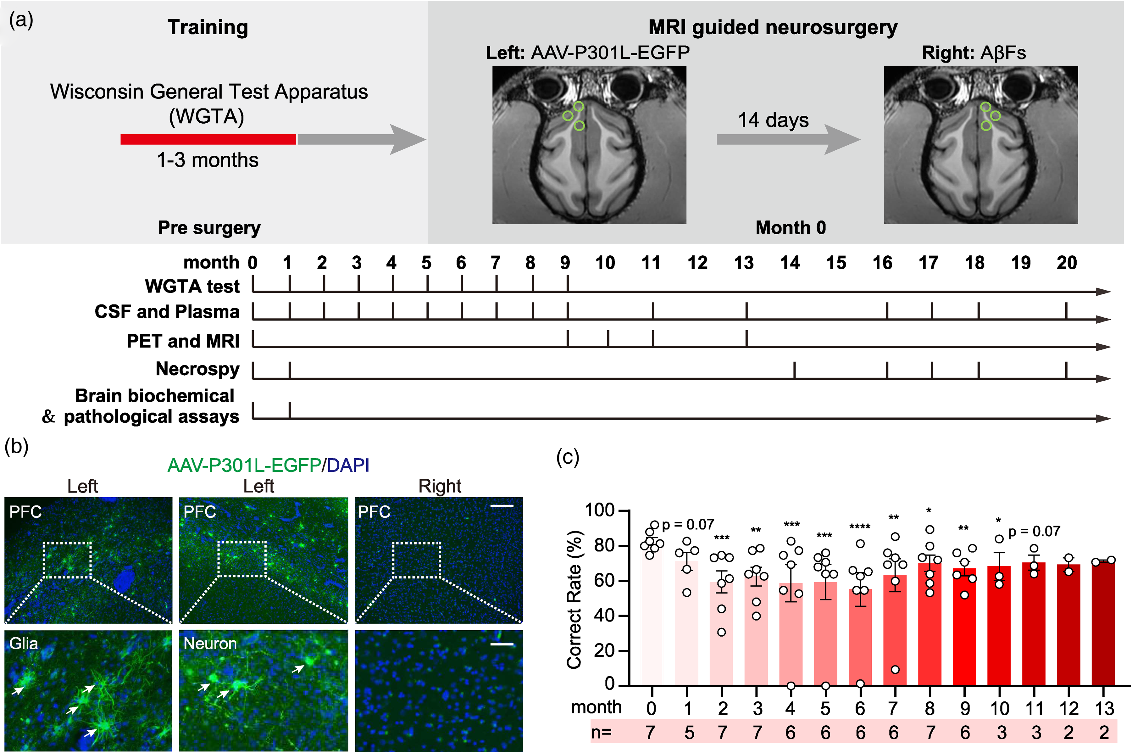
Bilateral PFC infusion of AAV-P301L-tau and AβFs induced a durable cognitive deficit in cynomolgus monkeys. (a) Schematics show experimental procedure. The male cynomolgus monkeys (15∼18 years old) were trained by Wisconsin General Test Apparatus (WGTA) for 1∼3 months to remember the slots where the foods were hidden, and then AAV-P301L-EGFP and AβFs (Aβ fibers) were respectively infused into the left and right prefrontal cortex (PFC) using MRI-guided stereotaxic apparatus. The following behavioral tests, CSF and plasma collection, PET/MRI evaluation, euthanasia and necropsy, and brain metabolism and pathology measurements were executed as indicated. (b) Representative images show robust expression of GFP throughout the PFC measured at 1 month after the infusion. Scale bar, 250 μm (upper), 50 μm (lower). (c) An enduring cognitive deficit was detected by WGTA at different time-points after brain PFC infusion of AAV-P301L-EGFP and AβFs. Each dot represents an individual animal. *p < 0.05; **p < 0.01; ***p < 0.001; ****p < 0.0001. One-way ANOVA with Fisher LSD test. N = 5∼7 monkeys per group.
PFC or PFC/hippocampal or hippocampal infusion of AAV-P301L-tau and AβFs or AβOs simulated Ad-like brain tau accumulation with increased tau in CSF and plasma
We first explored whether bilateral PFC infusion of AAV-P301L-tau plus AβFs could produce AD-like brain and CSF/plasma tau pathologies by using 18F-T807 PET scan, western blotting and MSD assays. 18F-T807 is a widely used radioactive tracer in brain PET scans to identify the presence and distribution of tau pathology, the salient features of AD.33,34
The linear color scale of the standardized uptake value ratio (SUVr) of 18F-T807 PET scan exhibited a significant tau accumulation in multiple brain regions of the monkeys measured from 9 to 13 months after the bilateral PFC AAV-P301L and AβFs-infusion (Figure 2(a) and (b)). The increased levels of total tau (tTau) probed by HT7, and pTau at pTau217, pTau181, pTau396 in the brain tissue were also detected at one month after the modeling by western blotting in the bilateral PFC and the hippocampus in the modeled monkeys (Figure 2(c)–(e) and Supplemental Figure 1). Interestingly, the tTau level probed by Tau5 was not significantly changed in the AAV-Tau-treated PFC and hippocampus (Figure 2(c)) though the increase was detected in the AβF-treated PFC and hippocampus (Figure 2(d), (e) and Supplemental Figure 1); six and three isoforms of tau proteins were detected respectively in the PFC and hippocampus of the control monkeys by HT7, an antibody produced specifically against human tau, and a significant mobility upshift was shown in the model monkeys (Figure 2(c)–(e) and Supplemental Figure 1); a significantly increased pTau404 was detected in the hippocampus of the model monkeys but the increase in the PFC was not as significant as the hippocampus (Figure 2(c)–(e) and Supplemental Figure 1); as for pTau181, an increased high molecular weight band (∼140 kDa) was detected in the AβF- but not in AAV-Tau-treated PFC or hippocampus (Figure 2(d) and Supplemental Figure 1). These data together demonstrate that bilateral PFC infusion of AAV-P301L-tau plus AβFs could induce AD-like tau pathologies. Moreover, the isoforms of primate tau proteins are more similar to those of humans, and the expression and phosphorylation patterns of tau are different in distinct brain regions of the monkeys.
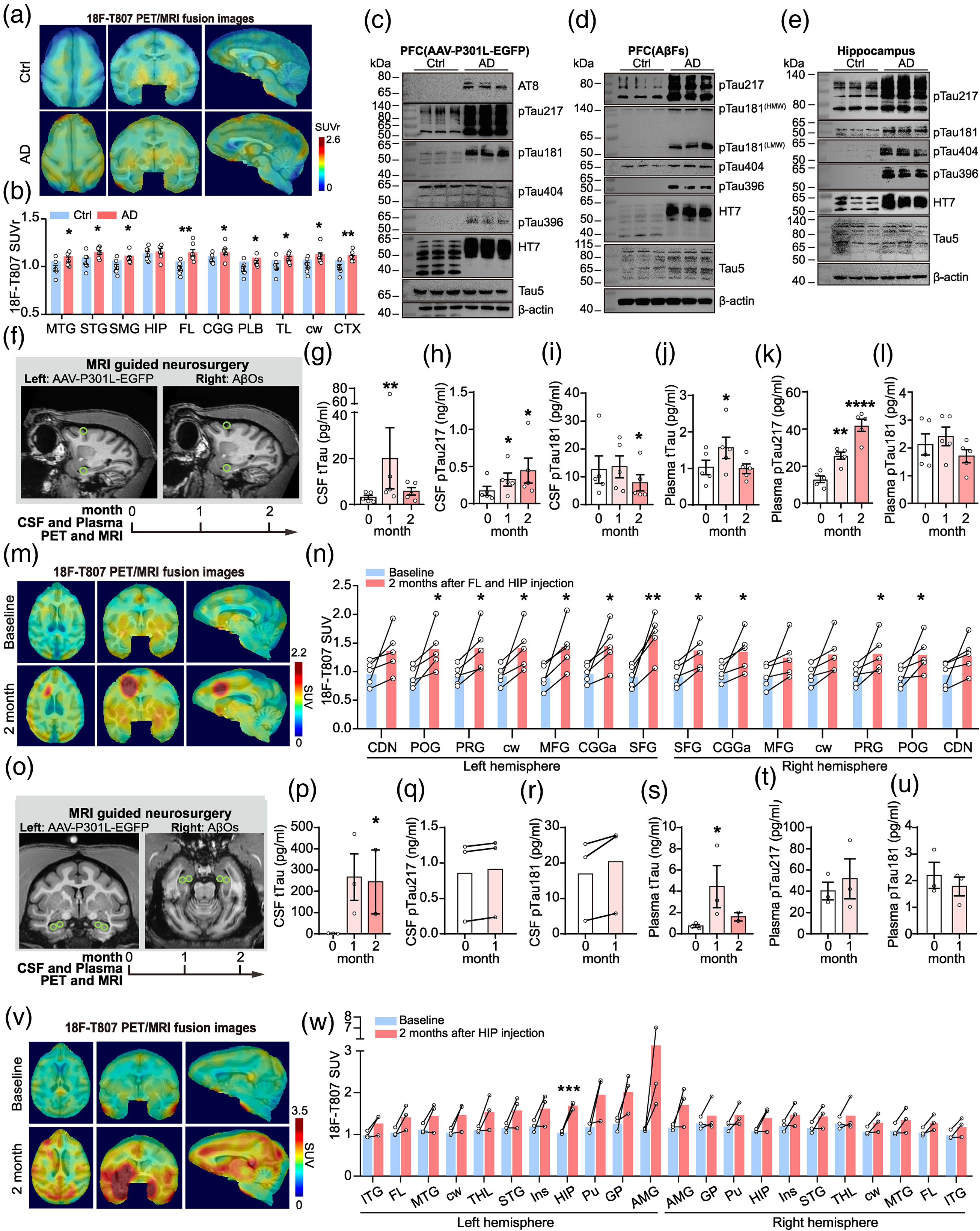
Bilateral PFC or PFC/hippocampal or hippocampal infusion of AAV-P301L-tau and AβFs/AβOs increased brain tau accumulation with an increased tau release into the CSF and plasma. (a, b) Representative 18F-T807 PET/MRI fusion images indicated an increased tau accumulation in the monkey brain measured at 9∼13 months after bilateral PFC infusion of AAV-P301L and AβFs. The linear color scale of the standardized uptake value ratio (SUVr) at selected brain regions (referred to pons) and the changes of 18F-T807 SUVr in the selected brain regions were shown. MTG: middle temporal gyrus; STG: superior temporal gyrus; SMG: supramarginal gyrus; HIP: hippocampus; FL: frontal lobe; CGG: cingulate gyrus; PLB: parietal lobe; TL: temporal lobe; cw: cerebral white matter; CTX: cerebral cortex. *p < 0.05. **p < 0.01. Student's t test. N = 7 monkeys per group. (c, d) The increased total tau (tTau) and the pTau levels in the AAV-P301L or AβFs-infused PFC extracts measured at 1 month after bilateral PFC infusion of AAV-P301L and AβFs by western blotting. Tau5 probes total tau, and HT7 reacts with human tau proteins. (e) The increased tTau and pTau levels in the hippocampus measured at 1 month by western blotting after bilateral PFC infusion of AAV-P301L and AβFs. (f) The coordinates for stereotaxic PFC plus hippocampal infusion of AAV-P301L and AβOs guided by T1-weighted MRI, and the procedure schematics. (g–l) The increased CSF and plasma tTau and pTau levels detected by MSD at the indicated time points after PFC plus hippocampal infusion of AAV-P301L and AβOs. Each dot represents an individual animal. (m, n) An increased tau accumulation detected at 2 months after PFC and hippocampal infusion of AAV-P301L and AβOs measured by 18F-T807 PET/MRI. The linear colour scale with the standardized uptake value range and the changes of 18F-T807 SUV in the selected brain regions were shown. CDN: caudate nucleus; POG: postcentral gyrus; PRG: precentral gyrus; cw: cerebral white matter; MFG: middle frontal gyrus; CGGa: anterior cingulate gyrus; SFG: superior frontal gyrus. (o) The coordinates for stereotaxic hippocampal infusion of AAV-P301L and AβOs guided by T1-weighted MRI, and the procedure schematics. (p–u) The increased CSF and plasma total tau and pTau proteins detected by MSD at the indicated time points after hippocampal infusion of AAV-P301L and AβOs. Each dot represents an individual animal. (v, w) The increased tau deposition measured by 18F-T807 PET/MRI at 2 months after hippocampal infusion of AAV-P301L and AβOs. The linear color scale with the standardized uptake value range and 18F- T807 SUV changes in the selected brain regions were shown. ITG: inferior temporal gyrus; FL: frontal lobe; MTG: middle temporal gyrus; cw: cerebral white matter; THL: thalamus; STG: superior temporal gyrus; Ins: insula; HIP: hippocampus; Pu: putamen; GP: globus pallidus; AMG: amygdala.
Then, we tested whether simultaneous infusion of AAV-P301L plus AβOs into PFC and hippocampus could also induce AD-like pathologies. Guided by T1-weighted MRI (Figure 2(f)), we performed brain infusion, and then we tested tau level in CSF and plasma at 1 and 2 months after the infusion. An increased tTau and pTau217 but not pTau181 was detected in both CSF and plasma by MSD assay (Figure 2(g)–(l)), with a simultaneous tau accumulation in multiple brain regions measured by 18F-T807 PET/MRI at 2 months after infusion (Figure 2(m) and (n)). These data suggest that simultaneous infusion of AAV-P301L plus AβOs into PFC and hippocampus could increase pTau217 at one and two months with an increased tTau at one month in both CSF and plasma, with simultaneous brain tau accumulation after the modeling.
As hippocampus is the involved brain region in early stage of AD, we then studied whether infusion of AAV-P301L and AβOs into the hippocampus alone could induce AD-like pathology. By MRI-guided neurosurgery, we infused AAV-P301L into the left hippocampus and AβOs into the right hippocampus (Figure 2(o)). A significantly increased tTau was detected in both CSF and plasma at 1 or/and 2 months, whereas the changes of pTau217 and pTau181 were not consistent after the modeling (Figure 2(p)–(u)). Simultaneously, hippocampal tau accumulation was also detected by 18F-T807 PET MRI at 2 months after the infusion (Figure 2(v) and (w)). These data suggest that infusion of hippocampal alone could induce tau accumulation at multiple brain regions within 2 months with an inconsistent CSF and plasma tau changes.
Together, these data demonstrate that bilateral PFC, or PFC plus hippocampus, or hippocampus alone infusion of AAV-P301L-tau and AβFs or AβOs can efficiently induce the AD-like brain tau accumulation with increased tTau and pTau levels in the CSF and plasma.
PFC or PFC/hippocampal or hippocampal infusion of AAV-P301L-tau and AβFs or AβOs simulated Ad-like brain Aβ accumulation with altered Aβ level in CSF and plasma
The increased brain Aβ deposit forming extracellular plaques with reduced Aβ42 and Aβ42/Aβ40 ratio in CSF have been applied for clinical diagnosis of AD, 35 therefore, we measured the levels of Aβ40, Aβ42 and the ratio of Aβ42/Aβ40 in the monkeys CSF from 0 to 20 months after bilateral PFC infusion of AAV-P301L-tau and AβFs. No significant change was induced in Aβ42 and Aβ40 levels, although an increased trend in Aβ42/Aβ40 ratio was detected in CSF at 4, 5, and 6 months after bilateral PFC AAV-P301L-tau and AβFs infusion (Figure 3(a)–(c)). By using 18F-AV45, an amyloid tracer developed to map the burden of Aβ plaques in the living human brain, 36 we found a stable Aβ accumulation in multiple brain regions of the monkeys at 9 to 13 months after bilateral PFC AAV-P301L-tau and AβFs-infusion (Figure 3(d) and (e)). Additionally, the models also showed a significantly increased staining by thioflavin-S (ThioS) (Figure 3(f)), a dye specifically binds to β-flaky structures.37,38
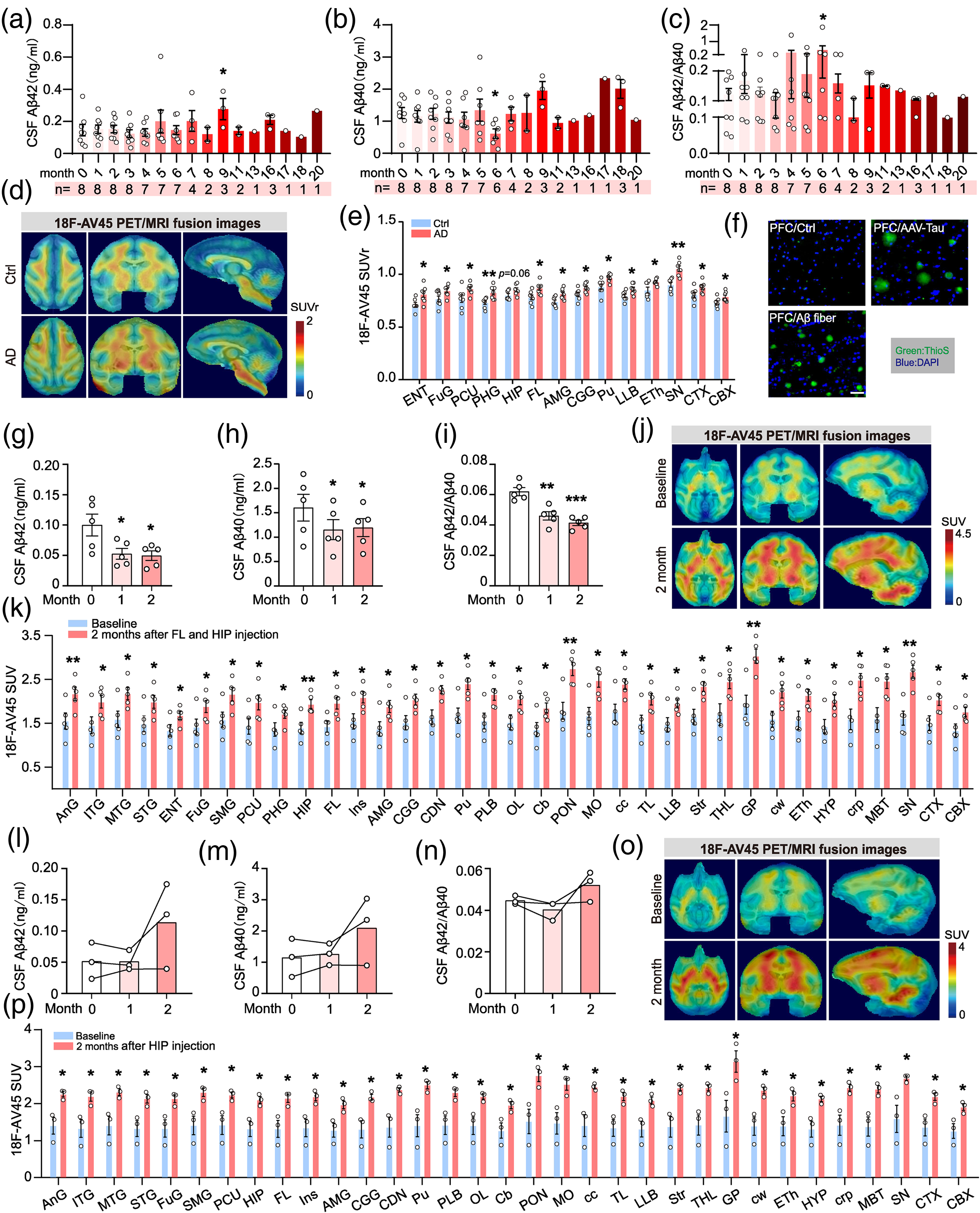
Bilateral PFC or PFC/hippocampal or hippocampal infusion of AAV-P301L-tau and AβFs/AβOs increased brain Aβ accumulation with altered Aβ levels in CSF and plasma. (a–c) The altered total levels of Aβ42 (a) and Aβ40 (o), and the ratio of Aβ42/Aβ40 (c) in monkey CSF after bilaterally infused with AAV-P301L and AβFs in PFC for 0∼20 months. N = 1∼8. (d, e) The increased Aβ deposition in the monkey brain bilaterally infused with AAV-P301L and AβFs in PFC for 9∼13 months. The linear color scale with SUVr range (referred to pons) and changes of 18F-AV45 SUVr in the selected brain regions were shown. ENT: entorhinal area; FuG: fusiform gyrus; PCU: precuneus; PHG: parahippocampal gyrus; HIP: hippocampus; FL: frontal lobe; AMG: amygdala; CGG: cingulate gyrus; Pu: putamen; PLB: parietal lobe; LLB: limbic lobe; ETh: epithalamus; SN: substantia nigra; CTX: cerebral cortex; CBX: cerebellar cortex. N = 5. (f) The fibrillar Aβ deposits in the monkey PFC detected by Thioflavin-S (ThioS) staining at 1 month after bilateral PFC infusion of AAV-P301L and AβFs. Scale bar, 50 μm. (g–i) The altered total levels of Aβ42 (h) and Aβ40 (i), and the ratio of Aβ42/Aβ40 (j) in the monkey CSF after AAV-P301L and AβOs infusion in PFC and hippocampus, respectively. N = 5. (j, k) The increased Aβ deposition in the monkey brain measured 2 months after AAV-P301L and AβOs infusion in PFC and hippocampus, respectively. The linear color scale with SUV range and changes of SUV for 18F-AV45 in the selected brain regions were shown. AnG: angular gyrus; ITG: inferior temporal gyrus; MTG: middle temporal gyrus; STG: superior temporal gyrus; ENT: entorhinal area; FuG: fusiform gyrus; SMG: supramarginal gyrus; PCU: precuneus; PHG: parahippocampal gyrus; HIP: hippocampus; FL: frontal lobe; Ins: Insula; AMG: amygdala; CGG: cingulate gyrus; CDN: caudate nucleus; Pu: putamen; PLB: parietal lobe; OL: occipital lobe; Cb: Cerebellum; PON: pons; MO: medulla; cc: corpus callosum; TL: temporal lobe; LLB: limbic lobe; Str: striatum; THL: thalamus; GP: globus pallidus; cw: cerebral white matter; ETh: epithalamus; HYP: hypothalamus; crp: cerebral peduncle; MBT: midbrain tegmentum; SN: substantia nigra; CTX: cerebral cortex; CBX: cerebellar cortex. N = 5. (l–n) The altered total levels of Aβ42 (l) and Aβ40 (m), and the ratio of Aβ42/Aβ40 (n) in CSF of monkey bilaterally infused with AAV-P301L and AβOs in hippocampus for 0∼2 months. N = 3. (o, p) The increased Aβ deposition in the monkey brain measured by 18F-AV45 PET/MRI at 2 months after the hippocampal infusion. The linear color scale with the standardized uptake value (SUV) range and the changes of treated monkeys in SUV for 18F-AV45 in the selected brain regions were shown. AnG: angular gyrus; ITG: inferior temporal gyrus; MTG: middle temporal gyrus; STG: superior temporal gyrus; FuG: fusiform gyrus; SMG: supramarginal gyrus; PCU: precuneus; HIP: hippocampus; FL: frontal lobe; Ins: Insula; AMG: amygdala; CGG: cingulate gyrus; CDN: caudate nucleus; Pu: putamen; PLB: parietal lobe; OL: occipital lobe; Cb: cerebellum; PON: pons; MO: medulla; cc: corpus callosum; TL: temporal lobe; LLB: limbic lobe; Str: striatum; THL: thalamus; GP: globus pallidus; cw: cerebral white matter; ETh: epithalamus; HYP: hypothalamus; crp: cerebral peduncle; MBT: midbrain tegmentum; SN: substantia nigra; CTX: cerebral cortex; CBX: cerebellar cortex. N = 3.
In the model with PFC and hippocampal infusion of AAV-P301L-tau and AβOs, a significant reduced CSF Aβ42, Aβ40, and Aβ42/Aβ40 was detected at 1 and 2 months after the infusion (Figure 3(g)–(i)). Simultaneously, multiple brain Aβ accumulation was shown by 18F-AV45 PET/MRI (Figure 3(j) and (k)).
In the model with hippocampal alone infusion of AAV-P301L-tau and AβOs, we observed a slightly increased Aβ42, Aβ40, and Aβ42/Aβ40 in CSF of the monkeys measured at 2 months by MSD assay (Figure 3(l)–(n)). Simultaneously, a significant brain Aβ deposition was shown by 18F-AV45 PET/MRI (Figure 3(o) and (p)).
These findings suggest that bilateral PFC, or PFC plus hippocampal, or hippocampal alone infusion of AAV-P301L-tau and AβFs or AβOs can efficiently induce the AD-like brain Aβ accumulation, while the changes of Aβ in CSF were depended on the paradigms used for modeling.
PFC or PFC/hippocampal or hippocampal infusion of AAV-P301L-tau and AβFs or AβOs simulated Ad-like neurodegeneration shown by the increased NfL in CSF and plasma
Recently, an increased NfL level in CSF and plasma has been recommended as a quantitative biomarker of neurodegeneration.39,40 Therefore, we tested the NfL level in the brain tissue, CSF and plasma of the monkeys after bilateral PFC infusion of AAV-P301L-tau and AβFs at different time points. A significantly increased NfL in CSF was already detected at 1 month after the modeling, and this increase was lasted for at least 8 months (Figure 4(a)). On the other hand, the significantly increased NfL in the plasma was only detected at 2 months after the modeling (Figure 4(b)). In brain tissue, western blotting detected a significant decrease in NfL at the PFC (AAV-P301L-tau-infused site) and a slight increase in NfL at the PFC (AβFs-infused site), but the changes in the PFC of the AAV-P301L-tau infused site and the hippocampus were not significant (Figure 4(c)–(h)). Interestingly, the level of NfH (heaving chain of neurofilament) was significantly increased in the bilateral PFC and hippocampus of the model monkeys (Figure 4(c)–(h)).
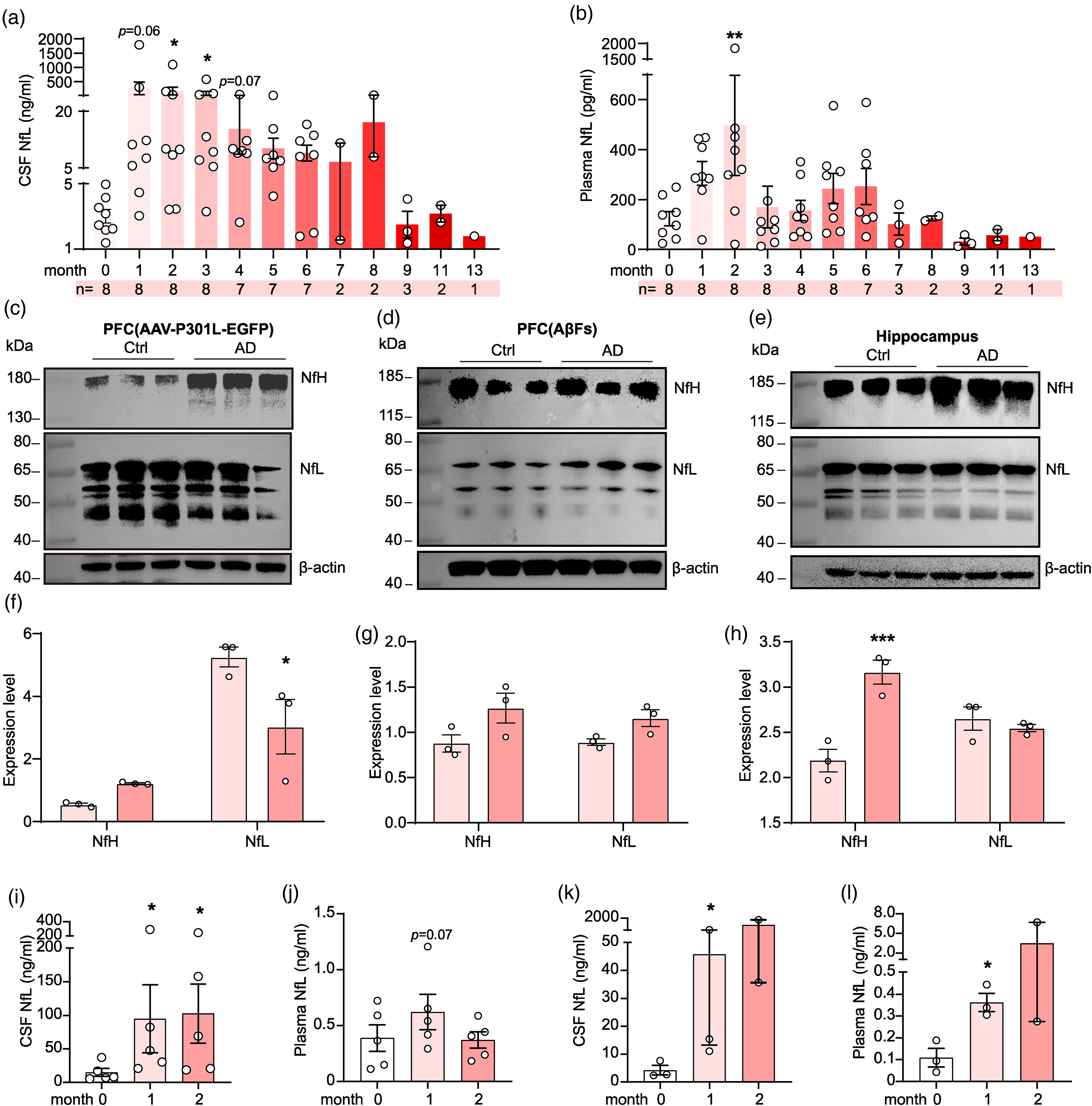
PFC or PFC/hippocampal or hippocampal infusion of AAV-P301L-tau and AβFs/AβOs induced a consistent increase of NfL in CSF and plasma. (a, b) Bilateral PFC infusion of AAV-P301L and AβFs induced a constant increase of NfL in CSF and plasma of the monkeys measured by MSD. Each dot represents an individual animal. *p < 0.05; **p < 0.01. One-way ANOVA with Dunnett's multiple comparisons test. N = 1∼8 monkeys per group. (c–h) Bilateral PFC infusion of AAV-P301L and AβFs slightly increased NfL in the PFC and hippocampus of the monkeys measured at 1 month by western blotting. (i, j) PFC plus hippocampal infusion of AAV-P301L and AβOs significantly increased CSF but not plasma NfL level in monkeys measured by MSD at 2 months. Each dot represents an individual animal. *p < 0.05; One-way ANOVA with Dunnett's multiple comparisons test. N = 5 monkeys per group. (k, l) Bilateral hippocampal infusion AAV-P301L and AβOs significantly increased NfL level in monkey CSF and plasma measured by MSD at the indicated time points. Each dot represents an individual animal. *p < 0.05; One-way ANOVA with Dunnett's multiple comparisons test. N = 2∼3 monkeys per group.
In the model with PFC and hippocampal infusion of AAV-P301L-tau and AβOs, a significant increase in CSF NfL level was detected at 1 and 2 months after modeling, but no significant change in plasma NfL level (Figure 4(i) and (j)). Interestingly, a significantly increased CSF and plasma NfL was detected at 1 and 2 months in the model with only hippocampal infusion of AAV-P301L-tau and AβOs (Figure 4(k) and (l)).
These data together indicate that bilateral PFC, or PFC plus hippocampal, or only hippocampal infusion of AAV-P301L-tau and AβFs or AβOs can efficiently induce AD-like neurodegeneration demonstrated by the increased NfL in CSF, plasma with an increased NFH in the brain, though different models show slightly different results.
PFC or PFC/hippocampal or hippocampal infusion of AAV-P301L-tau and AβFs or AβOs induced an extensive glial activation
A strocytes and microglial activation, indicating inflammation, are commonly seen in the brains of AD patients and the AD transgenic mice. 41 Therefore, we measured the levels of glial fibrillary protein (GFAP, an astrocyte marker) and ionized calcium-binding adaptor molecule 1 (Iba1, a microglial marker) at 1 month after bilateral PFC infusion of AAV-P301L-tau and AβFs by immunohistochemical staining. 42 Significant activation of astrocytes and microglia was detected in both gray and white matter of PFC at 1 month after the infusion (Figure 5(a)–(d)). We also measured GFAP levels in CSF and plasma in the monkeys with PFC and hippocampal infusion of AAV-P301L-tau and AβOs at 0, 1, or 2 months, a slightly increased GFAP was detected in CSF and plasma (Figure 5(e) and (f)). In the models with only hippocampal infusion of AAV-P301L-tau and AβOs, a significantly increased GFAP was detected in CSF and plasma of monkeys (Figure 5(g) and (h)).
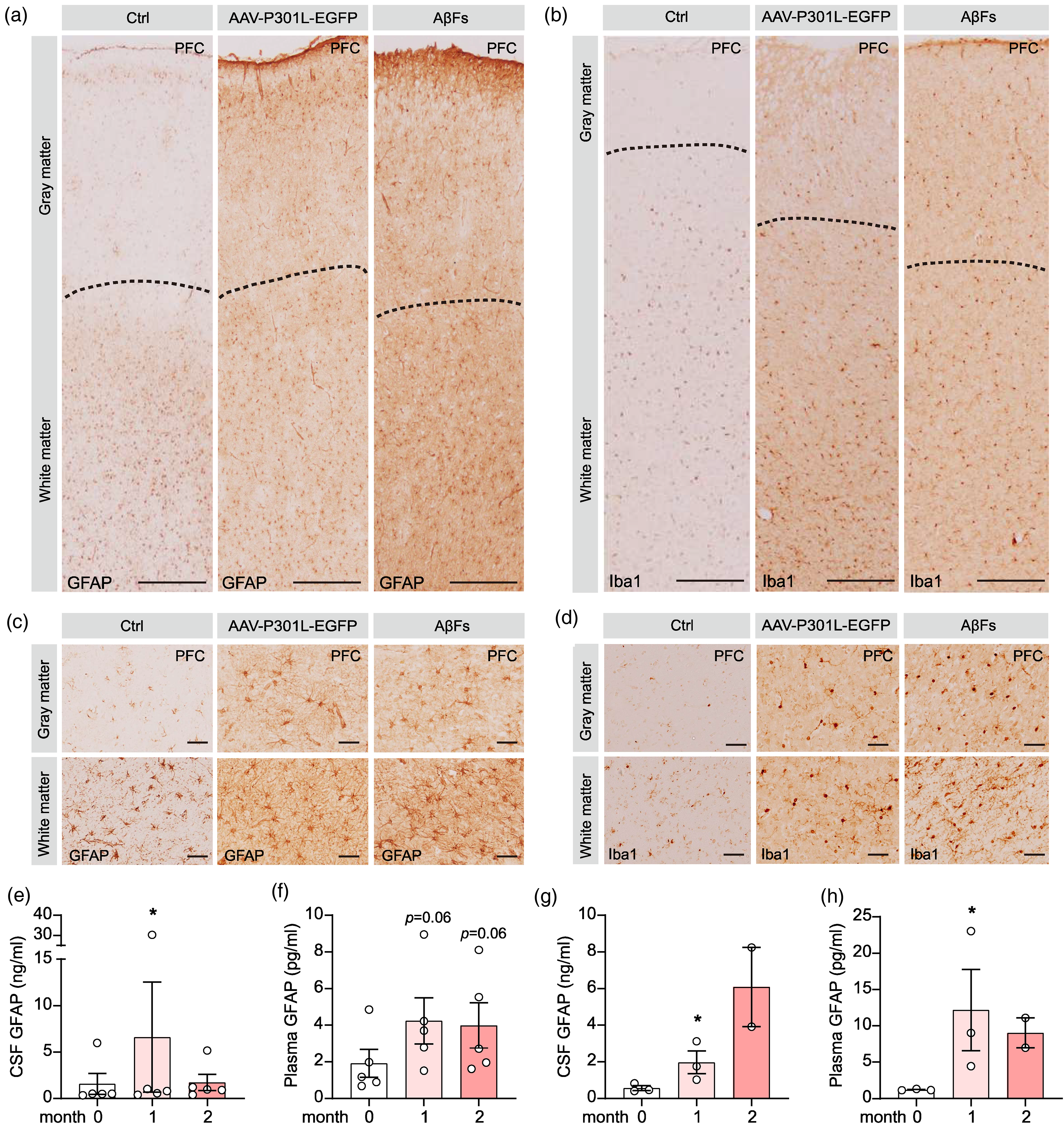
PFC or PFC/hippocampal or hippocampal infusion of AAV-P301L and AβFs/AβOs induced an extensive glial activation. (a–d) Robust activation of astrocytes and microglia was detected in the PFC subset of the monkeys by immunohistochemical analysis at 1 month after bilateral PFC infusion of AAV-P301L and AβFs. Scale bar, 500 μm (a), 50 μm (b). (e, f) The increased GFAP protein level in CSF and plasma of the monkeys measured by MSD at the indicated time points after PFC plus hippocampal infusion of AAV-P301L and AβOs. Each dot represents an individual animal. *p < 0.05; One-way ANOVA with Dunnett's multiple comparisons. N = 5 monkeys per group. (g, h) The increased GFAP protein level in CSF and plasma of the monkeys measured by MSD at the indicated time points after hippocampal infusion of AAV-P301L and AβOs. Each dot represents an individual animal. *p < 0.05; One-way ANOVA with Dunnett's multiple comparisons. N = 2∼3 monkeys per group.
These data together suggest that bilateral PFC or PFC plus hippocampal or hippocampal infusion alone of AAV-P301L-tau and AβFs or AβOs induced a widespread activation of astrocytes and microglia, as seen in the AD brains.
Discussion
In the present study, we constructed monkey models by simultaneous infusion of AAV-Tau, AβFs and/or AβOs into the bilateral PFC, or PFC plus hippocampus, or hippocampus alone of the monkeys. By measuring the AD-like cognitive deficits and hallmark pathologies at different time points after the modeling, we found that simultaneous infusion of AAV-Tau, AβFs and/or AβOs can synergically promote multiple AD-like neuropathic features, including Aβ, tau, NfL, and inflammations, with a lasting cognitive deficit. The previous AD-like monkey models made by transgenic could not achieve mass production, 28 or did not simultaneously induce all AD-like neuropathologies with behavioral deficits by separately brain injection of Aβ and tau.29–31 To our best knowledge, these are the first reported mass-producible monkey models showing a durable AD-like hallmark pathologies (Aβ, tau, NfL, inflammation) and cognitive deficit. As monkeys are genetically and metabolically the closest to humans, these models may bring about more effective AD drug discovery and development.
Rodents as disease models have been widely used in biomedicine- and drug-associated research because of their low price, wide resources, high survival rate, clear genetic background, mature gene operation and low feeding cost. 43 The current laboratory used AD rodent models include APP Tg2576, APP/presenilin 1 (APP/PS1), 3Tg x AD, hTau-Tg, non-Tg mice treated with injections of Aβ or tau, and models of aging. These rodent models have made great contributions for understanding the pathogenesis of AD onset and the progression. However, the clinical drug development based on these rodent AD models have been disappointing. One of the major reasons causing this situation may be due to the great genetic and metabolic differences between rodents and humans, therefore, they cannot simulate the real situation of human patients in many aspects.44–47
Non-human primates have similar brain structures to humans, with developed prefrontal cortex and hippocampus, advanced brain functions and high interoperability, therefore, they can complete complex behavioral tests and display human-like brain pathologies. For instance, the amino acid sequences of Aβ and tau in non-human primates are respectively 100% and 98% consistent with those of humans, and the human-like precipitation of Aβ and pTau have been detected in the brain of old monkeys. 48 Although the old age-dependent spontaneous monkey model has shown great potential for AD research, the procedures have several insurmountable problems, such as time-consume, individual differences, high costs, and unable to be mass-produced, etc.24–27 Therefore, scientists have been trying to experimentally simulate the monkey models by intracranial injection of Aβ or tau. Limited by the reproductive characteristics of monkeys, transgenic technology cannot achieve mass production. 28 On the other hand, the monkey models produced by intracranial injection of Aβ or tau independently did not show simultaneous Aβ and tau pathology with cognitive deficits.28–31 To remedy these deficiencies, we successfully developed monkey models showing simultaneously AD-like cognitive deficits and all kinds of hallmark neuropathological features, including Aβ, pTau, neurodegenerations, and neural inflammation. As these monkey models meet all clinical diagnosis criteria of AD, they can be used for AD drug development.
Hippocampus is the early involved brain region in AD onset, and the hippocampal damage will cause spatial cognitive dysfunction in AD patients. PFC is closely related to long-term memory, and PFC disruption will lead to AD-like memory deficits. Therefore, we chose to infuse Aβ and tau simultaneously through PFC and/or hippocampus in the present study. Although three different paradigms, i.e., bilateral PFC infusion of Aβ and tau, PFC plus hippocampal infusion of Aβ and tau, and only bilateral hippocampal infusion of Aβ and tau, could efficiently produce the AD-like pathologies, they showed slight differences. For instance, bilateral PFC and PFC plus hippocampus infusion of Aβ and tau induced a reduced CSF Aβ level, which was similar to that observed in AD patients.49,50 However, an increased CSF Aβ was detected by only hippocampal infusion of Aβ and tau. Though the reason for this discrepancy is currently not fully understood, this particular monkey model is valuable for evaluating the efficiency of Aβ-targeting drugs.
Correlated with the intracellular accumulation of the hyperphosphorylated tau, cognitive impairment is an important behavioral feature of the AD patients. 51 Therefore, cognitive improvement is a major indicator for evaluating drug efficiency regardless its underlying pathological mechanisms. WGTA test has been used to assess primate learning and memory ability. 52 In the present study, we also used WGTA to measure the cognitive functions of the monkeys. We found that bilateral PFC overexpressing AAV-P301L-Tau and infusing Aβ successfully induced cognitive impairment in moneys, and the induced cognitive deficit persisted for at least 10 months which is more than enough for evaluating drug efficiency. The cognitive dysfunction in the monkey models produced by hippocampal infusion alone or hippocampus plus PFC infusion was expected based on the observed Aβ and tau pathologies, which will be confirmed in future studies. In addition to WGTA, we also developed touch screen test to measure the cognitive functions for our future studies. 53
PET/MRI can dynamically monitor non-invasively tau and Aβ aggregation inside the brains with high accuracy. In the current study, we also employed PET/MRI to detect precipitation of Aβ and tau proteins. By measuring SUV and SUVr as reported from the previous studies,54–56 we could also detect Aβ and tau accumulation in different brain regions inside and beyond the infusion sites. The long-term PET/MRI monitoring, Aβ/tau and behavioral tests for the monkey models produced by PFC plus hippocampal infusion of and hippocampal infusion alone of AAV-P301L-Tau and AβOs will be continued in future studies.
In conclusion, we produced in the present study monkey models showing AD-like cognitive deficits and hallmark neuropathologies, including an increased level of total tau and pTau, an increased NfL in the brain, CSF and plasma, and an increased brain Aβ accumulation with altered Aβ levels (increase or decrease) in CSF. As these monkey models are mass-producible, they can be used for evaluating AD drug efficiency.
Supplemental Material
sj-docx-1-alz-10.1177_13872877251334316 - Supplemental material for A mass-producible macaque model displays a durable Alzheimer-like cognitive deficit and hallmark amyloid-β/tau/neurofilament light chain pathologies
Supplemental material, sj-docx-1-alz-10.1177_13872877251334316 for A mass-producible macaque model displays a durable Alzheimer-like cognitive deficit and hallmark amyloid-β/tau/neurofilament light chain pathologies by Feng He, Wen-Jiao Shi, Wen Liu, Jing-Xin Fan, Zhi-Gang He, Ya-Qi Zhang, Jing Xiao, Wei-Wei Ruan, Yong-Kang Gai, Hong-Li Zhang, Bin-Bin Yang, Yao Qin, Hao Wang, Jia Li, Jun-Li Wang, Sha Liu, Li-Ping Shi, Zhong-Xu Chen, Wei-Jie Jiang, Ni An, Peng-Jing Xue, Zi-Hao Wang, Rui-Jie Yang, Peng-Yu Tian, Zhu Chen, Ling Xiao, Zheng-Sheng Yang, Kang-Bo Feng, Wei-Ye Tan, Zhan-Meng Sun, Wei Xu, Huaqing Shu and Jian-Zhi Wang in Journal of Alzheimer's Disease
Footnotes
Acknowledgments
The authors have no acknowledgments to report.

Ethical considerations
The study has been approved by the Institutional Animal Care and Use Committee of Hubei Topgene Biotechnology (IACUC-YJY-2022-001).
Consent to participate
Not applicable.
Consent for publication
Not applicable.
Author contributions
Feng He (Investigation; Writing – original draft); Wen-Jiao Shi (Resources); Wen Liu (Data curation; Formal analysis; Investigation; Methodology; Software); Jing-Xin Fan (Data curation; Formal analysis); Zhi-Gang He (Formal analysis; Investigation); Ya-Qi Zhang (Formal analysis; Investigation; Methodology); Jing Xiao (Investigation; Methodology); Wei-Wei Ruan (Investigation; Methodology); Yong-Kang Gai (Investigation; Methodology); Hong-Li Zhang (Investigation; Methodology); Bin-Bin Yang (Investigation; Methodology); Yao Qin (Investigation; Methodology); Hao Wang (Investigation; Methodology); Jia Li (Investigation; Methodology); Jun-Li Wang (Investigation; Methodology); Sha Liu (Investigation; Methodology); Li-Ping Shi (Investigation; Methodology); Zhong-Xu Chen (Investigation; Methodology); Wei-Jie Jiang (Investigation; Methodology); Ni An (Investigation; Methodology); Peng-Jing Xue (Investigation; Methodology); Zi-Hao Wang (Investigation; Methodology); Rui-Jie Yang (Investigation; Methodology); Peng-Yu Tian (Investigation; Methodology); Zhu Chen (Investigation; Methodology); Ling Xiao (Investigation; Methodology); Zheng-Sheng Yang (Investigation; Methodology); Kang-Bo Feng (Investigation; Methodology); Wei-Ye Tan (Investigation; Methodology); Zhan-Meng Sun (Investigation; Methodology); Wei Xu (Investigation; Methodology); Huaqing Shu (Conceptualization; Validation; Visualization); Jian-Zhi Wang (Conceptualization; Methodology; Project administration; Supervision; Validation; Visualization; Writing - review & editing).
Funding
The author(s) disclosed receipt of the following financial support for the research, authorship, and/or publication of this article: This study was supported by the National Natural Science Foundation of China (No. 82372081; No. 82230041; No. 91949205) and the Science and Technology Innovation 2030-Major Projects (STI2030-Major Projects-2022ZD0211800).
Declaration of conflicting interests
The author(s) declared no potential conflicts of interest with respect to the research, authorship, and/or publication of this article.
Data availability
The data supporting the findings of this study are available on request from the corresponding author.
Supplemental material
Supplemental material for this article is available online.
References
Supplementary Material
Please find the following supplemental material available below.
For Open Access articles published under a Creative Commons License, all supplemental material carries the same license as the article it is associated with.
For non-Open Access articles published, all supplemental material carries a non-exclusive license, and permission requests for re-use of supplemental material or any part of supplemental material shall be sent directly to the copyright owner as specified in the copyright notice associated with the article.
