



Abstract
Background
No effective drug treatment is currently available for Alzheimer's disease (AD), highlighting the urgent need to develop efficient therapeutic options. We have developed a formula based on medicine and food homology (MFH) consisting of egg yolk oil, perilla seed oil, raphani seed oil, cinnamon oil, and noni puree (EPRCN), and demonstrated that it can treat AD by alleviating neuroinflammation and oxidative stress. However, whether EPRCN can improve AD by regulating gut microbiota remains unknown.
Objective
The current study aimed to evaluate the effect of EPRCN on regulating gut microbiota and neuroprotection.
Methods
16S rRNA sequencing was used to assess the structure of gut microbiota. Hematoxylin-eosin (HE) staining, qRT-PCR, and ELISA were used to evaluate gut inflammation. Detected indexes associated with cholinergic dysfunction and neuronal damage to investigate the neuroprotective effects of EPRCN.
Results
16S rRNA gene analysis revealed that EPRCN remodeled the gut microbiota, inhibited gut metabolic disorders, and promoted CoA biosynthesis in scopolamine-induced mice. EPRCN can ameliorates gut inflammation by activating the cholinergic anti-inflammatory pathway. The results further indicated that EPRCN improved cholinergic dysfunction by inhibiting the activity of acetylcholinesterase and restoring cholinergic receptors. Additionally, EPRCN administration suppressed the neuronal loss and elevated brain derived neurotrophic factor expression in hippocampus. Correlation analysis found that alteration of several gut microbes was associated with indexes improved by EPRCN.
Conclusions
These findings suggest that EPRCN may serve as a promising dietary intervention for treating AD by regulating the microbiota-gut-brain axis and exerting neuroprotective function.
Keywords
Introduction
Alzheimer's disease (AD) is the most prevalent manifestation of dementia, and its clinical presentation is progressive cognitive dysfunction. 1 The clinical manifestations of AD include dysfunction and eventual failure of both neurochemical and structural neural networks, including the cholinergic dysfunction and neuronal damage. 2 Disorders in hippocampal cholinergic transmission impair the encoding of information and memory. 3 Acetylcholine (ACh) deficiency disorders extratelencephalic projection neurons in the cortex and further induced cognitive deficits in AD mice. 4 Together with cholinergic dysfunction, neuronal damage plays an important role in AD pathogenesis. Evidence from both AD patients and corresponding animal models indicates that neuronal death is an early AD-related processes. 5 Emerging evidence indicates that the homeostasis of intestinal immune environment plays a crucial role in the bidirectional communication between the gut and the brain. 6 Chronic gut inflammation can induce disorders in the intestinal barrier, leading to increased intestinal permeability. This condition allows for the invasion of bacteria, viruses, and their neuroactive products, which can trigger neuroinflammatory reactions in the central nervous system (CNS). 7 Gut microbiota are considered to contribute to homeostasis of intestinal immune environment through the production of microbial metabolites. 8 An increase in the abundance of butyrate-producing bacteria, such as Lachnospiraceae_NK4A136_group was associated with the inhibition of gut inflammation. 9 Furthermore, research has demonstrated that gut microbiota possess the ability to produce ACh, which can regulate the cholinergic anti-inflammatory pathway, thereby influencing the intestinal immune environment. 10
Intraperitoneal injection of scopolamine is capable of causing learning and memory disorders in mice. 11 Scopolamine administration was be considered as a psychopharmacological model of AD, which can induce the loss of cholinergic neurons and a reduction in ACh. 12 Therefore, the scopolamine-induced mice model could partly imitate symptoms in AD patients, which is suitable for quickly evaluating the therapeutic effect of compounds on learning and memory function. In addition, recent studies have shown that scopolamine can induce intestinal flora disturbance and participate in the process of causing cognitive impairment, which is consistent with the current trend in clinical research on AD.13–15
The current drugs approved by the Food and Drug Administration (FDA) merely offer temporary relief of AD-related symptoms. They neither prevent the degeneration nor restore the neuronal loss associated with the disease. 16 Aducanumab, the newly approved drug in 2021 also has considerable controversy regarding its side effects. 17 Therefore, prior to seeking an effective monotherapy strategy for AD, we turn to dietary interventions for disease prevention. Nowadays, therapies based on the concept of medicine-food homology (MFH) are emerging as an alternative to chemical drugs for patients. 18 A variety of MFHs have been identified to provide neuroprotective benefits and improve cognitive deficits by suppressing neuropathological indicators, such as anti-inflammatory responses, oxidative stress reduction, inhibition of autophagy and apoptosis. 19 In our previous work, our group developed a MFH formula consisting of egg yolk oil (Gallus gallus domesticus), perilla seed oil (Perilla frutescens), raphani seed oil (Raphanus sativus), cinnamon oil (Cinnamomum spp.), and noni puree (Morinda citrifolia) (EPRCN), which has been shown to prevent cognitive deficits in scopolamine-induced AD mice. 20 Combining in vivo and in vitro experiments, that study utilizes multi-omics approaches to elucidate that EPRCN attenuates neuroinflammation and oxidative stress in microglia through the anandamide (AEA)-Trpv1-Nrf2 pathway.
However, it remains unknown whether EPRCN as a dietary intervention can improve AD by regulating gut microbiota. Therefore, the current study focused on the regulation of gut microbiota and the neuroprotective potential of EPRCN against AD in scopolamine-induced mice. Our findings further validated the treatment effect of EPRCN for AD.
Methods
Drug preparation, animal model, and experimental procedure
Drug preparation and the animal model were conducted as described in our previous study. 20 Briefly, EPRCN is composed of egg yolk oil 58% egg yolk oil (G. gallus domesticus), 18.8% perilla seed oil (P. frutescens), 18.8% raphani seed oil (R. Sativus), 4% noni puree (M. citrifolia), and 0.4% cinnamon oil (C. spp.). EPRCN was dissolved in a 0.5% CMC-Na solution before the experiment (cat no. HY-Y0703, MedChemExpress). The mice used in this study were the same as those in the previous study. 20 Scopolamine was used for the construction of AD model in mice. This study was approved by the Ethics Committee on Laboratory Animals of Shanghai Institute for Biomedical and Pharmaceutical Technologies (protocol code 2023-30, approval date 3/28/2023). The experimental procedure is illustrated in Figure 1. Mice were randomly assigned to four groups and treated differently. Briefly, mice in the Vco group were given saline solution through oral gavage and intraperitoneal injection; those in the Sco group received saline via oral gavage along with intraperitoneal injections of scopolamine; Mice assigned to the Don group were administered donepezil orally and also received scopolamine via intraperitoneal injection; finally, the mice in the EPRCN group were treated with EPRCN through oral gavage while receiving scopolamine by intraperitoneal injection.

Experimental timeline of this study. Oa: orally administered; Ip: intraperitoneal injection.
Mice sacrifice and sample collection
Mice were subjected to decapitation and their brain were divided into two hemispheres. The right hemispheres of eight mice were placed in 4% paraformaldehyde and used for Nissl staining and NeuN+ immunofluorescence, and the left hemispheres were frozen in liquid nitrogen and placed at −80 °C and used for qRT‒PCR, western blotting and biochemical marker analysis. Colon tissues from 32 mice were quickly divided into two parts. One placed in 4% paraformaldehyde for hematoxylin and eosin (H&E) staining and the other frozen in liquid nitrogen and placed at −80 °C for qRT‒PCR. Feces were collected before decapitation and frozen in liquid nitrogen and placed at −80 °C for 16S rRNA gene sequencing.
Histological analysis of the colon
Histological analysis of the colon was conducted as described in previous study. 21 Briefly, histological analysis of the colon was conducted using H&E staining and the pathological sections were analyzed with a Nikon confocal microscope (Nikon, USA).
16S rRNA gene sequencing
16S rRNA gene sequencing were conducted as described in previous study. 21 Briefly, genomic DNA was extracted from the fecal samples from mice using a Bacteria Genomic DNA Kit (cat no. CW0552, CWBIO). 16S rRNA sequencing was performed using an Illumina system (Illumina Corp., CA, USA). Sequencing and bioinformatics analysis were performed by Majorbio Bio-Pharm Technology Co., Ltd (Shanghai, China). USEARCH software (https://www.drive5.com/usearch) was used to identified operational taxonomic units (OTUs). The microbial diversity was assessed by OPLS-DA and nonmetric multidimensional scaling (NMDS) analysis. Linear discriminant analysis effect size (LEfSe) was conducted to evaluate the differences in species abundance across the groups.
Nissl staining
The brain slices were placed in an oven at 60 °C for 2 h, dewaxed with Environmental Friendly Dewaxing Transparent Liquid (cat no. G1128, Servicebio), and dehydrated through a graded alcohol series. The slices were immersed in Nissl's staining solution (cat no. CO117, Beyotime), and the staining time (10 min) was adjusted. Afterwards, the slices were washed clean in running water and double-distilled water, dehydrated through a graded alcohol series (70%, 95%, and 100% alcohol) and made transparent by xylene. The distribution of neurons and staining of Nissl bodies were observed. Images were captured and analyzed at 200× and 400× magnification with an upright optical microscope (Nikon Eclipse E100, Nikon, USA).
Immunofluorescence assay
Immunofluorescence assay was conducted as described in previous study with slight modification. 22
Briefly, brain sections were preserved in 4% paraformaldehyde for 20 min, washed with PBS, treated with 1% Triton X-100 for 40 min, followed by another wash with PBS, and then blocked using 5% bovine serum albumin (BSA) for a duration of 40–60 min. The sections were respectively incubated with the rabbit polyclonal anti-NeuN (1:100), at 4 °C overnight. Following triple washes with PBS, the sections were incubated for 1 h with suitable fluorescein-conjugated secondary antibodies (1:200) linked to fluorescein isothiocyanate (FITC), counterstained with DAPI for 10 min, and then mounted using a fluorescence quencher.
Real-time quantitative PCR
Total cellular RNA was isolated according to the manufacturer's guidelines using TRIzol reagent (cat no. G3640, Servicebio). Following this, cDNA was synthesized by reverse transcribing RNA using the SweScript All-in-One RT SuperMix (cat no. G3337, Servicebio). The reaction was carried out with Universal Blue SYBR Green qPCR Master Mix (cat no. G3328, Servicebio). The primer sequences used were (5′-3′): S100a8 (Calprotectin coding gene) (forward: 5′- GTCCTCAGTTTGTGCAGAATATAAA −3′, reverse: 5′- GTCCTCAGTTTGTGCAGAATATAAA- 3′), Tnf (TNF-α coding gene) (forward: 5′- GATCGGTCCCCAAAGGGATG-3′, reverse: 5′-CCACTTGGTGGTTTGTGAGTG-3′), Il1b (IL-1β coding gene) (forward: 5′- TGCCACCTTTTGACAGTGATG-3′, reverse: 5′- TGCCACCTTTTGACAGTGATG-3′), Chrna7 (α7nAChR coding gene) (forward: 5′- CCGTGCCCTTGATAGCACA-3′, reverse: 5′- GGCATTTTGCCACCATCAGG- 3′), Chrm1 (forward: 5′- CTCTCCGGTCCCATGGTGATG-3′, reverse: 5′- CGGTGATGTTGGGACTGACA-3′), and Chrm3 (forward: 5′- TCTCCTCTTGAAGTGCTGCG-3′, reverse: 5′- TGGGAAACAAAGGCGAGGTT-3′). The primer sequence of GAPDH is referred to previous study. 23
The primer sequence was sourced from Primer Bank (https://pga.mgh.harvard.edu/primerbank/index.html) and synthesized by Shanghai Sangon Biotech Co., Ltd (Shanghai, China). The expression level of the target gene was calculated using the 2−ΔΔCt method.
Western blot analysis
Total protein of brain samples was extracted using the RIPA lysis buffer (cat no. G2002, Servicebio) supplemented with 1 mM phenylmethanesulfonyl fluoride (PMSF, cat no. G2008, Servicebio). A BCA protein assay kit (cat no. G2026, Servicebio) was used for determining the protein concentration. After separating within 10% SDS-PAGE gel electrophoresis (cat no. G2043, Servicebio), the proteins of each sample were transferred onto a polyvinylidene fluoride (PVDF, cat no. G6015-0.45, Servicebio) membrane. After blocked with 5% skim milk for 30 min, the membrane was incubated with BDNF primary antibodies (cat no. PA5-85730, Thermofisher) at 4 °C for 18 h, respectively. The membrane was washed with Tris Buffered Saline Tween (TBST) and then incubated with anti-rabbit IgG antibody conjugated to horseradish peroxidase at a dilution of 1:2000 (Bioss, China) for 1 to 2 h. The intensity of each band from various samples was analyzed using Image J software and normalized against GAPDH. This experiment was conducted three times.
Biochemical analysis
The activities of choline acetyltransferase (ChAT), acetylcholinesterase (AChE), and ACh were detected using assay kits (cat no. A079, A024, and A105-1, Nanjing Jiancheng Institute of Biotechnology) according to manufacturer's instructions.
Statistical analysis
GraphPad Prism version 9.5 (GraphPad Software, La Jolla, CA, USA) was used for statistical analysis. The correlation coefficient analysis was performed using the R package with the Spearman correlation method, and the parameters for correlation were established as follows: a coefficient greater than 0.35 or less than −0.35, along with a p-value of less than 0.05. The data are presented as the mean ± SEM, with each hollow dot on the bar indicating an individual sample measurement. Group comparisons were performed using one-way or two-way ANOVA, applying Tukey's correction for multiple comparisons when necessary. # and * represent p < 0.05, which was considered to indicate statistical significance.
Results
EPRCN regulated the gut microbiota structures in scopolamine-induced mice
Dysbiosis of the intestinal microbiome also plays an important role in the development of AD. The Shannon‒Wiener curve of 16S rRNA gene sequences remained almost constant after a rapid increase (Figure 2(a)). According to the β diversity analysis, the OTUs varied among the Vco, Sco, Don, and EPRCN groups according to the PCoA and NMDS analyses (Figure 2(b) and (c)), and they were significantly different among the groups according to the A generalized analysis of similarities (ANOSIM) analysis (p = 0.001). The 50 most abundant genera are shown in Figure 2(d). Lactobacillus, Enterorhabdus, Ruminococcus, and others, which have been reported to be associated with AD recovery, were restored in AD mice treated with EPRCN. At the phylum level, the OTUs identified in the feces of the mice were predominantly composed of Bacteroidota and Firmicutes (Figure 2(e)). However, at the genus level, Muribaculaceae_unclassified was the predominant genus in most samples (Figure 2(f)). EPRCN treatment restored the relative abundance of Lachnospiraceae_NK4A136_group but suppressed the increase in Desulfovibrionaceae_unclassified in AD mice. Furthermore, six OTUs were significantly different among the four groups (Figure 2(g)). The abundance of three OTUs belonging to Muribaculaceae, which has been reported as an anti-inflammatory bacteria, decreased in the Sco group but was close to the Vco group after treatment with EPRCN. The other three OTUs were significantly increased in the model group but inhibited in the EPRCN group. Two of these OTUs belonged to Clostridia, and one belonged to Desulfovibrionaceae.

EPRCN regulated the gut microbiota in scopolamine-induced mice. (a) Shannon–Wiener rarefaction curve of 16S rRNA sequencing. (b, c) OPLS-DA and NMDS analyses of the Vco, Sco, Don, and EPRCN groups. (d) The 50 most abundant genera in the feces of the four groups of mice. Species composition analysis at the phylum (e) and genus levels (f). (g) Six OTUs that were significantly different among the four groups. The data are expressed as the mean ± SEM, and each hollow dot represents a sample. *p < 0.05; **p < 0.01; ***p < 0.001; ns: no significant difference.
LEfSe analysis was further performed to estimate the relative abundance of bacteria in the four groups (Figure 3(a)). Desulfobacterota, Desulfovibrionia, Desulfovibrionales, Desulfovibrionaceae and Desulfovibrionaceae_unclassified functioned in the Sco group but differed from the other groups (Figure 3(b)). This means that Desulfovibrionaceae, which is highly abundant in AD mice, is closely related to gut microbiota disorders caused by scopolamine. In the EPRCN group, the short-chain fatty acid (SCFA)-producing bacteria Lachnospirales, Lachnospiraceae, and Lachnospiraceae_NK4A136_group may play a role and differ from the other groups.
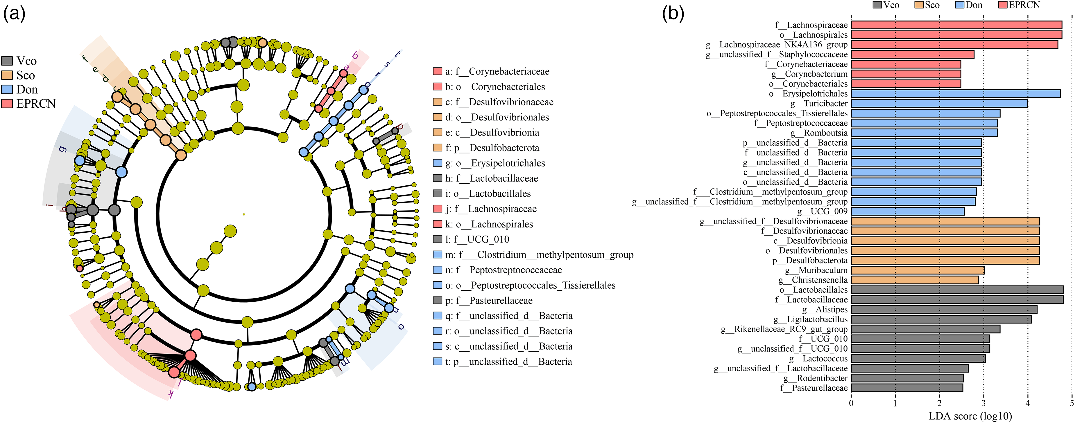
The predominant gut microbiota in the four groups. (a) Evolutionary branching diagram for LEfSe analysis. (b) Histogram of the LDA distribution for LEfSe analysis.
EPRCN decreases gut metabolic disorders and increases CoA biosynthesis of gut microbiota in scopolamine-induced mice
To further investigate whether EPRCN improves cholinergic dysfunction in the brain by affecting gut microbiota in mice induced with scopolamine, we compared the alternative pathways based on KEGG orthologs among the Vco, Sco, and EPRCN groups. The predicted functions of the gut microbiota, which were similar to those of the Vco group after EPRCN treatment, were associated with three metabolic pathways and one biosynthesis process (Figure 4(a)–(d)). Compared to the Vco group, the proportions of microbiota related to purine metabolism significantly increased in scopolamine-induced mice, while those related to nitrogen metabolism and sulfur metabolism significantly decreased. EPRCN treatment significantly inhibited this change. In addition, compared to the Vco group, pantothenate and CoA biosynthesis was impaired following exposure to scopolamine, but this impairment was reversed in the EPRCN group.

Functional prediction analysis of gut microbiota in Vco, Sco and EPRCN groups based on KEGG orthologs. (a) Group comparison of purine metabolism among Vco, Sco and EPRCN with Tukeyramer multiple comparison test. (b) Group comparison of Sulfur metabolism among Vco, Sco and EPRCN with Tukeyramer multiple comparison test. (c) Group comparison of Nitrogen metabolism among Vco, Sco and EPRCN with Tukeyramer multiple comparison test. (d) Group comparison of Pantothenate and CoA biosynthesis among Vco, Sco, and EPRCN with Tukeyramer multiple comparison test.
EPRCN rescues gut inflammation and activates cholinergic anti-inflammatory in scopolamine-induced mice
In the Vco group, the structure of the colonic mucosal epithelial cells remained intact with no obvious injury or inflammation (Figure 5(a)), and a small amount of focal inflammatory cell infiltration was found in the Sco group. While the Don group displayed signs of inflammation and cellular infiltration, it demonstrated notable improvement when compared to the Sco group. In contrast, the EPRCN group showed more preserved crypt and mucosal structures, with only slight indications of inflammation and cellular infiltration (Figure 5(b)–(d)). Moreover, the mRNA expression of inflammatory markers was downregulated in AD mice treated with EPRCN or donepezil, indicating the anti-inflammatory effects of EPRCN on the gut. Specifically, its inhibited effect on Calprotectin, Tnf-α, and IL-1β was greater than that of donepezil.

Effects of EPRCN on gut inflammation and cholinergic anti-inflammatory in scopolamine-induced mice. (a) Representative histological analysis of colons from the indicated groups of mice. qPCR analysis showing the (b) Calprotectin, (c) Tnf-α and (d) IL-1β levels in the colon from the indicated groups of mice (n = 3 per group, three repetitions per sample). (e) ACh level in the colon from indicated group mice (n = 6 per group). (f) α7nAChR levels in the colon from indicated group mice (n = 3 per group, two repetitions per sample). The data are expressed as the mean ± SEM, and each hollow dot represents a sample. *p < 0.05; ns: no significant difference.
We further explore the effects of EPRCN on cholinergic anti-inflammatory pathways. The results showed that scopolamine significantly reduced ACh and α7nAChR levels in the colon compared to the Vco group (Figure 5(e) and (f)). After treatment with EPRCN and donepezil, the intestinal ACh and α7nAChR levels of scopolamine-induced mice was significantly increased.
EPRCN restores cerebral cholinergic dysfunction in scopolamine-induced mice
We first tested the activities of AChE and ChAT as well as the levels of ACh in the hippocampus. Compared with Vco group mice, Sco group mice showed greater AChE activity and lower ChAT activity and ACh levels (Figure 6(a)–(c)). EPRCN and donepezil increased ChAT activity and ACh content but decreased AChE activity to regulate this abnormal situation, suggesting that EPRCN could reverse cholinergic dysfunction in the brains of scopolamine-induced mice. We also examined the mRNA levels of the cholinergic receptor muscarinic 1 (Chrm1), muscarinic 3 (Chrm3), and nicotinic alpha polypeptide 7 (α7nAChR). We found that EPRCN had a recovery effect on Chrm1 and Chrm3 similar to that of donepezil but was more effective at improving α7nAChR expression (Figure 6(e)–(g)).

EPRCN restores cholinergic dysfunction in scopolamine-induced mice. (a) AChE and (b) ChAT activities, and (c) ACh level in the hippocampus from indicated group mice (n = 5 per group). qPCR analysis showing the (d) Chrm1, (e) Chrm3, and (f) α7nAChR levels in hippocampus from indicated group mice (n = 3 per group). The data are expressed as the mean ± SEM, each hollow dot represents a sample. *p < 0.05, ns no significant difference.
EPRCN restores neuronal damage in scopolamine-induced mice
The number of Nissl's bodies can represent the neuronal activity of hippocampal cells. Compared with those in the Vco group, the number of Nissl's bodies was lower in the hippocampal cornu Ammonis 3 (CA3) of mice within Sco group (Figure 7(a), Supplemental Figure 1). This symptom was significantly changed after treatment with EPRCN or donepezil. The histological features of the hippocampus of mice in each group were shown in Figure 7(b). In comparison to the Vco group, the arrangement of cells in the hippocampal CA3 region of mice from the Sco group appeared disorganized and loose, with a reduction in cytoplasm and blurred cell nuclei. Additionally, there were extensive areas of nuclear pyknosis and necrosis of pyramidal cells. Importantly, treatment with EPRCN significantly mitigated neuronal damage in the hippocampal CA3 region, resulting in a more compact cell arrangement and reduced neuronal injury. Specifically, the morphology of hippocampal cells in the EPRCN group was regular, with clearly defined nuclei and cytoplasm, along with a notable decrease in nuclear pyknosis.

EPRCN suppressed neuronal damage induced by scopolamine. (a) Statistics of the number of Nissl bodies in the hippocampal CA3 region in 400 fields (n = 5 per group). Scale bar is 50 μm. (b) Representative Nissl staining of the hippocampal CA3 region in the Vco, Sco, Don, and EPRCN groups (200 and 400 ×). The black arrows indicate Nissl body. (c) Quantification of NeuN+ cells in the hippocampus. (d) Representative immunostaining for NeuN+ (green) in the hippocampal areas. scale bar is 200 μm. (e) Semi-quantification of BDNF level compared with GAPDH. The data are expressed as the mean ± SEM, and each hollow dot represents a sample. *p < 0.05; ns: no significant difference (colors are visible in the online version).
In addition, we use NeuN to label mature neuronal cells in hippocampus. Results showed that There were fewer mature neurons marked by NeuN+ in the hippocampal regions in Sco group (Figure 7(c)). Representative images exhibited that the scopolamine-induced decrease in the number of mature neurons was most obvious in the hippocampal dentate gyrus (DG) region (Figure 7(d)). Howerer, the number of NeuN+ positive cells were increased in the hippocampal regions of EPRCN- and donepezil-treated mice. Given the essential function of brain derived neurotrophic factor (BDNF) proteins in regulating neuronal cell survival, we evaluated the protein expression levels of BDNF to explore the possible mechanisms through which EPRCN may exert its anti-neuronopathy effects. The findings demonstrated that EPRCN treatment effectively restored the abnormal protein levels of BDNF (Figure 7(e)). Similarly, the hippocampal expression of BDNF were increased in both the EPRCN and donepezil groups.
Correlation analysis between the gut microbiota, and improved indexes after EPRCN treatment
Spearman's correlation analysis was performed (Figure 8) to clarify the correlation between gut microbiota and indexes that improved after EPRCN treatment, including level of ACh in colon, number of Nissl body per mm2 in hippocampus, and AChE activity in hippocampus. Results showed that the relative abundances of 10 genera, such as Lachnospiraceae_NK4A136_group (r = 0.6461, p = 0.0014), Ligilactobacillus (r = 0.6261, p = 0.04396), Oscillibacter (r = 0.6362, p = 0.0108), etc., were significantly correlated with level of ACh in colon. For the number of Nissl bodies per mm2 in the hippocampus, more than seven genera were significantly correlated, including Lachnospiraceae_NK4A136_group (r = 0.7173, p = 0.0026), Alloprevotella (r = 0.6388, p = 0.0016), Rikenella (r = 0.5295, p = 0.0424), Muribaculum (r = 0.6383, p = 0.0104), etc. The relative abundances of 11 genera, such as Lachnospiraceae_NK4A136_group (r = –0.6607, p = 0.0073), Dubosiella (r = –0.6272, p = 0.0073), Rikenellaceae_RC9_gut_group (r = 0.5362, p = 0.0393), and Muribaculum (r = -0.6202, p = 0.0136) were significantly correlated with AChE activity in mice form Vco, Sco, and EPRCN groups.

Spearman's correlation analysis for indexes improved by EPRCN (ACh in colon and Number of Nissl body, and AChE activity) and top 50 abundant gut microbiota in mice. Data were acquired from mice in Vco, Sco, and EPRCN groups. *p < 0.05, **p < 0.01, ***p < 0.001.
Discussion
A substantial body of research has demonstrated that extracts derived from MFHs have many biological characteristics and diverse functions, which contribute positively to the enhancement of cognitive impairment. 24 Previous studies have indicated that EPRCN improves cognitive deficits in mice by inhibiting neuroinflammation and oxidative stress. 20 However, whether EPRCN can improve AD by regulating gut microbiota remains unknown. In order to further explore the effect and mechanism of EPRCN's ameliorative effect on AD, this study focused on the effect of its regulatory effect on gut microbiota and neuroprotective effect and compared with the traditional drug donepezil. Results revealed that EPRCN can remodel gut microbiota structure, inhibited gut metabolic disorders, and promoted CoA biosynthesis. It also can improve gut inflammation and exert neuroprotection which is similar to donepezil in effect.
The disorder of the gut microbiota induces gut inflammation, affects the intestinal permeability, and further leads to the development of neurodegenerative diseases. 25 In this study, EPRCN treatment increased the abundance of beneficial bacteria, such as Lactobacillus, Enterorhabdus, Ruminococcus, and Muribaculaceae, which contributed to the improvement of intestinal inflammation. Conversely, it decreased the abundance of harmful bacteria, such as Desulfovibrionaceae which was increased in high-fat diet-induced mice exhibiting intestinal barrier damage and inflammation.26,27 The remodeling of these microbiota may play a role in EPRCN's ability to ameliorate gut inflammation in scopolamine-induced mice. Additionally, LEfSe analysis revealed that the butyrate-producing bacterium Lachnospiraceae_NK4A136_group was predominant in the gut microbiota of scopolamine-induced mice following EPRCN administration. Previous studies have demonstrated that the recovery of Lachnospiraceae_NK4A136_group in rats was associated with the restoration of cognitive function. 28 Lachnospiraceae_NK4A136_group was also found to be inhibited in the gut of APP/PS1 mice but showed recovery after probiotic intervention. 29 The upregulation of Lachnospiraceae_NK4A136_group induced by EPRCN may mediate the improvement of gut inflammation through the production of butyrate. Butyrate has been shown to inhibit the proliferation of neutrophils and macrophages while reducing the expression of inflammatory factors in the gut. 30 Research suggests that butyrate can improve gut innate immune function via G-protein-mediated signaling pathways, while also reducing excessive inflammatory responses by inhibiting histone deacetylase. 31
Functional prediction analysis indicated that EPRCN effectively restored the intestinal metabolic disorders induced by scopolamine, which included alterations in purine metabolism, sulfur metabolism, nitrogen metabolism, and pantothenate and CoA biosynthesis. In mice subjected to a high-fat diet, these intestinal metabolic disorders were associated with intestinal inflammation. 32 Evidence suggests that overactive purine metabolism can trigger intestinal inflammation. 33 Additionally, both sulfur and nitrogen metabolism have been associated with intestinal inflammation.34,35 Furthermore, enhanced pantothenate and CoA biosynthesis may indicate an increase in ACh synthesis by gut microbiota.
Gut inflammation may play an important role in the pathogenesis of AD. 6 Emerging evidence from observational studies has indicated that gut inflammation may be associated with an increased risk of AD. 36 Cinnamon essential oil can alter the gut microbiota structure in a mouse model of dextran sodium sulfate-induced colitis. 37 Perilla seed oil can alleviate gut inflammation in obese, insulin-resistant rats. 38 Therefore, EPRCN may has beneficial effects on gut inflammation. In this study, we found that chronic intraperitoneal injection of scopolamine induced intestinal inflammation in ICR mice, whereas EPRCN inhibited scopolamine-induced immune cell infiltration and the expression of inflammatory-related factors in these mice. In terms of the transcription of Calprotectin and IL-1β, EPRCN demonstrated a stronger inhibitory effect compared to donepezil. Increased expression of Calprotectin indicates the activation of intestinal immune cells. A recent study showed that the level of Calprotectin was higher in the stool of AD patients than in that of their cognitively intact peers. 39 IL-1β has the ability to drive chronic intestinal inflammation by facilitating the buildup of IL-17A-secreting innate lymphoid cells and CD4 (+) Th17 cells. 40 The improvement of both calprotectin and IL-1β is a primary manifestation of EPRCN's ability to ameliorate gut inflammation. We further found that EPRCN counteracted the scopolamine-induced reduction in ACh and α7nAChR levels in the colon, whereas donepezil did not exhibit this effect. Although ACh plays a crucial role in improving cholinergic dysfunction, there is insufficient evidence to support that acetylcholine derived from the gut can cross the blood-brain barrier and access the CNS, even in cases of blood-brain barrier injury. Therefore, ACh in the gut may activate the cholinergic anti-inflammatory pathway (CAP) to reduce inflammatory factors and improve immune cell infiltration in the gut. The CAP functions as a neuro-immunomodulatory pathway where ACh, released from the interaction between vagal nerves and α7nAChR, inhibits the production and release of pro-inflammatory cytokines, thereby modulating the local or systemic inflammatory response in a feedback loop. 41 Recent research has revealed that alleviating gut inflammation is a crucial prerequisite for effectively reversing cognitive deficits in mice with AD. 13 This provides preliminary evidence that EPRCN improves cognitive deficits through the gut-brain axis. In addition, correlation analysis revealed a significant positive correlation between Ligilactobacillus and level of ACh in colon. Lactobacillaceae members family have been found in previous study to raise gut ACh levels. 42 However, the association between Ligilactobacillus and intestinal ACh levels has been found for the first time in this paper. Since EPRCN may improve gut inflammation by regulating the abundance of Ligilactobacillus, it is necessary to further explore the specific mechanism.
The enhanced biological activity of the mixture of MFH has been widely reported. 43 Compared to individual MFHs, the combination of MFHs may alleviate symptoms in a synergistic and multi-target manner. Cholinergic system dysfunction in the brain is one of the key factors underlying AD. 44 Cholinergic lesions can result in decreased levels of ACh and cognitive decline in the early stages of AD. 45 The levels of ACh are dependent on the balance between its synthesis and degradation, which is strongly associated with the levels of ChAT and AChE. 46 AChE degrades ACh, while ChAT synthesizes ACh. The disruption of these two enzymes is the primary manifestation of cholinergic dysfunction in patients with AD, particularly concerning AChE. 47 Consequently, the development of AChE inhibitors has long been a strategy for treating AD. 48 Among the components that constitute EPRCN, the extract of perilla seed (P. frutescens) has been reported to exhibit inhibitory effects on AChE activity in previous studies. 49 Similarly, cinnamon oil (C. spp.), as studied by Mutlu et al., 50 and noni fruit extract (M. citrifolia), as investigated by Pachauri et al., 51 have demonstrated significant inhibitory effects on AChE activity. In this study, we found that a daily supplement of 20 mg/kg EPRCN significantly inhibited AChE activity more than donepezil (3 mg/kg/day) in scopolamine-induced mice. This significant inhibitory effect has not been documented in all studies investigating the individual impacts of the components found in EPRCN. This also explains how EPRCN restored ACh levels and ChAT activity in the hippocampus of scopolamine-induced mice. Donepezil is a well-known drug for AD that inhibits AChE activity. 52 Clinically, donepezil is currently used to treat symptomatic patients with mild to moderate AD. 53 However, many clinical studies have found that donepezil is short-acting and associated with gastrointestinal side effects. 54 While the safety and prolonged efficacy of MFHs have been acknowledged in recent years for their significant potential in the development of AChE inhibitors, limited AChE inhibition has hindered further progress. This study found that it is possible to develop a medication with AChE inhibitory activity that exhibits greater efficacy and safety than current clinical drugs by combining diverse MFHs, even if they do not act on a single target. Additionally, correlation analysis showed that gut microbiota might be related to EPRCN regulating AChE activity. Disruption of cholinergic signaling via muscarinic and nicotinic receptors has been associated with the pathogenesis of AD. 55 Our findings indicated that EPRCN increased the expression of Chrm1 and Chrm3, similar to the effects of donepezil in scopolamine-induced mice. The mRNA level of the α7nAChR was significantly increased after EPRCN administration compared to donepezil administration in scopolamine-induced mice. Consistent with the findings on AChE inhibitory activity, previous studies have not identified any components of EPRCN that demonstrate α7nAChR activation activity.
The activation effects of α7nAChR demonstrate the distinct mechanisms through which EPRCN and donepezil enhance cholinergic function. α7nAChR plays both neurotrophic and neuroprotective roles in maintaining the cholinergic phenotype. 56 Evidence indicates that activation of the α7nAChR can reverse behavioral changes in mice induced by scopolamine. 57 Clinical studies have also reported promising results with α7nAChR agonists, including improvements in memory and cognitive function. 58 Importantly, activation of α7nAChR has been associated with reduced cell death. 59
Apart from amyloid-β deposition and tau tangles, neuron loss is one of the hallmark neuronal pathologies of AD.60,61 Neuronal cell loss in the hippocampus is a common feature in patients with AD.62,63 The loss of neurons contributes to the development of cognitive disorders associated with AD. 64 In this study, EPRCN demonstrates the ability to attenuate hippocampal neuronal cell damage induced by scopolamine and exhibits efficacy comparable to that of donepezil. Donepezil can attenuate hippocampal neuronal damage by preventing reductions in the levels of phosphorylated CaMKII (p-CaMKII) and phosphorylated CREB (p-CREB) proteins in the hippocampus. 65 However, EPRCN did not alter the levels of these two proteins in the hippocampus, indicating that the mechanism through which EPRCN enhances neuronal damage is distinct from that of donepezil. This finding is consistent with the above conclusion that EPRCN may reduce hippocampal neuronal cell death by activating α7nAChR. Our subsequent correlation analysis found that EPRCN may inhibit neuronal damage through modulating the abundance of Lachnospiraceae_NK4A136_group, Alloprevotella, Rikenella, and Muribaculum partly. Among them, Lachnospiraceae_NK4A136_group and Alloprevotella have been found to be associated with neuronal damage in several studies and may drive the recovery of neuronal damage.66–68
Donepezil is one of the primary treatments options for patients suffering from AD. 69 However, research showed that the use of donepezil could significantly increase the risk of multiple adverse reactions. 70 The most frequent adverse drug reactions with donepezil was bradycardia (10%), weakness (5%) and convulsions (4%). 71 Mechanically, donepezil inhibits the rapid hydrolysis of ACh in the peripheral nervous system, which ultimately leads to diarrhea, nausea, and vomiting. 72 In addition, the annual treatment cost of donepezil has reached around $31,000, although it is already one of the most cost-effective treatment for AD at present. 73 Therefore, although there is evidence suggesting that increasing the dosage of donepezil can lead to more significant improvement in the cognitive abilities of AD patients, the increased risk of adverse reactions and high-cost makes donepezil still not an optimal approach. 74 There is a need to develop effective, side-effect-free, and low-cost interventions to relief or prevent AD. Formulations based on MFH may be a promising path. In previous study, the behavioral performances of the scopolamine-induced AD mice treated with EPRCN and those treated with donepezil were reported to both superior to those of the model group. 20 In this study, we found that EPRCN was comparable to donepezil in restoring the disorder of the ACh system and was more effective than donepezil in alleviating the intestinal inflammation induced by scopolamine. Another major reason that hinders the application of donepezil is that AD patients often forget to take or resist the medication. 75 EPRCN, as a combination of a variety of extracted oils, can be directly added to foods such as oil and seasonings, thereby increasing the success rate of intervention intake for AD patients. More importantly, as a dietary intervention, EPRCN can also be used to prevent high-risk individuals from developing mild cognitive impairment (MCI) and even AD, which donepezil cannot achieve. Donepezil can only be prescribed after the develop of MCI or onset of the AD. Based on previous and current studies, the recommended daily intake of EPRCN for a person weighing 70 kg is around 1400 mg, which amounts to approximately 3 EPRCN capsules (each 0.5 g). 20 This results in an annual expenditure of only around $400 for EPRCN, which is only about 1.29% of donepezil.
Conclusions
This study indicates that EPRCN supplementation plays a neuroprotective role in mice induced by scopolamine, which is associated with the regulation of gut microbiota and the restoration of gut immune homeostasis. EPRCN can remodel the gut microbiota, inhibit gut metabolic disorders, and promote coenzyme A (CoA) biosynthesis in scopolamine-induced mice. The enhanced CoA biosynthesis following EPRCN treatment may explain the increase in intestinal ACh levels, which activates the cholinergic anti-inflammatory pathway. Further results demonstrate that EPRCN exhibits a neuroprotective effect in scopolamine-induced mice by maintaining the stability of the cholinergic system and inhibiting neuronal damage. Correlation analysis revealed that the gut microbiota is associated with EPRCN-mediated improvements. Therefore, EPRCN supplementation could serve as a promising strategy for treating AD by regulating the microbiota-gut-brain axis and exerting neuroprotective functions. As a potential dietary intervention, future studies should focus on elucidating the detailed mechanisms by which EPRCN exerts its neuroprotective effects through gut microbiota.
Supplemental Material
sj-docx-1-alz-10.1177_13872877251339762 - Supplemental material for EPRCN exerts neuroprotective function by regulating gut microbiota and restoring gut immune homeostasis in Alzheimer's disease model mice
Supplemental material, sj-docx-1-alz-10.1177_13872877251339762 for EPRCN exerts neuroprotective function by regulating gut microbiota and restoring gut immune homeostasis in Alzheimer's disease model mice by Ming-Jie Li, Meng-Ning Lan, Yao-Xuan Du, Yue Liu, Hua-Yue Zhang, Min Guo, Shi-Wei Liu, Hai-Yang Xia, Zheng-Jun Wu and Hua-Jun Zheng in Journal of Alzheimer's Disease
Footnotes
Acknowledgments
The authors are grateful to Dr Dan Hu (Bright Dairy & Food Co., Ltd) for her help with the modification of figures and tables in this paper.

Ethical considerations
This study was approved by the Ethics Committee on Laboratory Animals of Shanghai Institute for Biomedical and Pharmaceutical Technologies (protocol code 2023-30, approval date 3/28/2023).
Author contributions
Ming-Jie Li: Conceptualization, Formal analysis, Investigation, Writing – original draft, Writing – review & editing. Meng-Ning Lan: Formal analysis, Investigation, Writing – review & editing. Yao-Xuan Du: Formal analysis, Investigation, Writing – review & editing. Yue Liu: Conceptualization, Data curation, Writing – review & editing. Hua-Yue Zhang: Conceptualization, Writing – review & editing. Min Guo: Conceptualization, Writing – review & editing. Shi-Wei Liu: Resources, Writing – review & editing. Hai-Yang Xia: Writing – review & editing. Zheng-Jun Wu: Supervision, Writing – review & editing. Hua-Jun Zheng: Conceptualization, Supervision, Writing – review & editing.
Funding
The authors received no financial support for the research, authorship, and/or publication of this article.
Declaration of conflicting interests
The authors declared the following potential conflicts of interest with respect to the research, authorship, and/or publication of this article: Authors Ming-Jie Li and Zheng-Jun Wu was employed by Bright Dairy & Food Co., Ltd Author Shi-Wei Liu was employed by Shanghai Xizuo Biotechnology Co., Ltd The remaining authors declared no potential conflicts of interest with respect to the research, authorship, and/or publication of this article.
Data availability statement
The gut microbiota data presented in the study are deposited in the GenBank Sequence Read Archive (SRA) under accession number PRJNA1161189.
Supplemental material
Supplemental material for this article is available online.
References
Supplementary Material
Please find the following supplemental material available below.
For Open Access articles published under a Creative Commons License, all supplemental material carries the same license as the article it is associated with.
For non-Open Access articles published, all supplemental material carries a non-exclusive license, and permission requests for re-use of supplemental material or any part of supplemental material shall be sent directly to the copyright owner as specified in the copyright notice associated with the article.
