



Abstract
Background
Although poor sleep is widely assumed to impair cognitive function, the impact of sleep disturbances (SD) on language function and the underlying mechanisms of this relationship remains unclear.
Objective
This study aimed to investigate the association between SD and language function in non-demented elderly individuals, identify potential neuroimaging correlates, and analyze risk factors for SD.
Methods
We analyzed 784 non-demented elderly subjects from the Alzheimer's Disease Neuroimaging Initiative (ADNI), categorized into SD (n = 256) and normal sleep groups (n = 528) based on self-reported sleep status. Cognitive differences were assessed, and the findings were validated using the China Longitudinal Aging Study (CLAS) and the Chinese Longitudinal Healthy Longevity Survey (CLHLS). Diffusion tensor imaging (DTI) metrics were correlated with language function, and SD risk factors were examined.
Results
In the ADNI cohort, elderly individuals with SD exhibited worse language function compared to those with normal sleep, and this finding was validated in the CLAS and CLHLS cohorts. Meanwhile, the decline in longitudinal language function among elderly individuals with SD occurred at a faster rate. Differences in DTI metrics between the two groups were primarily observed in the limbic and prefrontal regions. Finally, the risk factors for elderly with SD mainly included years of education, physical and emotional conditions, lifestyles, living environment, and parental survival status.
Conclusions
SD correlates with language impairment in non-demented elderly, possibly due to limbic/prefrontal tract damage. Risk factors encompass demographic, health, lifestyle, and socio-environmental aspects. Effectively managing these factors and treating SD may improve language function.
Introduction
The aging population has become a major challenge for most countries worldwide due to declining fertility rates and increasing life expectancy in both developed and developing countries.1,2 As this population is frequently plagued by various chronic diseases which significantly impact their quality of life, the corresponding health care and social care services await enhancement. Sleep is vital for the body's recovery and memory consolidation. As individuals age, changes in physiological mechanisms can lead to increased sleep fragmentation and deteriorating sleep quality, making individuals more susceptible to sleep disturbances (SD).3,4 These disturbances commonly manifest as trouble falling or staying asleep, falling asleep at inappropriate times, excessive sleep, and abnormal behaviors during sleep. 5
Chronic SD are associated with an increased risk of depression, anxiety, substance abuse, suicide, immune dysfunction, and traffic accidents.6–10 The projected increase in the elderly population will further exacerbate sleep-related health problems, placing a significant burden on families and society. 11 Studies have shown that SD could accelerate Alzheimer’ disease (AD) pathophysiology and promote the accumulation of amyloid-β (Aβ) and phosphorylated tau. Therefore, abnormal sleep patterns are a major risk factor for the possible progression of mild cognitive impairment (MCI) to AD. 12 MCI typically manifests as a constellation of cognitive deficits, mainly characterized by memory impairment, decreased attentional capacity, and compromised linguistic abilities.13,14
While sleep has been well-documented to influence critical physiological functions, including cardiovascular health, metabolic regulation, and immune system performance, increasing scientific attention is being directed toward understanding its profound impact on cognitive function. 15 Existing research has demonstrated that insufficient sleep can cause damage to memory, executive function and learning, as well as to arithmetic calculation.16–18 However, there are relatively few studies on the impact of sleep on language function. Only a small number of studies have demonstrated that sleep problems are risk factors for language disorders in preschool children, and poor sleep is also associated with poorer language function in adults.19,20 Moreover, sleep-dependent neurophysiological activities have been proven to consolidate the meanings of new words and simple grammatical rules, and to aid in sentence comprehension.21,22 Therefore, current study focuses on the impact of SD on language function, the related mechanisms and risk factors. Diffusion tensor imaging (DTI) has been used to investigate the white matter tracts underlying the perisylvian cortical regions, which are known to be associated with language function. 23 Research has explored the effects of sleep deprivation on cognitive impairment and individual differences by assessing DTI metrics in the superior longitudinal fasciculus. 24
In recent years, an increasing number of elderly people have been suffering from SD. While previous studies have confirmed the negative impact of poor sleep on cognitive function, there remains a lack of research on how SD specifically affects various aspects of cognition. This retrospective study aims to fill this gap by comparing the cognitive function of the community-based elderly population with non-organic SD to that of elderly individuals with normal sleep. The findings will provide a reliable data basis for future interventions.
Methods
Study design and samples
The Alzheimer's Disease Neuroimaging Initiative (ADNI) cohort (http://adni.loni.usc.edu/) is a multisite dataset initiated in 2003 designed to study clinical symptoms, imaging, and genetic and biochemical biomarkers of AD. The data collection and sharing procedures in ADNI were reviewed and approved by the ethics committees at all participating sites, and all participants or their guardians provided written informed consent in accordance with the Declaration of Helsinki. Participants in this study were retrieved from the ADNI cohort of older adults aged 55–90 years. Each participant underwent face-to-face health and neuropsychological assessment interviews at baseline and during annual follow-ups. After conducting interviews with the elderly participants and their caregivers, 256 participants with SD lasting more than 1 year and 528 without SD were identified. Based on the interview results, individuals were classified according to whether they had sleep problems. Those with sleep problems were assigned to the SD group, while those without sleep problems were categorized into the normal sleep group.
Demographic data (age, gender, years of education, APOE, hypertension), physical examination results, and neuropsychological test scores were collected. Professional neuropsychiatrists conducted the neuropsychological assessments and evaluated the cognitive function of the participants through the Mini-Mental State Examination (MMSE), Montreal Cognitive Assessment (MoCA), and the Alzheimer's Disease Assessment Scale-cognitive (ADAS-cog). Furthermore, the attending psychiatrist conducted clinical interviews, assessed the Geriatric Depression Scale (GDS), and combined the participants’ medical histories and physical examination findings to make diagnoses, excluding anxiety and depressive disorders. Cognitive level was assessed with age, gender, educational level, and APOE4 as covariates.
A validation dataset was obtained from the China Longitudinal Aging Study (CLAS) cohort. Participants who reported no SD symptoms before cognitive decline were selected for the normal group (n = 469), while those who reported sleep symptoms before cognitive decline were selected for the SD group (n = 244). All subjects underwent cognitive assessments, including MMSE, the Neuropsychological Test Battery (NTB), and the Beijing version of MoCA. Only subjects who met the following criteria were included: (1) signed informed consent; (2) were ≥60 years old, with no restrictions on gender or education; (3) were able to understand and complete the tests; (4) showed no signs of dementia and had normal daily life and social functions; (5) had a Clinical Dementia Rating (CDR) score of 0 or 0.5; an MMSE score of ≥18 for uneducated subjects, ≥21 for elementary school-educated subjects, and ≥25 for subjects with education levels higher than middle school; a Geriatric Depression Scale (GDS) score of ≤10; and a Hachinski ischemia index of ≤4. The exclusion criteria were: (1) lack of stable caregivers; (2) positive syphilis serology or thyroid dysfunction; (3) a history of mental illnesses such as schizophrenia or emotional disorders; and (4) other serious physical diseases. Cognitive level assessment with age, gender, and educational level as covariates.
The SD were identified based on the doctor's inquiry about the presence of sleep abnormalities. Those who answered affirmatively were classified into the SD group, while those who answered negatively were placed in the normal group.
Another validation cohort was derived from the China Longitudinal Healthy Lifespan Survey (CLHLS 1998–2018, n = 44,620) (http://opendata.pku.edu.cn/), which is a multistage, stratified, whole-cluster sampling study. It recruited older adults from half of the counties and cities in 22 of China's 31 provinces between 1998 and 2018. The significant status of study participants was determined at follow-up, and all surviving participants were re-interviewed. For the CLHLS cohort, 530 individuals with missing demographic data, 90 individuals younger than 55 years old, and 13,713 individuals with a diagnosis of cognitive impairment were excluded. Subjects were given a sleep screening at baseline using questions such as “How is your sleep quality now?” and “How many hours do you usually sleep each day?”, which were used to assess the core symptom of SD. First, 1474 subjects who answered “very good” or “good” were categorized as having “good sleep quality,” while 410 who answered “bad” or “very bad” were categorized as having “bad sleep quality.” Additionally, 937 individuals answered “so-so,” 166 were “not able to answer,” and 27,300 had missing data on sleep quality. Second, according to sleep duration classification, individuals who sleep <7 h/day were categorized as “short sleep”; individuals (age ≥ 65 years) who sleep between 7–8 h and those (age between 55–65 years) who sleep between 7–9 h were categorized as “normal sleep”; individuals (age ≥ 65 years) who sleep >8 h and those (age between 55–65 years) who sleep >9 h were categorized as “long sleep.” In total, 677 individuals were categorized as “long sleep,” 1128 as “short sleep,” and 1067 as “normal sleep.” Finally, 2522 individuals were included in this analysis, including 1861 in the SD group who had “bad sleep quality” as well as either “short sleep” or “long sleep” duration, and 661 in the normal group who had “good sleep quality” and “normal sleep” duration. The Chinese version of the Mini-Mental State Examination (CMMSE), consisting of 24 items across 7 cognitive domains, was used. The total score of the CMMSE ranges from 0 to 30 points. Cognitive impairment (or moderate-severe cognitive impairment) was defined as a CMMSE score below 18 points, as previously validated. Language function was assessed by summing the scores from two named entity tasks and one sentence repetition task through the CMMSE. Cognitive level assessment with age, gender, and educational level as covariates. We have classified the risk factors contributing to SD into the following categories: familial and social support factors (e.g., whether the mother or father is alive, availability of home visit services), housing and environmental conditions (e.g., type of dwelling, use of air purifiers or activated carbon), mental and emotional health (e.g., loss of interest in activities, lack of energy, sadness, depression, nervousness, excessive worrying, difficulty concentrating), current lifestyle and activities (e.g., engagement in housework, playing cards/mahjong), and physical health conditions(e.g., suffering from respiratory diseases like bronchitis, emphysema, pneumonia, or asthma).
Analyses of DTI metrics
DTI data in ADNI were accessed (https://ida.loni.usc.edu/pages/access/studyData.jsp?categoryId=14&subCategoryId=30, and “Microsoft Word- DTI-ADNI_Methods-Thompson-Oct2012.docx”). Anatomical images were linearly aligned to a version of the Colin27 brain template using FSL's flirt32 with 6 degrees of freedom. Axial diffusivity (AD), fractional anisotropy (FA), mean diffusivity (MD), and radial diffusivity (RD) values were extracted from 45 tracts (detailed tracts in Figure 3) from 224 subjects. DTI metrics were used for differential analysis and to examine their associations with cognitive function.
Thirty percent of the participants were utilized as the test set, while seventy percent were randomly assigned to the training set. Following feature selection, the training data underwent 10-fold internal cross-validation using the glmnet package in R, and binary classification between groups was performed. Discriminative power was evaluated by measuring the area under the curve (AUC) of the receiver-operator characteristic (ROC). Significant DTI tracts were visualized using DSI Studio software, and for each subject, the connection between cognitive and SD-prone areas was examined.
Data analysis
The Kolmogorov–Smirnov test was used to assess the normality of continuous data. Most of the continuous variables in this study followed a non-normal distribution. Therefore, we used the Mann–Whitney U test to perform cross-sectional intergroup difference analyses. All categorical variables were analyzed using the chi-square test.
A linear mixed-effects model was employed to explore the longitudinal effects of SD on language function in ADNI and CLHLS cohorts. To identify risk factors for SD, LASSO regression—an extension of generalized linear regression—was utilized. This method reduces the variance of regression coefficients and the prediction error by adding a term to the log-likelihood to penalize model complexity. 25 All categorical variables were analyzed using the chi-square test.
Statistical significance was set at a p-value of < 0.05. All analyses were performed using SPSS version 17.0 or R version 4.2.1.3.
Results
Participants’ characteristics
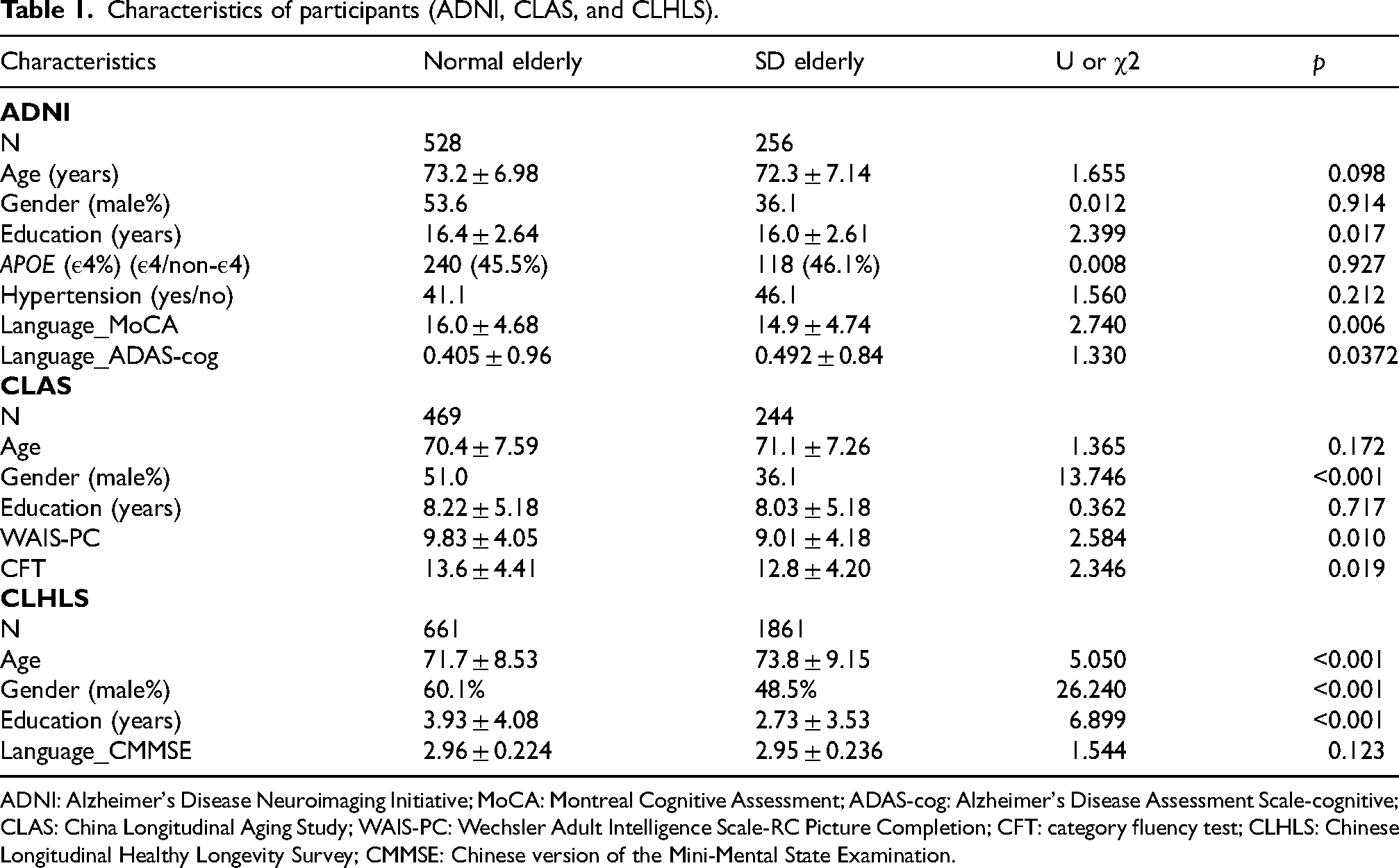
In the ADNI dataset, a total of 784 individuals without dementia were included in the present study. The participants were in middle-aged elderly, with an average age of 72.9 ± 7.04 years, and had a moderate level of education (M = 16.3 years). Male participants accounted for 46.2%. In this dataset, we also compared general information and language function between SD and normal sleep subjects. The results showed significant differences in years of education and language function, as assessed by the language subscales of the MoCA and ADAS-cog (Table 1, Figure 1A1-A3). The SD population has a lower educational level and poorer language function, as reflected in MoCA-language and ADAS-cog-language.

Group differences analysis of the language subscales of MoCA, MMSE, and ADAS-cog scales in the ADNI database (A1-A2). Group differences analysis of the WAISPC and CFT scores in the CLAS cohort (B1-B2). Group differences analysis of the CMMSE language subscale in the CLHLS cohort (C).
Characteristics of participants (ADNI, CLAS, and CLHLS).
ADNI: Alzheimer's Disease Neuroimaging Initiative; MoCA: Montreal Cognitive Assessment; ADAS-cog: Alzheimer's Disease Assessment Scale-cognitive; CLAS: China Longitudinal Aging Study; WAIS-PC: Wechsler Adult Intelligence Scale-RC Picture Completion; CFT: category fluency test; CLHLS: Chinese Longitudinal Healthy Longevity Survey; CMMSE: Chinese version of the Mini-Mental State Examination.
A total of 713 participants (45.9% male, N = 1299), with an average age of 73.2 ± 7.04 years, were included in the CLAS dataset. After interviewing the elderly and their caregivers, 244 participants were identified as having SD lasting more than 1 year, while 469 had no SD. Significant impairment (decrease) in SD population were identified in NTB scale, particularly in the category fluency test (CFT) scores (p < 0.05) and Wechsler Adult Intelligence Scale-RC Picture Completion (WAIS-PC) (p < 0.05) (Table 1, Figure 1B1-B2).
A total of 2522 participants (53.8% male, N = 327), with an average age of 70.6 years (SD = 7.48), were included in the CLHLS dataset. We also conducted cross-sectional comparisons of demographic and language. There was no significant difference in language function tests (CMMSE) between participants with SD and those without (Mann–Whitney test, p = 0.12) (Table 1, Figure 1C).
Longitudinal effects of SD on language function
To clarify the longitudinal changes in language function between the SD and normal sleep groups, we conducted a follow-up assessment of language-related measures from the ADNI and CLHLS cohorts. In the ADNI dataset and MoCA-language scores declined over time, and the rate of decline was significantly greater in the SD group compared to the normal sleep group (Figure 2A1-A2). Correspondingly, scores on the language component of the ADAS-cog scale increased over time, with a significantly greater increase in the SD group than in the normal sleep group (Figure 2A3). In the CLHLS cohort, scores on the language subscale of the CMMSE declined over time in both groups, with a faster rate of decline observed in the SD group compared to the normal group (Figure 2B). These longitudinal results suggested that the SD group experienced faster decline in language function.

The linear mixed-effects models were used to estimate the changes in MoCA and ADAS-cog scores across groups over time in the ADNI cohort (A1-A2). The linear mixed-effects model was used to estimate the changes in verbal function across groups over time in the CLHLS cohort (B).
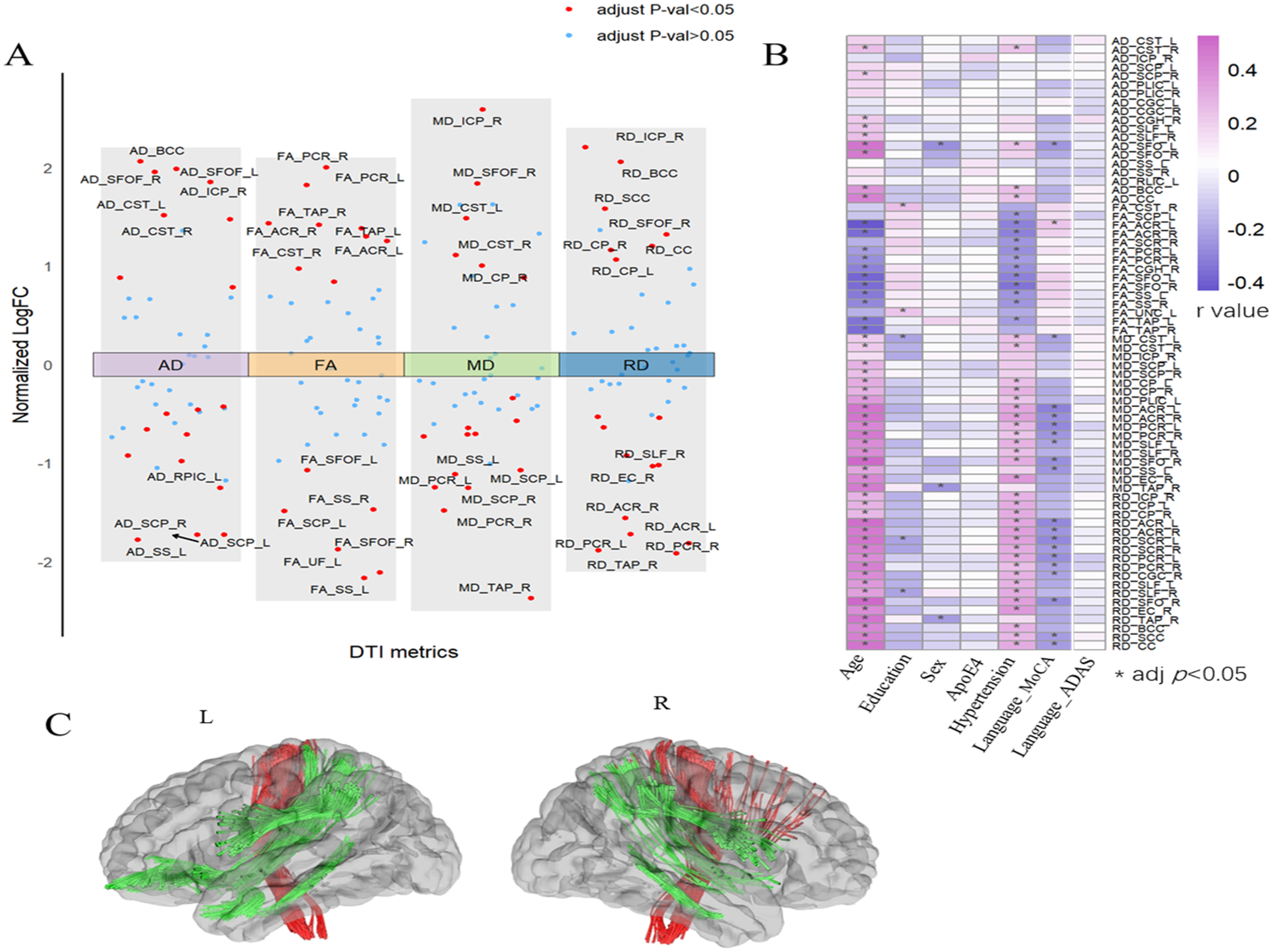
Diffusion tensor imaging (DTI) metrics included axial diffusivity (AD), fractional anisotropy (FA), mean diffusivity (MD), and radial diffusivity (RD). Differences between the sleep-normal and SD groups were analyzed within the ADNI cohort (A). Spearman correlation analyses examined the relationships between differential DTI metrics and language function, as well as demographic variables, with results presented in heat maps. The value within each box represents the correlation coefficient (r). * adj p < 0.05 (B). The DTI-derived metrics associated with sleep disturbances were visualized using the DSI Studio program (C).
DTI data analysis
DTI data of 224 individuals were included from the ADNI cohort, comprising 170 with normal sleep and 54 with SD. The demographic characteristics of the two groups with available MRI data were compared, with no statistically significant differences observed (p > 0.05) (Table 2). The DTI metrics were analyzed using Spearman correlation with language function. After FDR correction, multiple DTI measures related to language function were identified, including the bilateral superior longitudinal fasciculus (SLF), bilateral cingulate gyrus (CGC), bilateral posterior limb of the internal capsule (PLIC), bilateral sagittal stratum (SS), bilateral superior cerebellar peduncle (SCP), bilateral corticospinal tract (CST), bilateral cerebral peduncle (CP), and corpus callosum (CC), left retrolenticular limb of the internal capsule (RLIC-L), bilateral superior fronto-occipital fasciculus (SFO), left uncinate fasciculus (UNC-L), right inferior cerebellar peduncle (ICP-R), (Figure 3A, B). These regions were labeled using DSI Studio software (Figure 3C).
Characteristics of participants (ADNI).

Analysis of risk factors for SD
To further identify the risk factors that may influence SD, we conducted LASSO regression analyses in the CLHLS cohort. Participants were grouped based on the presence or absence of SD. In LASSO regression, the dataset was divided into a training set (80%) and a test set (20%). The model was fitted using minimum lambda value (lambda. min). In our study, lambda. min = 0.019. The statistically significant variables in the model and their regression coefficients are as follows: education (−0.100), depressed (−0.094), nervous (−0.062), worry about small things (−0.042), bronchitis (−0.020), difficult to concentrate (−0.007), use air purifier (0.023), play cards now (0.038), father alive (0.042), community home visit service (0.047), house type (0.048), house work now (0.096), mother alive (0.165), energetic (0.133). This analysis identified 6 major factors categories, including demographics, health conditions, emotional conditions, lifestyle, living environment, and parental survival status. Factors that impaired sleep included low education level, depression, nervousness, worrying about small things, bronchitis, and difficulty concentrating. In contrast, protective factors for sleep included the use of air purifiers, playing cards, having a living father, home visits by community members, type of house, doing housework, feeling energetic, and having a living mother (Figure 4).
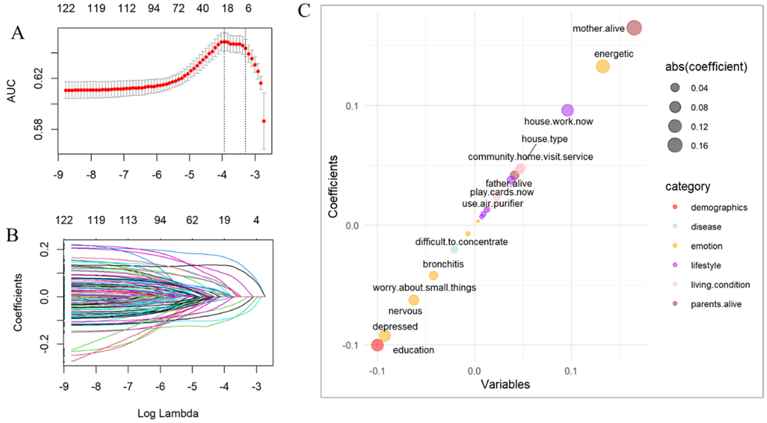
Risk factors were collated in the CLHLS cohort, and LASSO regression models were used to screen for sleep-related risk factors (A, B). Multivariate Cox regression analysis was used to estimate risk factors for cognitive progression after adjustment for age, education, gender, APOE genotype, hypertension, diabetes, and baseline MMSE. Positive coefficients indicate protective factors, while negative coefficients indicate risk factors; different colors were used to represent the various categories. Factors that impaired sleep included low education level, depression, nervousness, worrying about small things, bronchitis, and difficulty concentrating. In contrast, protective factors for sleep included the use of air purifiers, playing cards, having a living father, home visits by community members, type of house, doing housework, feeling energetic, and having a living mother (C).
Discussion
This study initially identified that non-demented elderly individuals with SD exhibited poorer cross-sectional language function compared to those without SD in the ADNI cohort, Similar results was observed in the CLAS and CLHLS cohorts. Longitudinal analysis demonstrated a more rapid decline in language function among elderly individuals with SD. Key differences in DTI metrics between the two groups were primarily observed in brain regions within the limbic and frontotemporal tracts, which are closely associated with language and emotional functions. Finally, the risk factors for SD in the elderly predominantly included personal factors, such as demographics, health conditions, emotional well-being, and lifestyles, along with socio-environmental factors, such as living environment and parental survival status.
Consistent with most prior studies, our findings indicate that sleep is crucial for learning, memory consolidation, and language integration, while SD negatively affects language function.26–28 In a longitudinal study, older adults with persistent difficulty initiating sleep showed cognitive impairments, including deficits in language function, over a 14-year period. 29 However, a longitudinal study on sleep in the elderly found that both long and short sleep durations were strongly associated with delayed recall but not with language function. 30 Discrepancies among studies may be attributed to variations in population characteristics, regions, observation periods, and assessment scales. Some studies have demonstrated a close relationship between sleep duration and the lymphatic system. The DTI analysis along the perivascular space (DTI-ALPS) index is a magnetic resonance imaging marker of glymphatic function, with higher DTI-ALPS values associated with better language function. 31 Therefore, SD may impair language function by disrupting an individual's glymphatic system.
Furthermore, SD has been shown to disrupt brain network connections associated with language. Resting-state functional magnetic resonance imaging (fMRI) studies have shown that sleep deprivation disrupts connectivity in language-related brain networks, particularly those involved in syntax processing. 32 Our study identified that differences in DTI metrics between non-demented elderly with and without SD were primarily concentrated in the limbic and prefrontal regions, both critical for language and emotion. This finding aligns with prior studies demonstrating similar effects of SD on the prefrontal regions. For example, Altena et al. reported reduced gray matter volume in the prefrontal cortex of individuals with chronic insomnia, a region crucial for language comprehension and emotional regulation. 33 Similarly, studies by Simon et al. and Yoo et al. highlighted the prefrontal cortex's vulnerability to sleep deprivation, particularly its role in higher-order cognitive processes and emotional regulation.34,35 Specifically, our findings indicate that the neural effects of SD extend beyond the prefrontal cortex, affecting regions such as the hippocampus and amygdala, which are crucial for language, memory, and emotional processing. 36 This finding highlights the limbic system's contribution to language function and bridges research on the relationship between language and memory, offering a broader perspective on brain regions impacted by SD.
Our study identified several factors contributing to SD, categorized into six groups: personal factors (demographics, health conditions, emotional conditions, and lifestyle) and socio-environmental factors (living environment and parental survival status). Protective factors for SD included using air purifiers, playing cards, having a living father, participating in community and home visits, housing type, and engaging in housework. Risk factors for SD included lower education, depression, nervousness, excessive worrying, bronchitis, and difficulty concentrating. Regarding personal factors, our finding that higher education is a risk factor aligns with previous studies. A cross-sectional survey of sleep status and risk factors in the NHANES database (2005–2018) found that lower educational attainment was an independent risk factor for SD in U.S. adults aged 40–69 years. 37 Regarding health-related risk factors, the link between depression and SD is well-established: chronic SD can lead to depression, and SD are often associated with depressive symptoms.38,39 Bronchitis, often marked by chronic coughing, disrupts sleep by causing insomnia or early-morning wakefulness. 40 The inflammatory response in bronchitis can cause systemic symptoms (e.g., fever, fatigue) and immune system activation, further impairing sleep quality. 41 Regarding emotional conditions, negative emotions mediate the relationship between structural and functional variations in emotion-related brain regions and sleep quality. 42 Additionally, lifestyles, such as daily activities or physical exercise, influence cognition by affecting sleep, which may mediate these effects. 43 Daily activities and physical exercise can improve quality of sleep by reducing inflammatory responses and increasing melatonin secretion.44,45 Good sleep, in turn, enhances memory consolidation, boosts focus, and improves executive function.46–48 A healthy lifestyle enhances sleep quality. For example, activities like playing bridge foster social interaction and improve mood, thereby supporting better sleep quality. 49
Regarding external factors, environmental conditions, including air pollution, noise, and other living circumstances, significantly affect sleep quality. 50 Improving these environmental conditions can enhance sleep quality. Studies have demonstrated that a favorable living environment, such as the presence of green vegetation, is associated with improved cardiovascular biomarkers and reduced sympathetic activation, both of which positively impact sleep. 51 Moreover, having a living father is a protective factor against SD, potentially due to the beneficial effects of a more complete family structure on sleep. 52 Furthermore, our findings align with previous research showing that supportive social relationships reduce negative emotions and enhance sleep quality. Supportive social ties may prevent SD by reducing excessive arousal, a major cause of chronic insomnia, and by mitigating negative emotional reactions while promoting emotional regulation. 53
This study has certain limitations. First, this study relied on self-assessment questionnaires to evaluate SD, which may introduce bias compared to objective methods. Additionally, other common SD, such as obstructive sleep apnea syndrome, were excluded from this study due to its focus on non-organic SD. Future research could integrate objective assessments to complement the current findings and facilitate comparisons with studies on organic SD. Finally, the CLAS and CLHLS databases lacked longitudinal data, with longitudinal validation only possible using ADNI data. To address this limitation, future work will include follow-up studies.
Conclusion
In conclusion, cross-sectional and longitudinal multidimensional studies suggest that SD is closely associated with language function impairment in the elderly non-demented population. The underlying mechanisms may involve damage to the limbic and prefrontal tracts. This study also identified risk factors for SD in the elderly, including internal factors (e.g., physical health, mental well-being, and lifestyle) and external factors (e.g., living environment and parental survival status). Addressing these risk factors and enhancing sleep quality may be effective strategies for preventing dementia, particularly with respect to language function.
Footnotes
Acknowledgements
Data collection and sharing for this project was funded by the Alzheimer's Disease Neuroimaging Initiative (ADNI) (National Institutes of Health Grant U01 AG024904) and DOD ADNI (Department of Defense award number W81XWH-12-2-0012). ADNI is funded by the National Institute on Aging, the National Institute of Biomedical Imaging and Bioengineering, and through generous contributions from the following: AbbVie, Alzheimer's Association; Alzheimer's Drug Discovery Foundation; Araclon Biotech; BioClinica, Inc.; Biogen; Bristol-Myers Squibb Company; CereSpir, Inc.; Cogstate; Eisai Inc.; Elan Pharmaceuticals, Inc.; Eli Lilly and Company; EuroImmun; F. Hoffmann-La Roche Ltd and its affiliated company Genentech, Inc.; Fujirebio; GE Healthcare; IXICO Ltd; Janssen Alzheimer Immunotherapy Research & Development, LLC.; Johnson & Johnson Pharmaceutical Research & Development LLC.; Lumosity; Lundbeck; Merck & Co., Inc.; Meso Scale Diagnostics, LLC.; NeuroRx Research; Neurotrack Technologies; Novartis Pharmaceuticals Corporation; Pfizer Inc.; Piramal Imaging; Servier; Takeda Pharmaceutical Company; and Transition Therapeutics. The Canadian Institutes of Health Research is providing funds to support ADNI clinical sites in Canada. Private sector contributions are facilitated by the Foundation for the National Institutes of Health ( ). The grantee organization is the Northern California Institute for Research and Education, and the study is coordinated by the Alzheimer's Therapeutic Research Institute at the University of Southern California. ADNI data are disseminated by the Laboratory for Neuro Imaging at the University of Southern California.
Data collection and sharing for the CLHLS data section was organized by Research Center for Healthy Aging and Development of Peking University and National Development Research Institute. Data collection and sharing for the CLAS data section was organized by Department of Psychiatry, Shanghai Mental Health Center.
Ethical considerations
For the ADNI and CLHLS data, all participants provided written informed consent approved by the institutional review board of each participating institution. Data collection in CLAS cohort was conducted in accordance with the recommendations of the Shanghai Mental Health Center Ethical Standards Committee on Human Experimentation (reference number: 2012-19).
Consent to participate
Written informed consent was obtained from all participants or their legal guardians in accordance with the Declaration of Helsinki.
Author contributions
Funding
The authors disclosed receipt of the following financial support for the research, authorship, and/or publication of this article: This study was funded by grants from the National Natural Science Foundation of China (grant number 82271607), the Special Sub Project of Strategic Leading Science and Technology of the Chinese Academy of Sciences (grant number XDA12040101), the National Science and Technology Support Program of China (grant number 2009BAI77B03), and the Excellent Youth (Yucai) Program of Baoshan District Health Commission (grant number BSWSYC-2024-20).
Declaration of conflicting interests
The authors declared no potential conflicts of interest with respect to the research, authorship, and/or publication of this article.
Data availability statement
ADNI data are available at: https://ida.loni.usc.edu/pages/access/studyData.jsp?project=ADNI. CLHLS data are available at: . CLAS raw data and all R codes can be requested via correspondence email.