



Abstract
Background
Activation of NLRP3 inflammasome has been implicated in cognitive impairment. Melatonin, known for its anti-inflammatory properties and traditional use in regulating circadian rhythms, is the focus of this study. This study intended to investigate the role of melatonin in diabetic cognitive impairment model.
Objective
The present study aimed to investigate the underlying mechanism of melatonin in alleviating diabetic cognitive impairment by suppressing NLRP3/Caspase 1 signaling pathway.
Methods
Cognitive function was assessed using Morris water maze test and Novel Object Recognition test. Apoptosis rate of hippocampal neurons was evaluated by TUNEL staining. Western blot was used to evaluate NLRP3/Caspase 1 pathway expression. Double immunofluorescence labelling of GFAP, Iba-1 or NeuN with NLRP3 respectively showed the localization of NLRP3 in hippocampus of db/db mice. In vitro, HT-22 cells treated with high glucose as cellular model were transfected with pc-DNA3.1-mNLRP3 or co-cultured with NLRP3 inhibitor MCC950 to elucidate NLRP3/Caspase 1 pathway in neuronal apoptosis regulation.
Results
Melatonin treatment improved cognitive function and morphologic abnormalities of hippocampal neurons. The double immunofluorescence labelling revealed melatonin inhibited NLRP3 inflammasome activation in hippocampal neurons rather than microglia or astrocytes. TUNEL staining and western blot showed melatonin markedly reversed the upregulation of NLRP3/Caspase 1 signaling pathway against neuronal apoptosis.
Conclusions
Melatonin attenuates diabetic cognitive impairment in db/db mice with down-regulation of NLRP3/Caspase 1 signaling pathway. In vivo and vitro studies supported that NLRP3 activation in hippocampal neurons was associated with diabetic cognitive impairment progression.
Introduction
Diabetes mellitus, characterized by elevated blood glucose levels, is a widespread chronic metabolic condition. The International Diabetes Federation (IDF) reports that the global diabetic population among adults aged 20–79 years numbered 537 million in 2021, with 90% suffering from type 2 diabetes mellitus (T2DM). Predictions suggest an increase to 643 million by 2030. 1 The risk of developing specific brain or mental disorders, particularly cognitive impairment, is increased among adults with T2DM. 2 Animal model studies suggest that the pathogenesis of diabetes-associated cognitive impairment involves a combination of vascular disease, defective central insulin signaling, neurodegeneration, and brain amyloidosis.3,4
Dysfunction in regulating inflammation, oxidative responses, and synaptic plasticity were observed in the hippocampus of diabetic model in previous study.5–7 The inflammatory response is increasingly recognized as a key role in the development and progression of diabetic cognitive impairment, with a particular focus on the involvement of inflammasome activation in neuroinflammation. Among the four inflammasome types in the CNS including NOD-like receptor pyrin domain-containing protein 1 (NLRP1), NLRP3, NLR family CARD domain-containing protein 4 (NLRC4) and absent in melanoma-2 (AIM2), CNS diseases were widely influenced by NLRP3. 8 NLRP3 response to exogenous and endogenous pathogen-associated molecular patterns (PAMPs) and damage-associated molecular patterns (DAMPs) contributes to recruitment of the adaptor protein apoptosis-associated speck-like protein containing a CARD (ASC) and the pro-caspase-1. 9 This leads to activation of caspase-1 and the cleavage of gasdermin D (GSDMD), which is a membrane pore forming protein. The N-terminal region of GSDMD binds to the membrane allowing the release of IL-1β and IL-1. 10 Excessive activation of NLRP3 inflammasome is tightly associated with many complications of diabetes. The activation of NLRP3/caspase-1/GSDMD pathway controls the neutrophil extracellular traps from neutrophils which delayed the wound healing of diabetic foot ulcer. NLRP3 gene silencing therapy ameliorated diabetic cardiomyopathy by inhibiting Caspase-1 and IL-1β activation.
Melatonin is synthesized and secreted by the pineal gland as well as other extrapineal organs, including immune system cells, brain, skin, and gastrointestinal tract. 9 Melatonin plays vital roles in various physiological activities including regulation of circadian rhythms, immune responses, oxidative processes, apoptosis or mitochondrial homeostasis. 10 Clinical studies demonstrated that serum melatonin is reduced in patients with chronic degenerative diseases like Alzheimer's disease, 11 depression, 12 and mild diabetes-associated cognitive impairment. 13 Melatonin interferes inflammatory process by downregulating GSK3β/Nrf2 level. 14 Melatonin reduced inflammatory cell aggregation and secretion of the inflammatory factors (IL-1β, IL-6) to attenuate rat intervertebral disc degeneration. 15 A research indicated that melatonin ameliorates spatial memory deficit via modifying neuroinflammation in mice with traumatic brain injury. 16 In addition, Melatonin inhibited inflammatory response of adipocytes and macrophages activation in obese mice. 17
However, studies about the involvement of NLRP3 inflammasome activation in the pathogenesis of diabetes-associated cognitive impairment are rare. The present study aimed to i) explore the protective effects of melatonin in diabetic cognitive impairment mice, ii) investigate the relationship between melatonin treatment and NLRP3 inflammasome activation both in vivo and vitro, iii) determine the underlying mechanism of NLRP3/Caspase 1 signaling pathway in anti-inflammatory effect of melatonin.
Methods
Drugs
Melatonin (HY-B0075, MedChemExpress, USA) was dissolved in anhydrous ethanol and diluted with physiological saline solution or Dulbecco's modified Eagle's medium (DMEM) to the final concentration.
MCC950 (HY-12815, MedChemExpress, USA) dissolved in DMSO was used as a specific inhibitor of NLRP3 to explore the effect of melatonin on NLRP3 inhibition.
Animals and treatment
Twelve-week male specific-pathogen free (SPF) homozygote db/db mice (C57BLKS/J-leprdb/leprdb, n = 20) and age-matched heterozygote male db/m mice with normal blood glucose (C57BLKS/ J-leprdb/+, n = 20) were obtained from Changzhou Cavans Laboratory Animal CO.,Ltd(Jiangsu, China). The justification of male rats was based on the relatively longer distance between the urethral orifice and genitals compared with female rats. The animals were housed in individual cages with a 12:12 h day/night cycle, temperature controlled between 22 ± 1°C and free access to food and water. All experimental procedures were conducted according to the Guide for the Care and Use of Laboratory Animals. All protocols for animal treatment were approved by the Animal Ethics Committee of The Second Hospital of Hebei Medical University. A period of 2 weeks was allotted for the animals to acclimate to the laboratory environment before commencing the experiment.
The 40 mice were randomly divided into four groups to investigate the impact of melatonin: db/m + saline solution (n = 10), db/m + melatonin (20 µM/kg)18,19 (n = 10), db/db + saline solution (n = 10), db/db + melatonin (20 µM/kg) (n = 10). Drugs were intraperitoneally administrated once daily for 6 weeks.
Behavioral tests
Morris water maze (MWM) test
The MWM test is a gold criterion for analyzing spatial learning and memory abilities in rodents.20–22 The experimental system consists of a circular metal pool (150 cm in diameter and 60 cm in height), an automatic image acquisition and processing system. The pool was filled with opaque water containing skimmed milk powder at a temperature of 22 ± 2°C. The pool was surrounded by various distinct visual cues, with the platform located in the center of the southwest quadrant (10 cm in diameter) below the water surface. Mice were released randomly from four starting points (east, west, south, and north) with their heads facing the pool wall. The time taken to find the hidden platform (escape latency) was recorded and analyzed over five consecutive days. Following the removal of the platform on day 6, the time spent in the target quadrant and the number of platform crossings were recorded and analyzed. Behavioral data were captured and analyzed using an automated image acquisition and processing system (SuperMaze software, Shanghai Xinruan Information Technology, Co., Ltd). Since MWM path varies remarkably, 10 mice in each group were chosen to test avoiding the deviation of results.
Novel Object Recognition (NOR) test
The NOR test is frequently utilized to assess the memory and cognitive abilities of animals, particularly in relation to object recognition and memory-related cognitive functions.23–25 The experiment was based on mice are more likely to investigate unfamiliar objects. Each mouse was individually placed in a plexiglass white box of dimensions 40 cm × 40 cm × 30 cm. The mouse was trained to distinguish objects with various shapes. The experimental process was divided into three stages: adaptation, familiarization, and testing. The relative identification index was used to evaluate the learning and memory of the mice. The index was calculated by formula N/(N + F) × 100%, where N (new) indicates the time to explore the novel object, and F (familiar) represents the time to explore familiar objects.
Hematoxylin-eosin staining and Nissl staining
The brain tissues (n = 5 in each group) were fixed in a 4% paraformaldehyde solution, then embedded in paraffin, and subsequently sliced into 5 µm thick sections of the hippocampus and cerebral cortex. Following this, the sections underwent hematoxylin and eosin staining (G1077 G1011, Servicebio, China) for 5 min each. 26
Nissl staining was carried out with 1% toluidine blue (G1086, Servicebio, China), for a duration of 30 min. Post-staining, the sections were washed, dehydrated, and sealed before being examined and imaged using an optical microscope from Zeiss, Germany. 27
Double immunofluorescence labelling
The frozen brain sections (5 µm) were fixed in 4% paraformaldehyde and ice-cold acetone respectively. After treating with 0.1% Triton X-100, and blocking with 5% goat serum (SL038, Solarbio, China), samples were incubated in primary antibodies for detecting NLRP3 (mouse, ABIN1169100, antibodies-online, Germany), NeuN (rabbit,ab177487,Abcam, dilution:1:500), GFAP (rabbit, ab7260, Abcam, dilution: 1:1000) and Iba1 (rabbit, ab178846, Abcam, dilution:1:1000) at 4°C overnight. After three washes with PBS, samples were incubated for 1 h in proper secondary antibodies at room temperature from light. After counterstaining with DAPI, immunofluorescence images were captured under a fluorescence confocal microscope (Zeiss, Germany).
Cell culture and treatment
Mouse hippocampal neuronal HT-22 cells obtained from Cell Resource Center, Peking Union Medical College were cultured in Dulbecco's modified Eagle's medium (DMEM) (11995, Solarbio, China) supplemented with 10% fetal bovine serum (11011–8611, Solarbio, China) and 1% penicillin-streptomycin (P1400, Solarbio, China) in a humidified incubator with 5% CO2 at 37°C. Seven experimental groups were established in 6-well plates: CON (25 mM glucose) group, CON + pcDNA3.1-vector group, CON + pcDNA3.1-mNLRP3 group, HG (75 mM glucose) group, HG + Mel (20 nM melatonin) group, HG + Mel + pcDNA3.1-mNLRP3 group, and HG + MCC950 (10 nM) group. Pharmacological agents were administered concurrently with glucose exposure: melatonin (prepared in 0.01% ethanol, final solvent concentration <0.001%) and MCC950 (dissolved in DMSO, final concentration 10 nM). Vehicle controls confirmed no solvent-induced effects. The pcDNA3.1-vector plasmid and pcDNA3.1-mNLRP3 plasmid were obtained from Shanghai Gengsi Biotechnology Co., Ltd After 24 h of high-glucose treatment, cells were exposed to Transfection Reagent (E2691, Promega, USA) for transient transfection experiments. Transfection reagent and DNA were sequentially introduced to the medium without antibiotics and incubated at room temperature for 5 min and 15 min respectively. Cells were maintained for 48 h with medium replenishment at 24-h intervals. Post incubation, cells were harvested for downstream analyses.
Quantitative real time polymerase chain reaction (QRT-PCR)
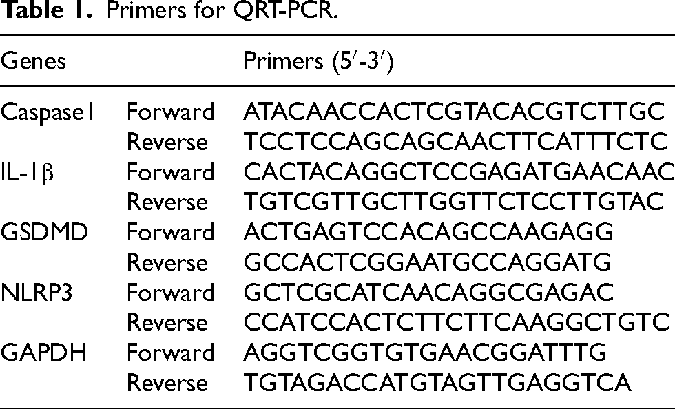
Total RNA was extracted by the 6 min high-quality RNA Extraction Kit (ZS-M11005, ZHONGSHI TONGCHUANG, China) and reversed by reverse-transcribed into cDNA with the PrimeScript RT reagent kit (ZHONGSHI TONGCHUANG, China). The quantity and quality of RNA were assessed by A260/A280 ratio. The primers utilized in this study are listed in Table 1.
Primers for QRT-PCR.
Western blot analysis
Hippocampal tissues were lysed in RIPA buffer (BB3201, Bestbio, Shanghai, China). A BCA kit (Beyotime) was used to determine protein concentration. Equal amounts of protein samples were isolated with SDS-PAGE gel, followed by transferred onto PVDF membranes. Then 5% skim milk was used to block the membranes for 1 h. The membranes were incubated at 4°C overnight with primary antibodies including mouse monoclonal anti-mouse NLRP3 antibody (ABIN1169100, antibodies-online,Germany,1:100), rabbit polyclonal anti-mouse Caspase1 antibody (ab138483, Abcam, USA; dilution: 1:1000), rabbit polyclonal anti-mouse IL-1b antibody (ab18955, Abcam, USA; dilution: 1:1000) or β-actin (AC043, Abclonal; dilution: 1:100000). Afterwards, the membranes were incubated with corresponding secondary antibodies for 1 h. Immunoreactive bands were visualized using the enhanced chemiluminescence detection system (Aplegen Gel Company, Inc., USA). The signal intensities were analyzed using ImageJ Software.
Enzyme-linked immunosorbent assay (ELISA)
Serum samples from mice and supernatants of cells (n = 5 in each group) were collected for the detection of inflammatory factor concentrations using IL-1β ELISA kit (KE10003, Proteintech, USA). The test was performed according to the manual instruction.
TUNEL staining
For TUNEL staining, HT-22 cells were incubated with TUNEL reaction mixture (MK1014-100, Boster Biological Technology, China) according to the manual instruction. DAPI was used to counterstain the sections for 4 min. The positive cells were observed using a fluorescence microscope.
Immunofluorescence staining for NLRP3 specks in HT-22 cells
HT-22 cells of each group were fixed using a 4% paraformaldehyde followed by permeabilization and blocking with 5% normal goat serum containing 0.3% Triton X-100. subsequently, the cells were incubated in primary antibody against NLRP3 (mouse-anti NLPR3, ABIN1169100, antibodies-online, Germany, 1:100) to detect the NLRP3 protein expression in HT-22 cells. One hour incubation at room temperature with a FITC-conjugated Goat Anti-Mouse IgG (H + L) Antibody (K1201, APEXBIO). DAPI was used to visualize cell nuclei. The images of the NLRP3 specks were observed by using a fluorescence microscope.
Statistical analysis
The data were presented as the mean ± standard error and analyzed using GraphPad Prism version 8 (GraphPad Software, La Jolla, CA, USA). Two-way repeated measures analysis of variance (ANOVA) was employed to demonstrate the statistical differences of means within each treatment group at the specific time points for the MWM tests. Group variations were assessed using one-way ANOVA followed by Turkey's multiple comparison tests. All p values <0.05 were considered statistically significant.
Results
Changes of body weight and blood glucose in db/db mice administered melatonin
To investigate the effect of melatonin on body weight and fasting blood glucose in diabetic mice, db/db mice were administrated melatonin treatment (20 mg/kg) for 6 weeks, and body weight and fasting blood glucose were measured on the 7th, 14th, 21st, 28th, 35th, and 42nd days. As expected, db/db mice showed significant increases in blood glucose and body weight. Blood glucose and body weight of db/db +20mel group were lower than those of db/db group (blood glucose: 29.67 ± 0.10 mmol/L versus 33.09 ± 0.14 mmol/L, p < 0.05; body weight: 53.7 ± 0.22 g versus 58 ± 0.46 g, p < 0.05), showing the advantages of melatonin in lowering blood glucose and weight in type 2 diabetic mice.
Melatonin treatment ameliorated diabetes-associated cognitive impairment
Cognitive function was evaluated by MWM test and NOR test. During the MWM assessment, a notable difference in escape latency time was observed in the db/db group starting from day 2 to day 5 compared with the db/m group (Figure 1A and B). The statistical analysis comparing crossing and swimming times in the platform quadrant on day 5 is illustrated in Figure 1C and D. Mice in the db/db group exhibited significantly shorter platform crossing times compared to the db/m group (p < 0.001). The db/db + MEL group exhibited a significantly higher number of crossing times compared to the db/db group (p < 0.05). Likewise, the db/db group demonstrated a significantly lower percentage of time spent swimming in the platform quadrant in comparison to the db/m group (p < 0.001). The db/db + MEL group displayed a significant increase in the percentage of time spent swimming in the platform quadrant as opposed to the db/db group (p < 0.05). Additionally, as illustrated in Figure 1E, a reduced discrimination index was evident in the db/db group (p < 0.05). Collectively, these findings indicate that melatonin administration restored cognitive function decline in db/db mice.
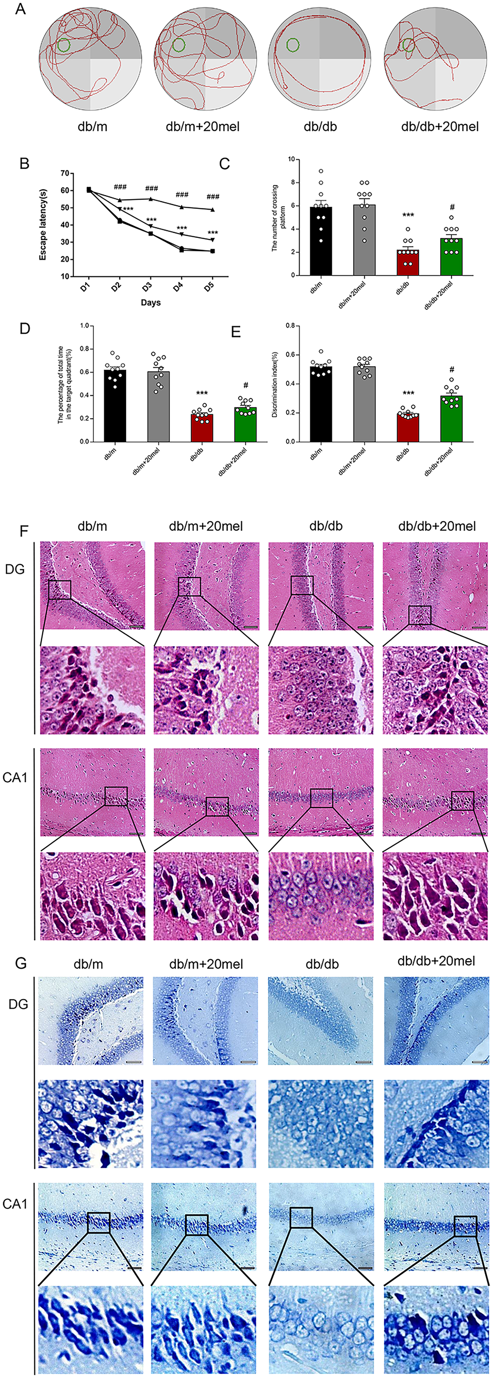
Melatonin improves cognitive inpairment in db/db mice. (A) representative swimming trace during the spatial probe test. (B)Escape latency of the hidden-platform test. (C) Number of target crossing in the probe trial. (D) The percentage of total time spent in target quadrant in the probe trial.(E) Discrimination index of the db/db mice.All data are expressed as means ± SEM for 10 mice in each group. *P < 0.05, compared with the db/m group. # P < 0.05, compared with the db/db mice.(C)HE staining in the hippocampal DG, CA1 rigions and cortex.(D)Nissl staining in the hippocampal DG, CA1 rigions and cortex. (n = 3,Scale bar = 50 μm).
Protective effect of melatonin against pathological deficits in the hippocampus of db/db mice
HE staining was performed to examine the effects of melatonin on neuronal loss and damage in the hippocampus of db/db mice. As shown in Figure 1F, well-arranged and intact hippocampal pyramidal cells were observed in hippocampal dentate gyrus (DG) region, CA1 region, CA3 region and cortex in db/m group. In contrast, deformed neurons, karyopyknosis and cavitation presented in the corresponding regions in db/db mice. After melatonin treatment, disarranged neuron cells were reduced in db/db mice. Nissl staining indicated a reduction in Nissl bodies, disrupted cellular structure, and neural impairment in the db/db group in contrast to the db-m group. Following MEL intervention, the count of Nissl bodies was restored in cortex and different regions of hippocampus (Figure 1G). Overall, these results proved that melatonin could protect hippocampal neurons from neuronal injuries in db/db mice (Table 2).
Comparisons of body weights and blood glucose levels in four groups of mice.
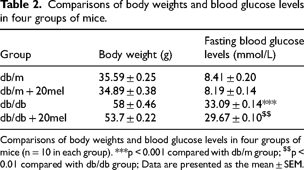
Comparisons of body weights and blood glucose levels in four groups of mice (n = 10 in each group). ***p < 0.001 compared with db/m group; $$p < 0.01 compared with db/db group; Data are presented as the mean ± SEM.
Melatonin inhibits the expression of the NLRP3/Caspase 1 signaling pathway in the hippocampus of db/db mice
Double immunofluorescence labelling in db/m group and db/db group showed that NLRP3 proteins were mainly colocalized in hippocampal neurons (Figure 2C) instead of astrocytes (Figure 2A and D) or microglia (Figure 2B and E), which was consistent with the results of previous study. 28 Melatonin treatment decreased the fluorescence intensity of NLRP3 in the neurons of DG (Figure 2F) in db/db mice. Furthermore, QRT-PCR and western blot results demonstrated that expression levels of NLRP3, GSDMD, Caspase-1 and IL-1β increased significantly in the hippocampus of db/db mice, compared with db/m mice (p < 0.001, Figure 3A, B and D). Melatonin prevented the aggravation of NLRP3/Caspase 1 signaling pathway in db/db mice (p < 0.001, Figure 3A, B and D). In addition, increased levels of IL-1β in the hippocampal tissues of db/db mice were reversed by melatonin treatment (p < 0.01, Figure 3C). These outcomes suggested that melatonin treatment could inhibited NLRP3/Caspase 1 pathway which mediated inflammasome activation and neuronal inflammation in db/db mice.
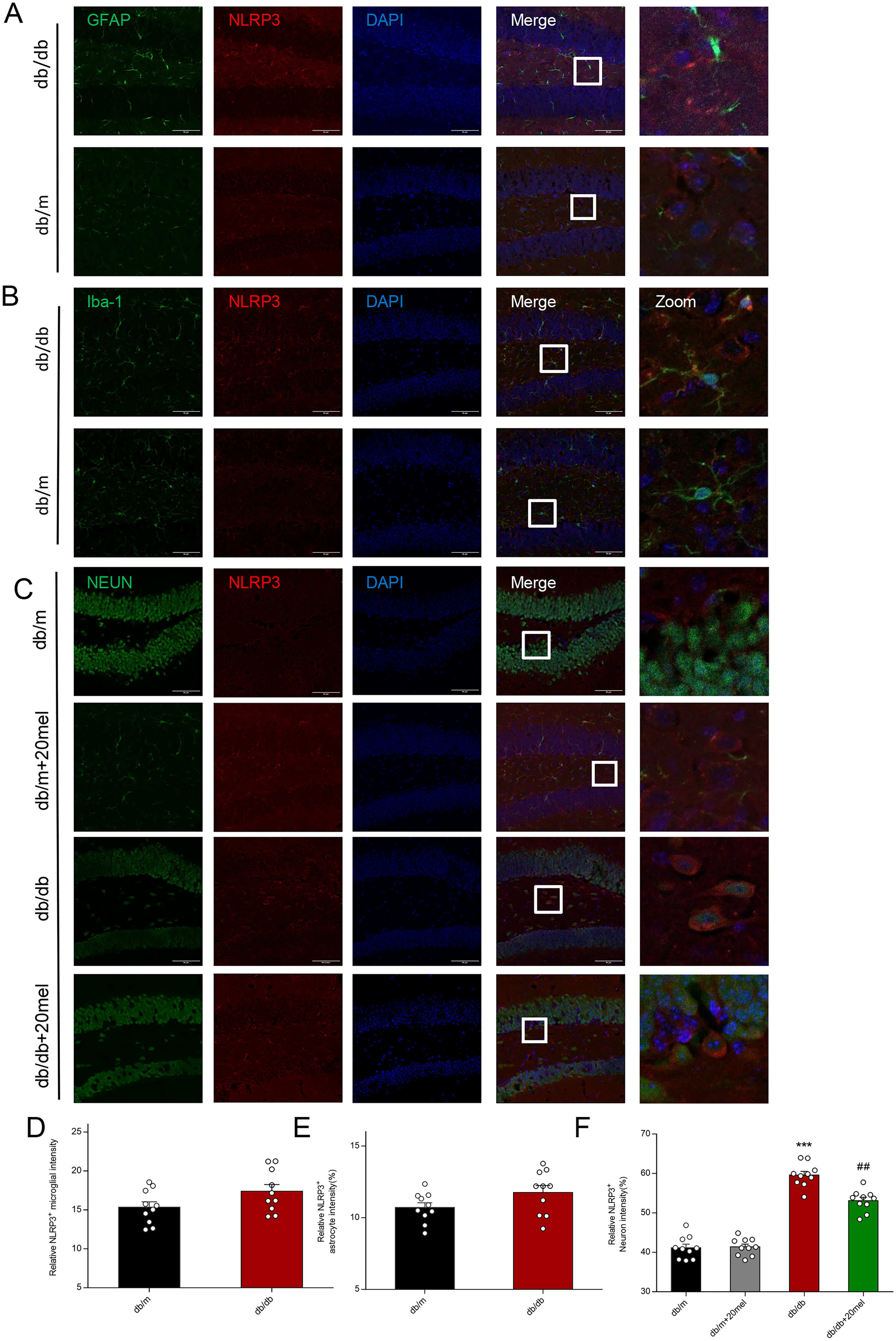
The cellular location of NLRP3 in the hippocampus of different groups for the double immunofluorescence labelling. (A) Immunofluorescence images of DG showed the expression of NLRP3 in GFAP-positive astrocyte. (B) Immunofluorescence images of DG showed the expression of NLRP3 in Iba-1-positive microglial. (C) Immunofluorescence images of DG showed the expression of NLRP3 in Neun-positive neuron.(n = 3 in each group;Scale bars =50 μm). (D) Relative NLRP3-positive microglial intensity.(E)Realative NLRP3-positive astrocyte intensity.(F)Relative NLRP3-positive neuron intensity. ***P < 0.001, compared with the db/m group. # #P < 0.01, compared with the db/db mice.
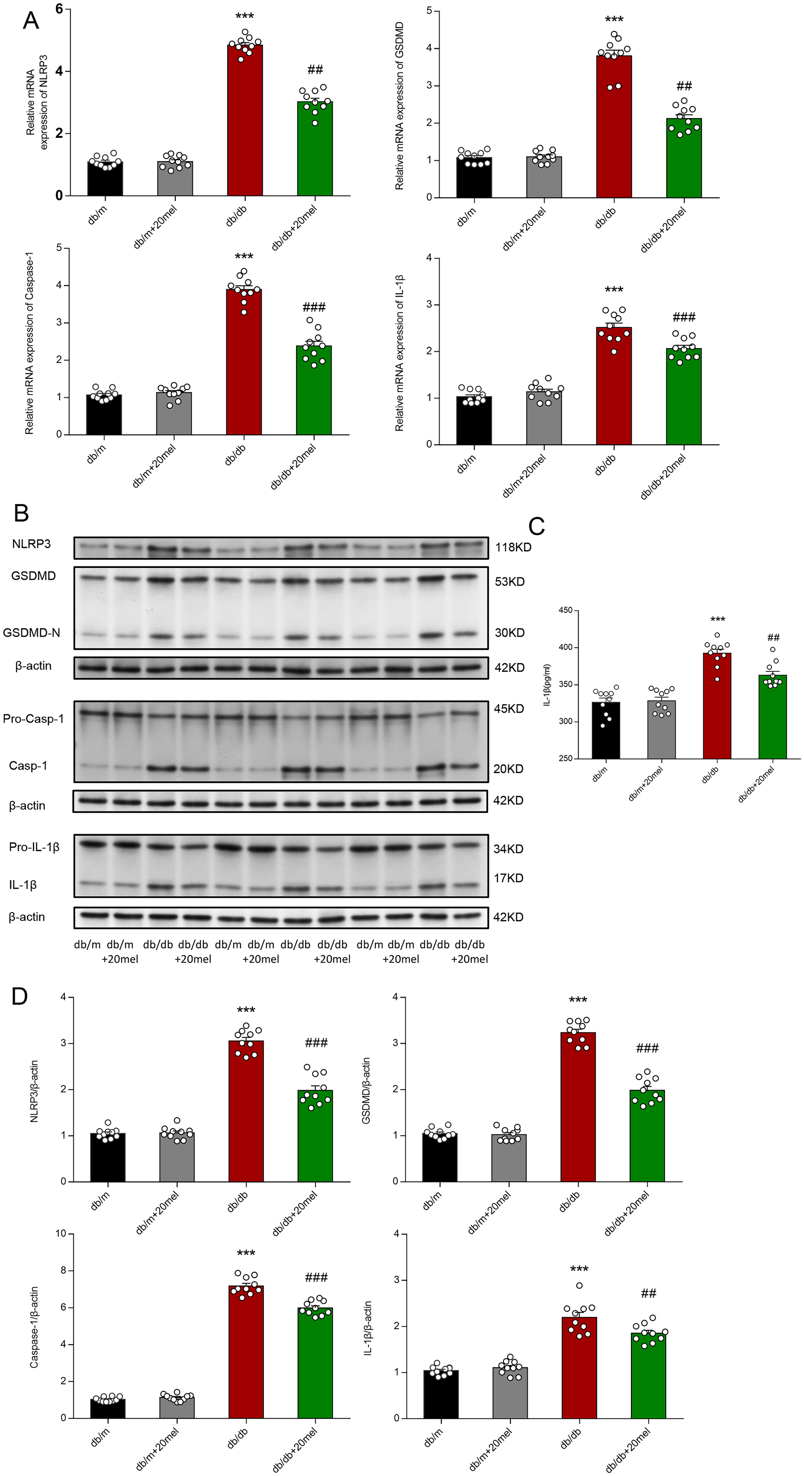
Melatonin inhibits NLRP3/Caspase 1 signaling pathway in the hippocampus of db/db mice. (A) QPCR analysis of NLRP3, GSDMD, Caspase-1 and IL-1b in the hippocampus of each group. (B) Representative protein bands and Western blot analysis of NLRP3, GSDMD, Caspase-1 and IL-1β in the hippocampus of each group. (C) Expression of IL-1β in the hippocampus of each group was measured by ELISA kit. (D) Western blot analysis of NLRP3, GSDMD-N, Caspase-1 and in the hippocampus of each group. Values are represented as means ± SEM for three mice in each group. *** P < 0.001, compared with the db/m group. ##P < 0.01, ###P < 0.001, compared with the db/db group.(n = 10 in each group).
Melatonin attenuated the activation of NLRP3/Caspase 1 signaling pathway in high-glucose exposure to HT-22 cells
Mouse hippocampal neuron cell line HT-22 exposed to high-glucose was used as cellular model of hippocampus in diabetic condition. The TUNEL assay confirmed that melatonin could ameliorate neuronal apoptosis in high-glucose group (Figure 4A). To evaluate whether melatonin suppressed NLRP3 activation in high-glucose exposed HT-22 cells, NLRP3 expression were analyzed by QRT-PCR and cell immunofluorescence. The outcomes showed that mRNA expressions of NLRP3, GSDMD, Caspase-1, and IL-1β were markedly enhanced by high glucose induction, which was significantly suppressed by melatonin treatment or NLRP3 inhibitor MCC950 (p < 0.001, Figure 4E). Melatonin treatment reversed the expression of key proteins in NLRP3/Caspase 1 signaling pathway in high glucose exposed HT-22 cells, which confirmed the effect of melatonin in suppressing neuronal inflammation (p < 0.05, Figure 4C and E). Additionally, ELISA assay indicated an increased IL-1β level in high glucose exposed HT-22 cells, in contrast to melatonin reducing IL-1β level in high glucose exposed group. In pcDNA-mNLRP3 group with NLRP3 overexpression, the anti-inflammatory effect of melatonin was diminished. The results proved that melatonin exerted anti-inflammatory effects through NLRP3/Caspase 1 signaling pathway (p < 0.05, Figure 4C–E). These findings suggested that melatonin inhibited high glucose-induced neuronal apoptosis and inflammation through inhibiting NLRP3/Caspase1 pathway.
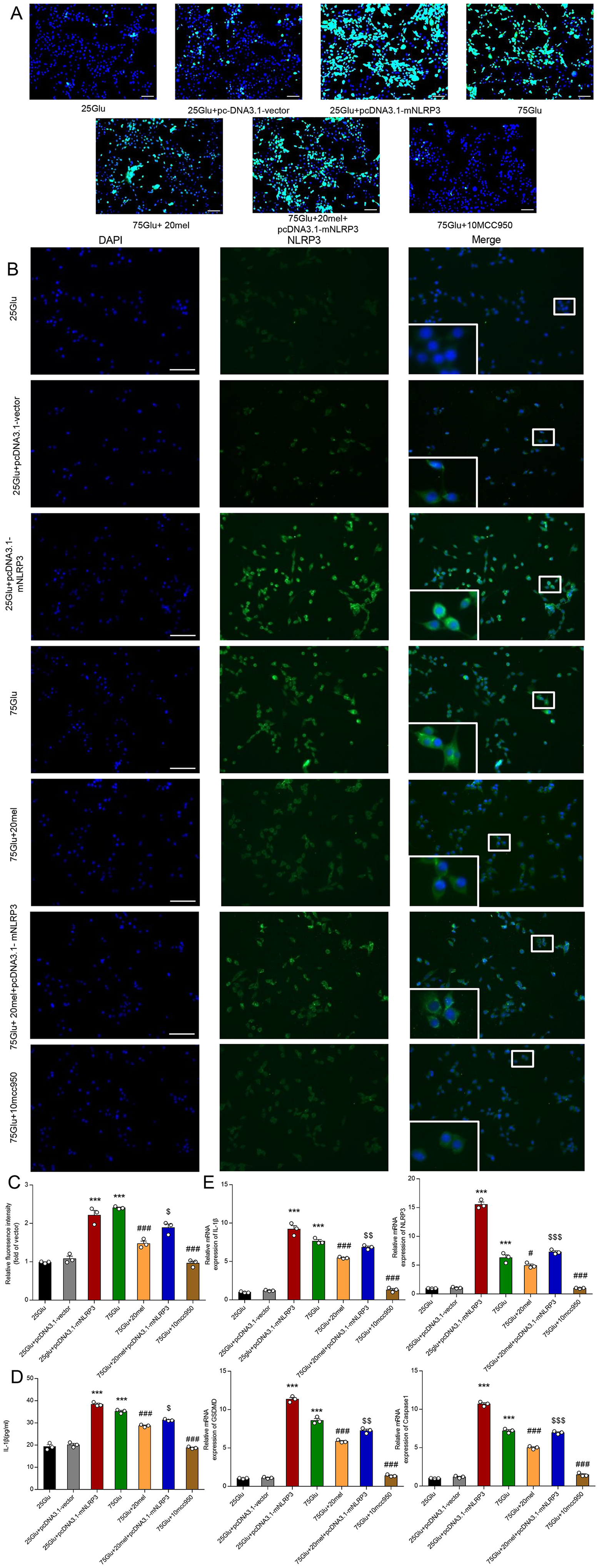
Melatonin inhibited cell apoptosis through inhibiting NLRP3/Caspase1 pathway. (A) Cell apoptosis was determined by TUNEL staining. (B) Immunofluorescence images of NLRP3 (green)/DAPI (blue) colocalization in hippocampal neuronal HT-22 cells at 48 h after treatment with 25 mmol/L glucose or 75 mmol/L glucose in the absence or presence of pc-DNA3.1-mNLRP3 (n = 3,Scale bar = 50 μm). (C) Relative fluoresence intensity of NLRP3 in each group. (D) Levels of IL-1β in each group was measured by ELISA kit. (E) QRT-PCR analysis of NLRP3, Caspase-1, GSDMD, IL-1β in hippocampal neuronal HT-22 cells in each group. Values are represented as means ± SEM .*** P < 0.001, compared with the 25Glu group. # P < 0.05, ## P < 0.01, ### P < 0.001, compared with the 75 Glu group.$ P < 0.05, $$ P < 0.01, $$$ P < 0.001, compared with 75Glu + 20mel.
Discussion
The present study demonstrated that (1) melatonin treatment significantly ameliorated diabetes-associated cognitive impairment, hyperglycemia, and overweight in db/db mice, (2) melatonin treatment significantly attenuated deformation and apoptosis of hippocampal neurons both in vivo and vitro, and (3) melatonin treatment suppressed the activation of NLRP3 inflammasome and attenuated hippocampal inflammatory responses. Overall, melatonin treatment significantly attenuated hippocampal neuron damage and alleviated cognitive impairment in diabetic db/db mice through inhibition of NLRP3/Caspase 1 signaling pathway (Figure 5). Hyperglycemia induced the activation and expression of NLRP3, which subsequently interacts with ASC and Caspase-1, forming the NLRP3 inflammasome complex. The NLRP3 inflammasome complex facilitates Caspase-1-mediated cleavage of GSDMD into two fragments: the N-terminal and C-terminal regions. Concurrently, active Caspase-1 initiates the processing of pro-IL-1β and pro-IL-18. The N-terminal region of GSDMD binds to the membrane, forming a pore essential for the release of IL-1β and IL-18, leading to neuronal apoptosis. Melatonin mitigates IL-1β and IL-18 production by suppressing the NLRP3/Caspase-1 signaling pathway activation in neurons, thereby conferring neuroprotection against diabetes.
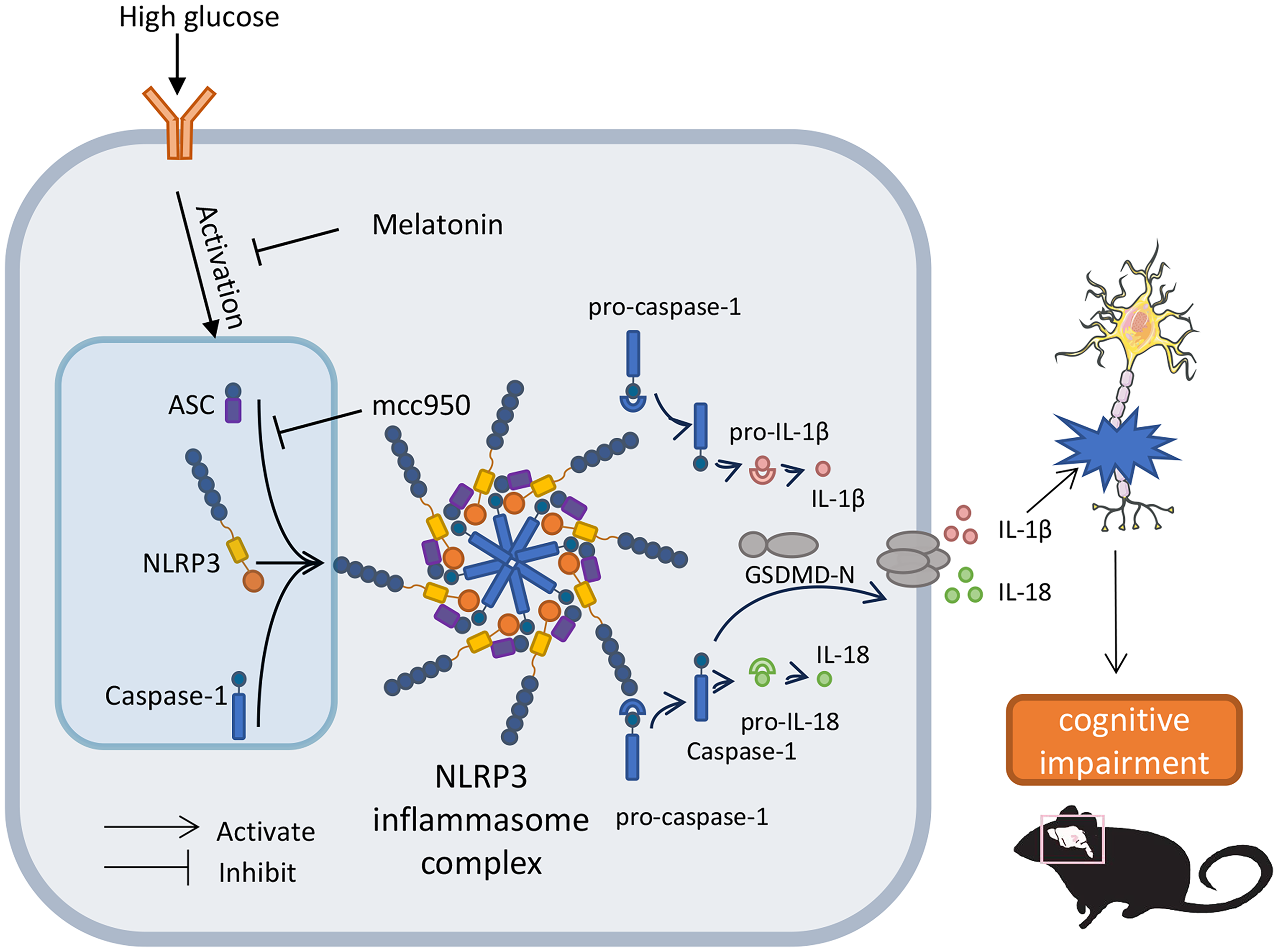
Schematic illustration of the mechanism of melatonin inhibition on NLRP3/Caspase-1 signaling pathway. High glucose strongly stimulates the activation and increased expression of NLRP3, which binds to ASC and Caspase-1 to assemble into NLRP3 inflammasome complex. NLRP3 inflammasome complex allows Caspase-1 to cleave GSDMD into two fragments: the N-terminal region and the C-terminal region. The active Caspase-1 also start the cleavage of pro-IL-1β and pro-IL-18. The N-terminal region of GSDMD binds to the membrane and form a pore. This pore is necessary for the release of IL-1β and IL-18, which induces neuronal apoptosis. Melatonin reduces the production of IL-1β and IL-18 by inhibiting the activation of NLRP3/Caspase-1 signaling pathway in neurons, thereby exerting neuroprotection against diabetes.
The present study clarified the neuroprotective effect of melatonin in db/db mice. Db/db mice as a type 2 diabetic model exhibited typical hippocampus-dependent memory impairment is widely used in diabetic cognitive decline research. Therefore, db/db mice were selected for investigating the pathogenesis of diabetes-related cognitive impairment and for screening potential drugs. 29 Female rodents exhibit cyclical hormonal fluctuations (estrous cycle) that may confound metabolic and inflammatory measurements. 30 And our lab's prior work established has already used male db/db model as diabetic cognitive impairment model. 31 Therefore we choose male db/db model to maintain consistency with our previous investigations.
One of the main chronic complications in T2DM is progressive deterioration of cognitive function. The available epidemiological data demonstrates a high estimated prevalence of cognitive impairment among individuals with T2DM worldwide. 32 Since the publication of the 2014 IDF guideline, 33 which was the first guideline to address the detection and management of diabetic cognitive dysfunction or dementia, diabetes-associated cognitive impairment has garnered increased attention from clinicians and researchers. While metformin and dulaglutide have exhibited promise in improving cognition in individuals with mild to moderate cognitive impairment in T2DM, their clinical efficacy remains to be fully elucidated.34,35 Our team is committed to investigating the pathogenesis of cognitive impairment in diabetes and exploring alternative medications to alleviate cognitive decline. It is imperative to identify potential drugs for treating diabetes-related cognitive deficits. The mechanisms underlying the efficacy of DL-3-n-butylphthalide (NBP) and troxerutin in ameliorating diabetic cognitive impairment have been elucidated.5,31
Melatonin, a substituted indoleamine derived from tryptophan, was first isolated from the pineal gland of a cow by Lerner and colleagues in 1958. 36 Melatonin is notably involved in regulating physiological functions such as sleep, circadian rhythms and neuroendocrine. Recently, melatonin has been employed in the delay of several degenerative processes. Melatonin has been suggested as a promising candidate for enhancing osteoarthritis and myocardial injury by inhibiting chondrocyte apoptosis and reducing inflammatory responses.37,38 Neuroprotective effects of melatonin have been revealed in Huntington's disease mice with neurodegeneration. 39 Melatonin inhibition of NLRP3 inflammasome in regulation of NF-κB Nucleocytoplasmic transport ameliorated postoperative cognitive dysfunction. 40 However, its role in preventing cognitive impairment associated with diabetes remains unclear. This led us to propose that melatonin may have a potential effect against cognitive impairment associated with diabetes.
MWM is the gold standard for assessing spatial learning and memory in rodents. Melatonin significantly reduced escape latency and enhanced the number of crossings in the target quadrant, aligning with prior research findings. 41 We demonstrated that db/db mice exhibited hippocampal neuronal deformation and decreased Nissl body number accompanied with increased neuronal apoptosis. Previous studies have identified several mechanisms of hippocampal damage in neurodegenerative diseases, including oxidative stress, mitochondrial dysfunction, and inflammatory responses. 42 The ELISA result of hippocampus tissue extracts revealed an increase in IL-1β level in db/db mice, which was reduced by melatonin treatment. As a key pro-inflammatory factor in vivo, IL-1β is involved in diverse autoimmune inflammatory responses and various cellular activities including cell apoptosis. The secretion of IL-1β is governed by exogenous PAMPs and endogenous DAMPs. PAMPs and DAMPs need to bind to membrane Toll-like receptors (TLRs) or intracellular NOD-like receptors (NLRs) for their action. Growing evidence has characterized that hyperactivation of NLRP3 inflammasome is involved in various inflammatory diseases. 43 PAMPs and DAMPs stimulate upregulation of NLRP3 expression, which contributes to the recruitment of ASC and pre-Caspase-1 to form a complex. 44 This leads to activation of Caspase-1 and cleavage of gasdermin D (GSDMD). As a membrane pore-forming protein, the N-terminal region of GSDMD binds to the membrane and allows the release of the pro-inflammatory cytokines IL-1β and IL-18. 45 MCC950, a recently identified inhibitor of the NLRP3 inflammasome, has shown promise in mitigating cognitive impairment and exerting neuroprotective effects. 28 MCC950 specifically inhibits NLRP3 inflammasome activation and IL-1β secretion by suppressing NLRP3-induced ASC oligomerization. 46 It has been reported that NLRP3 activation triggered by Aβ aggravated IL-1β releasing and pathogenesis of AD. 47 Consequently, we assumed that neuroinflammation in the hippocampus is associated with the development of cognitive dysfunction in db/db mice.
NLRP3/Caspase 1 signaling pathway activation was identified in the hippocampus of db/db mice, including upregulation of NLRP3, GSDMD, Cleaved-caspase-1 and IL-1β by western blot and QRT-PCR analysis. Melatonin administration for 6 weeks led to the suppression of the NLRP3/Caspase 1 pathway in db/db mice, along with an improvement in cognition. The findings highlight the significance of the NLRP3/Caspase 1 signaling pathway in the hippocampus regarding diabetes-related cognitive impairment. A recent study revealed that inhibition of NLRP3 inflammasome activation relieves motor deficits and nigrostriatal dopaminergic degeneration through Nrf2/NLRP3/Caspase 1 pathway in aged mice. 48 Isoflurane increased IL-18 and IL-1β secretion by activating the NLRP3/Caspase 1 pathway in aged mouse microglial cells. 49 These findings demonstrated NLRP3 is a critical factor concerning aged-related cognitive impairment.
In the cytoplasm, inflammasomes are detected in a broad range of cell types, comprising immune cells like macrophages, dendritic cells, T cells, and B cells, along with neuronal cells and glial cells such as microglia and astrocytes. Some studies have shown the predominant localization of the NLRP3 inflammasome in microglia within the central nervous system, whereas others have suggested its presence in astrocytes and neurons. In lipopolysaccharide challenged neuroinflammation-related cognitive impairment model, NLRP3 inflammasome activation was found in microglia of hippocampus. 9 Interestingly, we found NLRP3 colocalized with NeuN (neuronal marker) more than Iba1 (microglia marker) in high glucose group in vitro. In vivo, double immunofluorescence labelling of NeuN and NLRP3 revealed that NLRP3 expression level in neurons is parallel to the severity of hippocampal neural damage and cognitive decline in db/db mice. Melatonin reversed NLRP3/Caspase 1 signaling pathway in neurons, along with alleviated hippocampal neural damage and cognitive decline in vitro and vivo. A comparable investigation demonstrated that FGF21 mitigates neurodegeneration by diminishing neuroinflammation and oxidative stress through the regulation of the NF-κB pathway and the AMPKα/AKT pathway in neurons. 50 A previous study showed that Meisoindigo alleviated brain damage in ischemic stroke by suppressing NLRP3 inflammasome in neurons via TLR4/NF-κB signaling pathway. 45 Based on the present results, we speculated that NLRP3 upregulation and activation in hippocampal neurons may lead to neuron apoptosis and cognitive impairment in db/db mice. Melatonin exerted a protective effect on diabetic cognition through NLRP3/Caspase 1 signaling pathway in hippocampal neurons in T2DM.
In vitro, the protective effect of melatonin in high glucose HT-22 cells was reversed by NLRP3-overexpressed plasmid, which is confirmed by increased level of IL-1β. The specific inhibitor of NLRP3 MCC950 was employed as positive control in high glucose group, which indicated that the NlLRP3 inhibition was directly correlated with neuronal apoptosis and neuroinflammation reduction. Similarly, TUNEL assays showed more apoptotic cells in the high glucose group than in the control group, which was partly reversed by melatonin and MCC950. The increased expression of NLRP3, GSDMD, Cleaved-caspase-1 and IL-1β were observed both in high glucose group and NLRP3-overexpressed group. Melatonin reduced the expression levels of these molecules, consistent with NLRP3 inhibitor MCC950. Thus, the in- vitro experiments suggested that melatonin protected hippocampal neuronal damage and apoptosis through NLRP3 and its downstream pathway.
The present study has two potential limitations. Melatonin exerts many of its functions by binding to the MT1 and MT2 G-protein-coupled membrane receptors initiating diverse signaling mechanisms.51–53 These receptors are found in multiple organs of the body, comprising the central nervous system, retina, liver, gut, and skin. Since melatonin was administrated intraperitoneally, the involvement effects of other organs on cognitive function cannot be excluded, such as the microbiota-gut-brain axis.54,55 Secondly, we only examined the inhibitory effect of melatonin on the NLRP3/Caspase 1 signaling pathway. Further investigations are required to examine the impact of melatonin on additional signaling pathways, including TGF-β, MAPK, and NF-κB pathways. 37
Overall, our study demonstrated that melatonin exerted neuroprotective effects on diabetes-associated cognitive impairment by effectively inhibiting NLRP3/Caspase 1 signaling pathway. These results suggest that melatonin may be a promising candidate for combating cognitive dysfunction in diabetes.
Footnotes
Acknowledgements
We would like to thank Hebei Key Laboratory of Rare Disease and Hebei Key Laboratory of Neurophysiology for the technical support. The authors would like to thank professor Sheng Wang for his kind support in this project.

Ethical considerations
All experimental procedures were conducted according to the Guide for the Care and Use of Laboratory Animals. All protocols for animal treatment were approved by the Animal Ethics Committee of Hebei Medical University.
Consent to participate
Not applicable
Consent for publication
Not applicable
Author contributions
Funding
The authors disclosed receipt of the following financial support for the research, authorship, and/or publication of this article: This work was supported by the Natural Science Foundation of Hebei Province, Health and Medical Science Research Project of Hebei, (grant number nos. H2021206452, nos.H2021206187, no.20200908).
Declaration of conflicting interests
The authors declared no potential conflicts of interest with respect to the research, authorship, and/or publication of this article.
Data availability statement
Data will be made available on request.