



Abstract
Background
The rising prevalence of Alzheimer's disease (AD) highlights the urgent need for novel therapeutic approaches capable of suppressing further neurodegeneration. Aberrant aggregation of the amyloid-β peptide fragment of the amyloid-β protein precursor (APP) has long been considered to be a central feature of AD pathology. However, directly targeting the amyloid-β peptide is complicated by its conformational flexibility. Instead, reducing APP expression at the translational level represents a promising alternative. The highly structured 5′ untranslated region (UTR) of APP mRNA provides a targetable element for selective inhibition using RNA-binding therapeutics.
Objective
To develop and evaluate engineered protein-based RNA binders (PROTEIMERs) that selectively target the APP 5′-UTR to inhibit translation.
Methods
We applied high-throughput phage display screening techniques to identify two PROTEIMERs with high affinity for the APP 5′-UTR, confirmed via surface plasmon resonance. Domain engineering enabled the fusion of these binders to an RNase domain to facilitate catalytic degradation of APP mRNA.
Results
PROTEIMERs bound the APP 5′-UTR with nanomolar affinity. Structural modeling of the PROTEIMER–RNA complexes revealed that the engineered mutations within the binding pocket predominantly interact with the 5′-AGA-3′ cleft of the APP mRNA. RNase-fused PROTEIMERs mediated sequence-specific APP mRNA cleavage in vitro, demonstrating robust target engagement and degradation. The PROTEIMER ProAPPS3-11 effectively inhibited APP translation in SH-SY5Y cells, reducing protein levels by up to 60% in a dose-dependent manner.
Conclusions
These findings establish the feasibility of PROTEIMERs as novel RNA-targeting biologics with therapeutic potential to reduce APP mRNA and protein levels, mitigating downstream AD-related neurodegeneration.
Keywords
Introduction
Alzheimer's disease (AD) is the neurodegenerative disorder that constitutes the leading cause of dementia accounting for 60–80% of all cases in elderly patients over the age of 65. 1 The pathology of AD is characterized by a progressive decline in cognitive function and is primarily marked by the accumulation of amyloid plaques and neurofibrillary tangles in the brain. 2 This formation of amyloid plaques is highly toxic, with their deposition in the extracellular spaces of the brain shown to disrupt cell function leading to neuronal degeneration 3 often via activating tau protein dependent pathways. 4 These amyloid plaques are predominately formed by the aggregation of amyloid-β (Aβ) peptides that are highly amyloidogenic and are generated due to improper processing of the amyloid-β protein precursor (APP).5,6 Recent studies have also revealed that the APP protein contributes not only to this neurotoxic amyloid production but also to the accumulation of a more pathogenic C-terminal fragment (APP-CTF),7,8 which is strongly associated with lysosomal disruption and ferroptosis in the brains of AD patients.9–12 Work by Nixon and colleagues demonstrated that familial AD (fAD) mutations and APP gene duplications elevate APP-CTF levels, impairing lysosomal function and neuronal viability through both γ-secretase-dependent and independent mechanisms. 13 These disruptions lead to defective autophagic flux, accumulation of cellular waste, impaired endolysosomal acidification, iron dyshomeostasis, and mitochondrial dysfunction. 10 14–17
Previous studies demonstrated that iron enhances APP translation and that APP interacts with the iron exporter ferroportin to promote iron efflux from neurons. 18 This is consistent with observations in fAD patients carrying APP duplications, who often suffer from severe intracerebral hemorrhages. 19 Disruption of APP-mediated iron export can trigger ferroptosis—an iron-dependent form of oxidative cell death characterized by mitochondrial damage.14,20,21 In Down syndrome (DS) fibroblasts, 22 impaired lysosomal acidification has been demonstrated using pH-sensitive dyes. 23 Pharmacological inhibition of APP Tyr682 phosphorylation has been shown to reduce APP-CTF levels and restore lysosomal function in vivo. 10 Collectively, these findings support a model in which APP-CTF–mediated disruption of lysosomal v-ATPase activity, coupled with dysregulated APP translation, serves as an initiating mechanism in AD pathogenesis.11,20,24 Therefore, targeting excess APP expression represents a compelling therapeutic strategy to mitigate lysosomal damage, restore autophagic and pH balance, and prevent iron-induced neurotoxicity. 25 APP translation thus emerges as a promising and previously underexplored therapeutic target for AD and related neurodegenerative disorders.
However, directly targeting the expressed APP protein has proven to be difficult due to its intrinsically disordered nature and lack of a defined binding site.7,8,26 An alternative approach to reduce the expression of APP could lie in targeting the initial APP mRNA to reduce total APP protein production. 27 Targeting of mRNA is a much more straightforward process as high affinity binders can be easily designed around a variety of common drug design scaffolds such as proteins, nucleic acids, or small molecules.24,28,29 The 5′ untranslated region (UTR) of mRNA is of particular interest as a druggable target due to its ability to regulate translation and form unique stable structural motifs.30,31 The 5′-UTR of the APP mRNA is highly rich in GC content with many elements forming stable secondary structures that could be more easily targeted. 32 One element of note is an iron-responsive element (IRE) that plays a critical role in cellular iron homeostasis.20,31 The IRE is most commonly found in transcripts relating to cellular iron maintenance, such as Ferritin H/L chains. Interestingly, modified IREs have also been shown to be present in transcripts associated with neurodegenerative disorders such as SNCA in Parkinson's disease and the aforementioned APP, further highlighting the connection between these neurodegenerative proteins and cellular iron regulation. 31 To be more easily recognized, IREs can form stable stem loop structures that play a critical role in forming a larger internal ribosomal entry site (IRES) within the 5′-UTR. Under conditions of iron-mediated stress, the IRES is activated to maintain levels of critical proteins through cap-independent translation initiation. 33
Because the IRE forms a stable stem-loop structure within the 5′-UTR region of the APP mRNA, many studies using antisense oligonucleotides (ASOs) or small molecules have already explored it as a therapeutic target to modulate APP expression. 33 Designed ASOs targeting the APP mRNA have been shown to inhibit translation of the gene but have faced numerous difficulties due to the high polarity of nucleic acids that affect not only the stability of the drug, but also inefficient delivery and uptake to the target.34,35 The use of small molecules has also shown promise in inhibiting the translation of the APP RNA through site specific design to target the structural motifs.36,37 However, in both cases these molecules must bind to their target mRNA for the duration of their efficacy window. A different approach to target the IRE of the APP mRNA could be utilizing a protein-based therapeutic that can directly bind the unique stem loop structure. Utilizing a protein-based binder would not only allow for better sequence and structure specificity but could also be fused to an RNase active protein to form an RNA targeting chimera (RNATaC) that can directly degrade the APP mRNA upon binding. 30 This would allow for immediate regulation of APP translations as well as permit the designed RNATaC to affect multiple mRNA transcripts.
To explore a new avenue for a possible treatment for Alzheimer's Disease, we identified possible RNA-binding proteins, which we have termed PROTEIMERs (
Methods
Phage display library construction
NNK (N = any nucleotide, K = G, T) libraries, as described in US Patent 12,104,201, 38 were cloned into T7 bacteriophage using the T7Select10-3 Cloning Kit (Millipore Sigma, Burlington, MA). 30
Phage purification from lysate
Phages were purified from library lysates using polyethylene glycol (PEG) precipitation. Lysates were first filtered through a 0.45 µm filter, then mixed with 20% (v/v) PEG solution (20% PEG-6000, 2.5 M NaCl) and incubated for 1 h at 4°C. Phages were pelleted by centrifugation at 4500 RPM for 30 min at 4°C and resuspended in PBS. A second precipitation was performed by adding 20% PEG solution, incubating for 10 min at 4°C, and centrifuging again under the same conditions. Final phage pellets were resuspended in PBS and stored at 4°C for up to one month.
Synthesis of biotinylated 5′-UTR of APP mRNA
The 5′-UTR of APP mRNA containing structured stem-loops was synthesized by Integrated DNA Technologies (IDT). The 5′-terminus was modified to contain a biotin-TEG with an extended spacer arm. APP mRNA sequence used is as follows:
5′- BiotinTEG – GCGGCGGUGGCGGCGCGGGCAGAGCAAGGACGCGGCGGAUCCCACUCGCA-3′
Biopanning
10 µL of 100X concentrated phages was first pre-blocked in 90 µL of blocking buffer containing 10 mM Tris-HCl pH 8, 1 mM EDTA, 250 mM NaCl, 0.5% Triton X-100, 50 µg of tRNA and 20 U of RNase Inhibitor. This blocking was left to incubate shaking at RT for 20 min. 100 nM of the biotinylated 5′-UTR APP mRNA bait was folded by incubating at 80°C for 5 min. The newly folded mRNA was then added to the blocked phages and incubated for an additional 20 min with shaking at RT. 10 µL of Dynabeads M-280 Streptavidin (Invitrogen, 11206D) were enriched with the phage mixture after incubation and allowed to incubate shaking at RT for 30 min to promote capture. The magnetic beads with the captured phages were then washed extensively with a buffer containing 10 mM Tris-HCl at pH 8.0, 1 mM EDTA, 250 mM NaCl, and 0.5% Triton X-100 to remove all unbound phages. Parallel to this process, BLT5615rna cells were grown to an OD of 0.4 in LB + M9 media. The cells were induced with Isopropyl β-D-1-thiogalactopyranoside (IPTG) for 30 min to produce capsid 10A protein. The collected beads with the captured phages were then used to infect the capsid-producing 5616rna cells at 37°C for 1–2 h until lysis could be seen to occur. 39 The lysate was then centrifuged at 7100 xg for 20 min at 4°C to pellet the cellular debris. The resulting supernatant was then PEG purified with a solution containing 20% PEG 6000 and 2.5 M NaCl before resuspending the final pellet in PBS buffer to obtain the newly purified phage product for further biopanning. Subsequent biopanning rounds followed the same procedure as above with slight modifications where each round saw reductions in mRNA bait concentrations (from 100 nM to 10 nM) and the amounts of magnetic beads used for capture (from 10 µL to 1 µL). Biopanning was performed over several rounds until sequence convergence could be seen.
Creation of constructs
Potential candidates from biopanning studies were cloned into the pET-28b expression vector with an N-terminal 6xHis-tag and SUMO-tag to aid in downstream purification. Candidates that displayed successful binding to the target APP RNA were subsequently cloned into a pET-24 plasmid to generate a large fusion construct consisting of several protein domain linkages to produce the Ang2-PUM-CANDIDATE-PIN-ABD construct.
Protein purification of candidates
The cloned candidates were transformed into the Rosetta cell line and plated onto agar plates containing kanamycin and chloramphenicol. A single colony was inoculated into an overnight starter culture before being transferred into a larger 500 mL volume of LB medium to grow to an optical density of 0.6 at 37°C. The cells were then induced using 0.1 mM IPTG overnight at room temperature. The following day, the cells were pelleted at 4000 xg for 30 min and stored at −20°C until needed.
On the day of purification, the frozen pellets were resuspended in a lysis buffer that contained 50 mM Tris-HCl at pH 8.0, 150 mM NaCl, 1% Triton X-100, and 20 mM imidazole. The resuspended cells were left to incubate with shaking on ice for 45 min before undergoing 10 cycles of 10 s ON 30 s OFF sonication utilizing a Fisher Scientific Sonic Dismembrator Model 100 at power level 12. The cellular debris was spun down at 8000 x g for 40 min and the produced supernatant was filter sterilized with a 0.45 µm filter. The collected filtered lysate was then loaded onto a Ni-NTA Superflow Cartridge (Qiagen) under a mobile phase containing 50 mM Tris-HCl at pH 8.0, 150 mM NaCl, and 20 mM Imidazole. Elution of the candidate protein from the column was achieved with an elution buffer containing 50 mM Tris-HCl at pH 8.0, 150 mM NaCl, and 300 mM Imidazole. Fractions containing the protein of interest were buffer exchanged into PBS pH 8 and concentrated using a Vivaspin 20 MWCO 10,000 spin column. The final product was used immediately for binding experiments or stored at −80°C until needed.
Enzyme-linked immunosorbent assay (ELISA)
96-well ELISA plates were coated with the His-tagged APPS3-5 or APPS3-11 before blocking with PBS containing 0.05% TWEEN and 2% BSA. Washing steps were performed with PBS containing 0.05% TWEEN. The biotinylated APP mRNA bait was prefolded as done previously during biopanning. 30 The folded APP mRNA was then serially diluted to generate a concentration range from 33 nM to 1 µM and added to the appropriate coated wells to incubate for 1 h at 37°C. A streptavidin-horseradish peroxidase conjugate (ThermoFisher Scientific, 21130) was then added to each well and left to incubate for an additional hour at 37°C. Binding was measured using a tetramethylbenzidine substrate working solution (BioLegend, 421101). Optical density was measured at 450 nm against a reference wavelength of 600 nm using a Tecan Infinite M200 Pro plate reader.
Surface plasmon resonance (SPR)
The OpenSPR system (Nicoya) was primed with a running buffer that contained 10 mM Tris-HCl pH 8, 1 mM EDTA, 100 mM KCl and 0.1 mM DTT. 1 µM of biotinylated APP mRNA bait was then incubated as performed in initial biopanning. A high sensitivity biotin chip (Nicoya, SEN-HS-8-BIOTIN) was prepared by first surface conditioning with 10 mM HCl before activation by adding 0.5 M streptavidin. The newly folded biotinylated APP mRNA was then immobilized onto the chip through the biotin-streptavidin coupling.
The purified candidate protein was tested by making five dilutions ranging from 60 nM to 2 µM with the SPR running buffer. The diluted candidate proteins were run subsequently from lowest concentration to highest at a flow rate of 20 µL/min to allow for a contact time of 5 min before the subsequent injection. Single cycle analysis of the SPR data was performed using TraceDrawer under a 1:1 binding model.
Computational structure prediction
Structure prediction of each biopanning candidate was generated using the Alphafold3 webserver. 40 The amino acid sequence of each candidate was used as an input to generate a 3D model prediction in the presence of the 50 base-pair IRE motif of the APP mRNA bait that was used for initial biopanning testing. 3–5 replicates of these candidate-APP models were generated and used for comparison to verify binding predictions. The highest-scoring models were analyzed more thoroughly using ChimeraX. 41
Generation of T7-APP mRNA
A construct encoding the T7 promoter sequence, GG nucleotides to enhance transcriptional yield, the APP 5′-UTR hairpin sequence used for biopanning, and the 94 native nucleotides following the hairpin sequence was first synthesized by IDT, and then PCR amplified for use as an in vitro transcription template. APP RNA was generated using the HiScribe T7 High Yield RNA Synthesis Kit (NEB, E2040S) with the prepared template following kit instructions. The synthesized APP RNA was cleaned up using the Monarch Spin RNA Cleanup Kit (NEB, T2040) and analyzed on 15% TBE-Urea polyacrylamide gels (Invitrogen, EC6885BOX). Gel was stained with GelRed Nucleic Acid Gel stain (Biotium, 41001) for 30 min at room temperature before imaging with the E-Gel Imager Gel Documentation System (Invitrogen, 4466601) under UV transillumination.
In vitro degradation assay
In vitro degradation reactions contained 800 ng T7-APP RNA and 3 µM (APPS3-11) or 4.5 µM (APPS3-5) protein of interest in 10 µL total in reaction buffer containing 50 mM HEPES, pH 7.5, 200 mM NaCl, 0.1 mg/mL BSA, 1 mM MgCl2, and 1 mM DTT. They were incubated at 37°C for 0–120 min before the reaction was halted with addition of 10 µL RNA loading dye containing formamide. The samples were heated at 70°C for 10 min, cooled to 4°C for 5 min, then run on a 15% TBE-Urea polyacrylamide gel (Invitrogen, EC6885BOX) for 65 min. The gel was stained with GelRed Nucleic Acid Gel stain (Biotium, 41001) for 30 min at room temperature before imaging with the E-Gel Imager Gel Documentation System (Invitrogen, 4466601) under UV transillumination.
Cell culture and treatment
Human SH-SY5Y neuroblastoma cells were maintained in DMEM supplemented with 10% fetal bovine serum (FBS; Invitrogen, Carlsbad, CA) and penicillin/streptomycin (Bio-Whittaker, Walkersville, MD). Cells were seeded in 100 mm culture dishes and grown to ∼60% confluency before treatment. Cells were then transfected with 1, 2, or 5 μM of PROTEIMER ProAPP1 or DMSO control using Lipofectamine (Invitrogen) following the manufacturer's protocol. After 48 h of treatment, cells were washed twice with ice-cold PBS, detached, and lysed in PBS by sonication.
Western blotting
For analysis of APP expression, cytoplasmic protein lysates were prepared by homogenizing cells in midRIPA buffer (25 mM Tris-HCl, pH 7.4; 1% NP-40; 0.5% sodium deoxycholate; 15 mM NaCl) supplemented with protease inhibitors, RNase inhibitor, and 10 mM DTT. Proteins were separated by SDS-PAGE and transferred to membranes for immunoblotting. APP was detected using an N-terminal antibody (22C11, Millipore) and a C-terminal APP-specific antibody (A8717, Sigma). β-actin (Chemicon) served as the loading control. Signals were developed with enhanced chemiluminescence (ECL; Pierce, Rockford, IL) and visualized using a Bio-Rad PhosphorImager. Band intensities were quantified with Quantity One™ software (Bio-Rad).
LDH cytotoxicity assay
The cytotoxicity of SH-SY5Y neuroblastoma cell treatment with APP PROTEIMER was tested using the CyQUANT LDH cytotoxicity assay kit (Thermo Fisher, C20300). Media was collected from the cells and tested following kit instructions. The LDH positive control supplied with the kit was also tested. Absorbance at 490 nm and 680 nm (reference wavelength) was measured using a Tecan Infinite M200 Pro plate reader.
Results
Construction of TEXS3 library
The TEXS3 PROTEIMER library was constructed and utilized to screen for high affinity binders to the target APP RNA.30,38,42 The TEXS3 library was developed off of the gyrI-like domain of the human TEX264 protein that normally functions in promoting degradation of ER proteins as well as in TOP1cc DNA repair. 43 This gyrI-like domain shares similar homology to various bacterial gyrI-like proteins, such as CTR107 and Lin2189, that have been shown to possess well defined binding pockets capable of binding to a wide variety of small molecule targets. 38 The TEXS3 library was designed to attempt to take advantage of this binding capability and improve targeting to the APP RNA. Twelve amino acid residues at variable positions within the ligand binding pocket were selected for mutation to generate the library (Figure 1). NNK degenerate codons were introduced into these positions to allow for a high amount of diversity in species up to 5.58 × 10¹⁰. 30

Schematic representation of the phage display biopanning cycle. (a) Summary of the biopanning cycle. Upon generation of the initial TEXS3 phage library, the phages were incubated with the biotinylated 5′-UTR of the APP mRNA. Bound phages were captured using streptavidin-conjugated magnetic beads and separated from non-specific binder through multiple washes. The captured phages were then amplified to generate a new library with higher specificity to the 5′-UTR of the APP mRNA. This cycle is performed over multiple rounds for higher and higher specificity. (b) The predicted structure of the TEXS3 PROTEIMER in green. 12 amino acid residues were selectively mutated in purple to generate the TEXS3 library (Color figures available online).
Discovery of proteins that bind APP 5′-UTR by phage display
Biopanning utilizing the T7 phage display system in conjunction with the developed TEXS3 library was performed to identify any possible binders to the 5′-UTR containing the IRE motif of the APP mRNA (Figure 1). The T7 Phage display system was used with the 5615rna cell line to avoid degradation of RNA by native RNases. 39 A biotinylated version of the 5′-UTR APP mRNA was utilized as a bait to target the TEXS3 protein from the constructed library of 5.58 × 10¹⁰ species being expressed on the phage surface. The APP bait-phage mixture was enriched with streptavidin-conjugated magnetic beads to capture the successfully bound phages, while unbound phages were washed away. Upon 8 rounds of biopanning, two sequences emerged as potential candidates, namely APPS3-5 and APPS3-11 (Table 1).
Summary of biopanning candidates.

Alignment of the wild-type (WT) TEXS3 amino acid sequence with enriched variants identified through biopanning of a TEXS3 display library. Residues that were variable in the WT TEXS3 library are indicated in red. Residues that differ in each enriched candidate are highlighted in purple. Biopanning yielded two enriched clones, designated APPS3-5 and APPS3-11.
SPR results validate candidates
The two positive candidates from biopanning, APPS3-5 and APPS3-11, that demonstrated binding to the APP mRNA were cloned into a pET-28b expression vector with an N-terminal 6x-histidine and a SUMO tag to be purified through nickel affinity chromatography. The purification led to a highly purified product that could be utilized to study the binding and kinetics of each candidate with greater detail (Supplemental Figure 1).
Initial binding capabilities were re-evaluated using an ELISA where the purified APPS biopanning candidates were adhered into the wells of a 96-well plate. The biotinylated APP mRNA was then applied to each well at varying concentrations and binding was confirmed using a streptavidin-HRP conjugate. The results showed an increase in signal over the concentration gradient, confirming the high binding affinity of these candidates (Figure 2).

ELISA testing binding of APPS PROTEIMERS to 5′-UTR of the APP mRNA. ELISA verified initial binding seen through biopanning in a concentration dependent manner from 37 nM to 1 µM. All experiments were performed in duplicates.
Upon verification of binding affinity through ELISA, candidates were further tested for kinetics utilizing SPR. SPR can be used to measure the real-time binding interactions between a bound ligand and analyte pair. While multicycle kinetic analysis is the most common assessment utilized to measure kinetic binding through SPR, single cycle analysis has proven to be reliable in understanding the binding kinetics in systems where analyte dissociation and sensor regeneration are not possible.44–46
Single-cycle kinetic analysis of the two candidates, APPS3-5 and APPS3-11, displayed high affinities primarily to the folded 5′-UTR of APP mRNA bait. APPS3-5 displayed a KD of 176 nM and APPS3-11 displayed a KD of 7.3 nM (Figure 3(a)). Both candidates showed high binding affinity likely due to the extremely slow dissociation rates where little to no change in signal was observed over a 90-min time interval. However, this binding signal could only be seen when utilizing a chip bound with folded APP mRNA. When the APP mRNA bait is not folded prior to binding to the SPR chip, no signal is obtained for binding of the APPS proteins, which indicates the need for secondary structure to allow for binding to occur. Binding to the APP mRNA could also only be seen when utilizing the APPS PROTEIMERS when compared to non-APP mRNA PROTIEMERS. Previously designed TEXS3 based binders that were specific to other mRNA targets, such as the 5′-UTR of the SNCA mRNA, showed no binding signal (Figure 3(b)). These results along with the gathered ELISA data demonstrate the possibility of developing protein-based high affinity RNA binders capable of targeting the 5′-UTR IRE motif of APP mRNA.
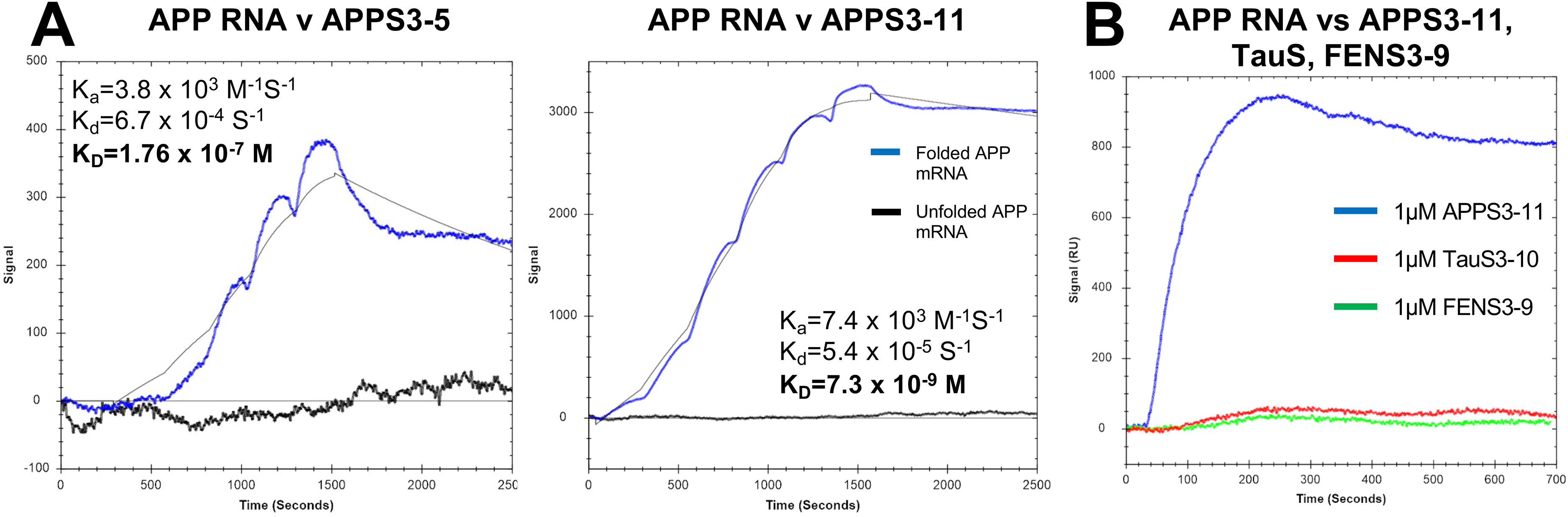
Kinetic analysis of APPS PROTEIMERS. (a) Single cycle kinetics of APPS PROTEIMERS binding to the 5′-UTR APP mRNA. Kinetic fit was performed with a 1:1 binding model. Injections were performed in order of increasing concentration (62 nM, 125 nM, 250 nM, 500 nM, and 1 µM) without dissociation or regeneration between each injection. (b) Binding of multiple PROTEIMERS to 5′-UTR of the APP mRNA. PROTEIMERS generated previously to target various other baits (Tau mRNA or Fentanyl molecule) were tested to show binding specificity to the APP mRNA. Only APPS3-11 could be seen to bind to the 5′-UTR APP mRNA.
Structure prediction of biopanning candidates binding to APP IRE mRNA motif
The SPR results had validated that each of the chosen biopanning candidates were capable of binding to the IRE domain of the APP mRNA. To further verify how this binding may occur, structure prediction modeling was performed to generate models of the protein-RNA interaction between each biopanning candidate and the IRE motif of the APP mRNA. These models were generated using the Alphafold3 webserver by inputting the amino acid sequences of each candidate and pairing them with the 50-bp IRE motif of the APP mRNA. Several rounds of each prediction were performed and compared against each other to determine where binding would most likely occur. Through these prediction models, both biopanning candidates APPS3-5 and APPS3-11 appeared to bind to the target APP within the canonical ligand-binding cleft of its GyrI-like domain. The designed mutations incorporated into the protein binding surface of the candidates predominantly form this interaction with the APP mRNA, where the 5′-AGA-3′ region can be seen to fold outward into the binding pocket (Figure 4). This 5′-AGA-3′ region coincides with a unique alternative RNA triloop formation as described by Cho et al. (2010). 21 This set of nucleotides coincides with key IRE motifs and is predicted to form a bulge-like structure with the 5′-UTR APP mRNA (Supplemental Figure 2).47,48 These structural predictions provide a general insight into understanding how these interactions may contribute to the affinity and specificity between protein and mRNA.
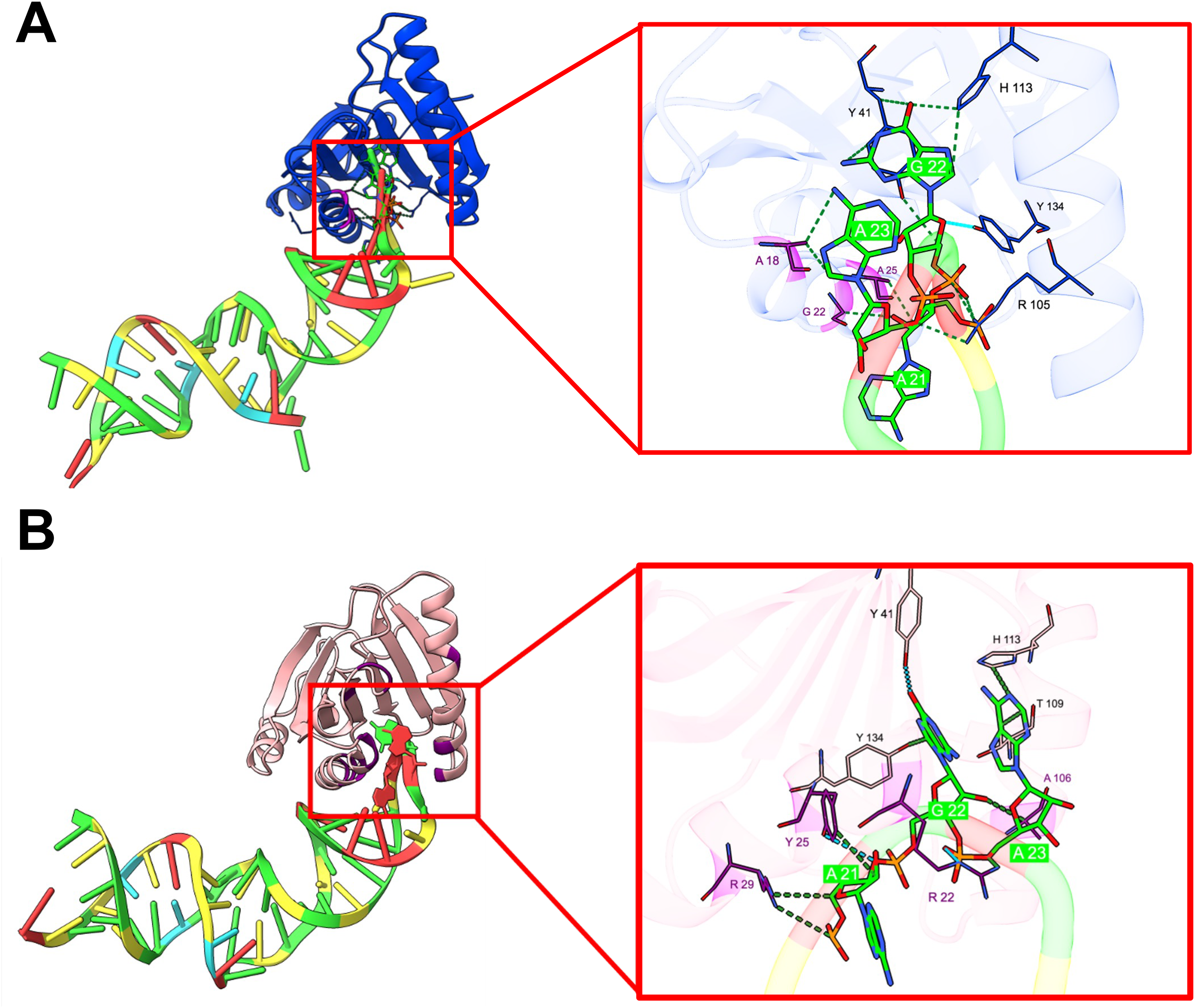
Predicted binding of APPS PROTEIMERS to the hairpin of the APP mRNA. (a) Binding predication between APPS3-5 (green) and the APP mRNA. The 5′-AGA-3′ region can be seen to unfold into the binding pocket of the APPS3-5 PROTEIMER where it could potentially be stabilized by multiple of the selected mutated amino acids (purple). (b) Binding predication between APPS3-11 (red) and the APP mRNA. The 5′-AGA-3′ region can be seen to unfold into the binding pocket of the APPS3-11 PROTEIMER where it could potentially be stabilized by multiple of the selected mutated amino acids (purple) (Color figures available online).
Candidates can target the degradation of RNA in vitro
After confirming that the biopanning candidates could bind to the APP RNA, we evaluated their capacity to promote RNA degradation. New large fusion proteins (ProAAPS3-5, ProAPPS3-11) were designed to incorporate these RNA-binding domains (APPS3-5, APPS3-11) with multiple secondary protein domains each providing unique functions to the fusion protein as a whole to aid in this role of targeted RNA degradation (Figure 6(a)). A C-terminal PIN RNA endonuclease was added to provide the catalytic activity needed to cleave the RNA upon binding. To enhance sequence specificity, a PUM1-derived PUF domain was incorporated to bind a region immediately downstream of the 5′-UTR hairpin targeted by the candidate constructs. For in vivo application, the design was further optimized by adding an N-terminal Angiopep-2 peptide to enable transcytosis across the blood–brain barrier and entry into neurons, along with two C-terminal albumin-binding domains (ABD) to prolong systemic circulation and improve pharmacokinetic stability (Supplemental Figure 3B). The delivery of protein-based therapeutics to the CNS remains a major challenge due to the protective nature of the blood–brain barrier (BBB) and limited uptake by neuronal cells.49,50 To overcome this, we appended Angiopep-2 peptide to the N-terminus of these constructs. These motifs exploit receptor-mediated transcytosis (RMT) pathways via LRP1—a well-characterized strategies for CNS delivery.49,50 As LRP1 is abundantly expressed at the BBB and in neurons, it offers an effective route for delivering therapeutics into the brain and neuronal cells via receptor-mediated endocytosis (RME).
The final constructs (ProAPPS3-5, ProAPPS3-11) were expressed, purified, and tested for their ability to degrade an in vitro-transcribed segment of APP RNA. The construct was incubated at a 3 µM (ProAPPS3-11) or 4.5 µM (ProAPPS3-5) concentration with the APP RNA for 0-120 min at 37°C and the RNA from the reactions was analyzed by polyacrylamide gel electrophoresis. Over time, the band for the full-length RNA diminished in intensity, then disappeared, while bands/a smear for RNAs of shorter lengths appeared, indicating that the RNA was being targeted by the RNase fusion construct (Figure 5). ProAPPS3-11 was able to degrade the APP RNA more quickly and completely than ProAPPS3-5, although both constructs were active. RNA incubated without protein did not degrade significantly over time, confirming that the construct with the RNase was responsible for degradation.
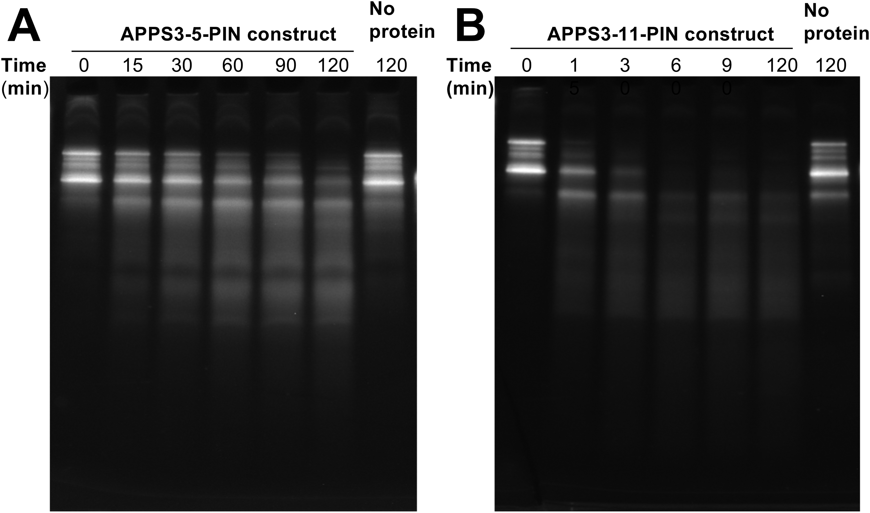
In vitro RNA degradation assay RNA gel electrophoresis. (a) ProAPPS3-5 (4.5 µM) and (b) ProAPPS3-11 (3 µM) were incubated with APP mRNA for specified amounts of time. The RNA was run on a 6% TBE-urea PAGE gel and imaged with GelRed stain under UV light.
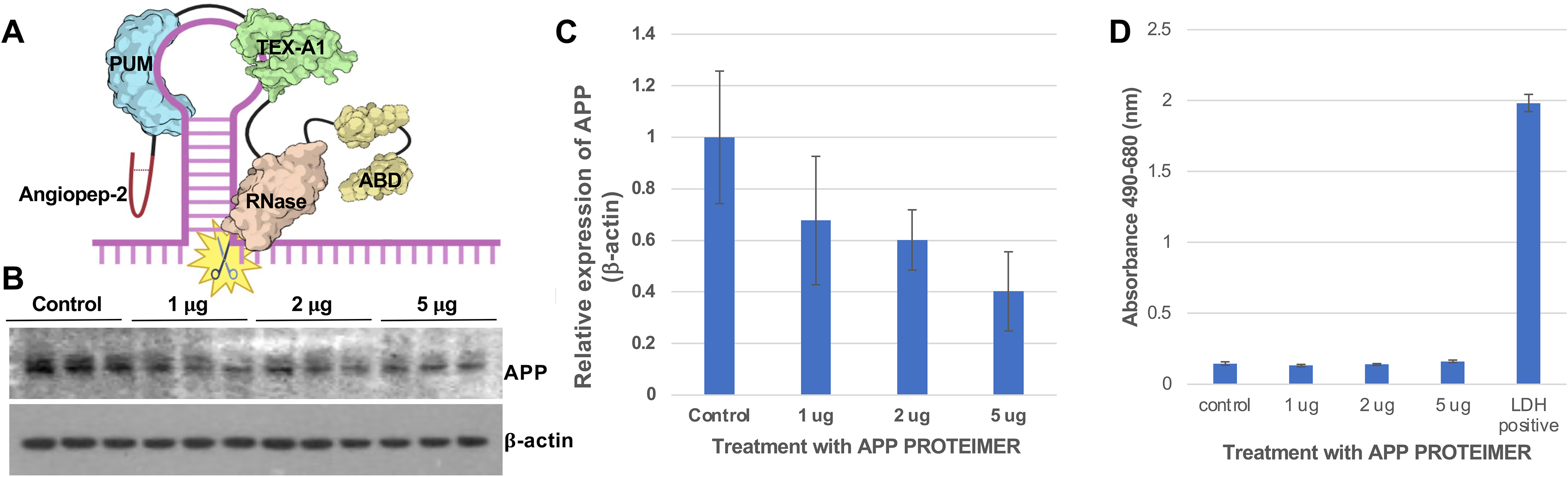
Quantification of a western blotting of PROTEIMER ProAPPS3-11 inhibiting APP protein expression in SH-SY5Y neuroblastoma cells. (a) The ProAPPS3-11 construct is composed of several functional domains: angiopep-2 (purple), PUM (blue), TEX-A1 (green), an RNase domain (orange), and an albumin-binding domain (ABD) (yellow). (b) Western blot analysis following 48 h of treatment with ProAPPS3-11 at 1, 2, and 5 μg revealed a dose-dependent reduction, with (c) APP protein levels decreasing to approximately 60% at the highest concentration. (d) Cytotoxicity of treatment with ProAPPS3-11 at different concentrations measured by CyQUANT LDH cytotoxicity assay (ThermoFisher) represented as the average absorbance at 490 nm minus the reference absorbance at 680 nm for 3 samples for each treatment (Color figures available online).
Confirmation of APP target engagement and silencing in SH-SY5Y neuroblastoma cells
To assess functional target engagement, SH-SY5Y neuroblastoma cells, which endogenously express APP, were treated with the multifunctional PROTEIMER construct, ProAPPS3-11. This engineered fusion protein incorporates the same modular domains used in the in vitro degradation assay as seen previously (Figure 6(a)).
As an initial evaluation of biological activity, we examined ProAPPS3-11’s ability to inhibit APP translation in a dose-dependent manner (Figure 6(a)–(c)). The construct was specifically designed to bind the A-bulge near the base of the IRE hairpin within the 5′-UTR of APP mRNA. Human SH-SY5Y neuroblastoma cells remained healthy throughout the experiment, comparable to untreated controls. Western blot analysis performed after 48 h of treatment with pcDNA3 control or increasing concentrations (1, 2, and 5 μg) of the pcDNA3-ProAPPS3-11 plasmid revealed a clear dose-dependent reduction in APP protein levels. APP expression was normalized to β-actin, which served as a loading control, confirming the specificity and consistency of the observed translational repression (Figure 6(a)–(c). At the highest concentration tested, APP expression was reduced by approximately 60%, confirming effective and selective translational repression of APP by ProAPPS3-11 (Figure 6(c)). In order to test whether ProAPPS3-11 treatment had a detrimental effect on cells, we used an LDH cytotoxicity assay. In the assay, all cell treatments had similar low corrected absorbance readings at 490 nm compared to the LDH positive control supplied by the kit. This indicated that, even at the highest dose of the PROTEIMER, there was minimal cytotoxicity (Figure 6(d)).
Discussion
The work presented here showcases two novel RNA binding proteins, termed PROTEIMERS, that were designed and tested to specifically target the modified IRE motif of the 5′-UTR of the APP mRNA. Both PROTEIMERs were derived from the TEX264 protein scaffold, taking advantage of its defined binding pocket within its gyrI-like domain. The mutations made on the TEX264 scaffold to generate PROTEIMERs APPS3-5 and APPS3-11 were identified by phage display enrichment for selective targeting of the IRE hairpin loop mRNA. Analysis through SPR identified that both PROTEIMERs had high affinity in the nanomolar range to the target IRE loop. Computational structure prediction was included to identify possible molecular interactions between the PROTEIMER and key nucleobases of the IRE mRNA loop. The prediction models offer some general ideas as to what may be occurring within the binding pocket. Over several cycles of model generation, similar interactions could be seen to occur with every model, primarily an interaction between the non-polar aromatic amino acid residues within the binding pocket and the 5′-AGA-3′ region of the 5′-UTR of the APP mRNA (Figure 4). The hydrophobicity of the inner cavity may allow for an even more advantageous binding as previous studies have shown the difficulties of utilizing highly charged IRE-targeting small molecules for therapeutic viability.
In contrast, the use of a protein-based repressor molecule would also overcome this liability by having the larger size of the protein be able to encapsulate the target IRE motif. 30 However, the nucleobases in which this binding actually occurred in the computational predictions provided an even greater validity to the structural interactions since great majority displayed binding centered around the 5′-AGA-3′ tri-loop region as described by Cho et al., (2010). 21 This set of bases coincides with multiple prediction models that have identified this region to be a key IRE motif that would form a distinct structural bulge. As the distinct secondary structure plays a key role in drug targeting, these computational predictions offer a great insight on how this interaction may possibly be occurring.
The 5′-UTR IRE motif of the APP mRNA has been the subject of many studies in an attempt to develop therapeutic targeting. 21 51–53 At its basis, the IRE region is well known to play a key role in iron-dependent cap-independent translation and the ability to inhibit the production of APP through its regulation. Because of this, extensive work has been performed to develop small molecules capable of targeting this region to provide some form of therapeutic effect. 51 A key takeaway coming from these studies has been the viability of targeting the stable secondary structure motifs in a similar fashion to structure-based inhibition of protein targets. A large obstacle when designing these small molecule inhibitors is the balancing of size, specificity, and stability while keeping the entire molecule in mind. However, protein-based binders benefit from the modularity that comes from incorporating and removing individual protein domains to confer unique functions to the fusion protein as a whole. For example, increased sequence specificity can easily be achieved by incorporating a secondary PROTEIMER that is capable of targeting an adjacent region of the single-stranded RNA. This could be done with the addition of a Pumilio (PUM) domain that can be easily fused to the designed PROTEIMER. 30 Enhanced circulation time through the blood and improved uptake through the blood-brain barrier of the PROTEIMER in cells can also be achieved by incorporating a multitude of protein stabilizer domains such as a transferrin receptor targeting protein domain, 31 an FcBP peptide, 32 or an albumin binding domain. 33 The easy access and incorporation of these multiple protein domains allows for a high amount of design flexibility for the PROTEIMERS that are not so easily replicated when producing small molecules or ASO conjugates. 28
The greatest aspect of designing with PROTEIMERs is the ease of incorporating a protein domain that is capable of performing a certain site-directed function to the target RNA. One function could be the fusion of a catalytically active RNase domain that would allow the PROTEIMER to degrade the targeted mRNA in vivo. Degradation and removal of the targeted mRNA before translation would allow for the cessation of harmful protein production before they could further exacerbate the disease in Alzheimer's patients. This targeted degradation has already been demonstrated before through variations utilizing small molecule chimeras fused with a chemically destructive payload (PINAD) 34 to induce degradation upon binding or through moieties capable of recruiting the native RNase as seen with ribonuclease targeting chimeras. 35 While the use of an RNase active PROTEIMER is another alternative to the targeted degradation of mRNA, their design offers a much more streamlined production. We demonstrate that RNase-active PROTEIMERs can selectively degrade SNCA 30 and APP mRNAs in vitro (Figure 5). This study identified two novel high-affinity PROTEIMERs that selectively bind the 5′-UTR of APP mRNA. These engineered proteins have been designed to reduce steady-state levels of APP in the brain, thereby lowering Aβ production and limiting the accumulation of the neurotoxic APP-CTF.54,55 By targeting both APP and APP-CTF, our PROTEIMERs aim to prevent lysosomal dysfunction, impair autophagy, 56 and offset ferroptosis16,57—key pathological features observed in the TgCRND8 mouse model of APP-CTF overproduction. These agents are being developed to mitigate APP-CTF–induced autophagic vacuolization, cellular waste accumulation, lysosomal pH elevation, and iron-dependent cell death.10,11
In conclusion, this study demonstrates the feasibility of reducing APP expression by selectively targeting its 5′-UTR using the PROTEIMER platform. In vitro treatment of SH-SY5Y neuroblastoma cells with the ProAPPS3-11 construct resulted in approximately 60% reduction in APP protein levels, validating its effectiveness in selectively repressing APP translation. These findings underscore the potential of PROTEIMER-based therapeutics as a novel and targeted strategy for modifying disease progression in APP-associated neurodegenerative disorders, including Alzheimer's disease. By suppressing upstream APP synthesis this approach offers a compelling therapeutic avenue to prevent toxic amyloid-β aggregation and slow disease progression of AD. Unlike strategies aimed at clearing existing amyloid plaques—which remain challenging due to their complex and dynamic nature—limiting de novo APP production may serve as a complementary mechanism to prevent further neurotoxicity and cognitive decline.
Finally, continued development is needed to optimize these therapeutic constructs for enhanced specificity, stability, and modular functionality. The intrinsic modularity of PROTEIMERs provides a versatile platform for engineering next-generation therapeutics with tailored properties and multifunctional capabilities.
Supplemental Material
sj-docx-1-alz-10.1177_13872877251393907 - Supplemental material for PROTEIMERs as catalytic inhibitors of APP mRNA translation: Toward a new therapeutic for Alzheimer's disease
Supplemental material, sj-docx-1-alz-10.1177_13872877251393907 for PROTEIMERs as catalytic inhibitors of APP mRNA translation: Toward a new therapeutic for Alzheimer's disease by Daniel von Salzen, Katherine Senn, Hannah K Cho, Catherine M Cahill, Jack T Rogers and HyunDae D Cho in Journal of Alzheimer's Disease
Footnotes
Acknowledgements
We thank all the CrossLife Technologies members for their continuous support in this study.
Author contribution(s)
Funding
The authors disclosed receipt of the following financial support for the research, authorship, and/or publication of this article: We gratefully acknowledge funding support for this study provided by CL funds.
Declaration of conflicting interests
The authors declared the following potential conflicts of interest with respect to the research, authorship, and/or publication of this article: CrossLife Technologies holds patents (US20210139958A1, and US3/713,768) covering both the PROTEIMER technology and the 5′ UTR mRNA of APP-targeting candidates utilized in this study. The authors declared no potential conflicts of interest with respect to the research, authorship, and/or publication of this article.
Data availability statement
The datasets generated during and/or analyzed during the current study are available from the corresponding author on reasonable request.
Supplemental material
Supplemental material for this article is available online.
References
Supplementary Material
Please find the following supplemental material available below.
For Open Access articles published under a Creative Commons License, all supplemental material carries the same license as the article it is associated with.
For non-Open Access articles published, all supplemental material carries a non-exclusive license, and permission requests for re-use of supplemental material or any part of supplemental material shall be sent directly to the copyright owner as specified in the copyright notice associated with the article.
