



Abstract
Alzheimer's disease (AD) is a complex neurodegenerative disorder characterized by progressive cognitive decline and multifaceted pathogenic mechanisms (including amyloid-β [Aβ] plaques, tau neurofibrillary tangles, synaptic dysfunction, and neuroinflammation). Importantly, no effective disease-modifying treatment is currently available for AD. Emerging evidence implicates dysregulated mammalian target of rapamycin (mTOR) signaling as a key contributor to AD pathogenesis. This review analyzes how aberrant mTOR signaling influences major aspects of AD pathology, including Aβ production and clearance, tau protein hyperphosphorylation, autophagy dysfunction, synaptic plasticity impairments, neuroinflammation, and oxidative stress. Notably, hyperactivated mTOR accelerates AD progression through multiple mechanisms. It promotes Aβ accumulation and tau pathology, suppresses autophagic clearance of toxic aggregates, and disrupts neuronal homeostasis, thereby exacerbating cognitive decline. Consequently, mTOR has gained attention as a therapeutic target. This review evaluates the therapeutic potential of various mTOR-targeted interventions, such as the mTORC1 inhibitor rapamycin and its analogues (rapalogs), second-generation ATP-competitive mTOR inhibitors, and certain natural compounds and traditional Chinese medicine approaches. These strategies have demonstrated promise in mitigating AD-related pathology by enhancing autophagy, reducing Aβ/tau burden, and preserving synaptic and cognitive function in preclinical studies. However, the clinical translation of mTOR-targeted therapies faces key challenges, including poor blood–brain barrier penetration of many mTOR inhibitors, potential systemic side effects, and limited clinical validation to date. Further research is needed to optimize brain delivery, dosing regimens, and target specificity to fully realize the therapeutic potential of mTOR modulation in AD.
Graphical Abstract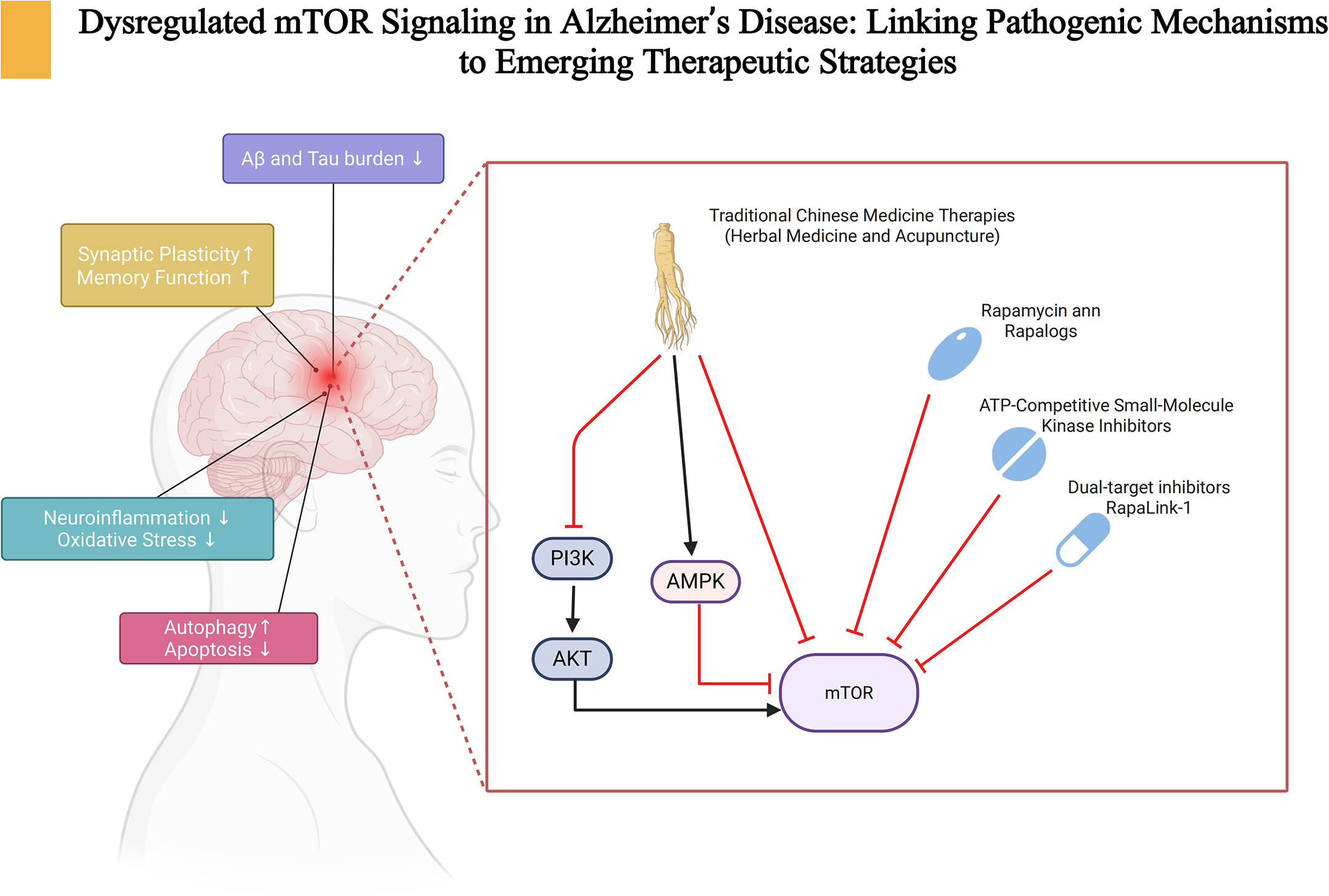
Introduction
Alzheimer's disease (AD) is a progressive neurodegenerative disorder characterized by cognitive decline and memory loss, and it is the most common cause of dementia in older adults. 1 Hallmark neuropathological features include extracellular amyloid-β (Aβ) plaques and intraneuronal neurofibrillary tangles (NFTs) composed of hyperphosphorylated tau; these lesions precipitate synaptic dysfunction, neuroinflammation, mitochondrial impairment, and metabolic dysregulation, ultimately driving cognitive deterioration.2,3 With global population aging, the number of people living with AD is projected to exceed 131 million by 2050.4,5 Current therapies—acetylcholinesterase inhibitors (donepezil, rivastigmine, galantamine) and the N-methyl-D-aspartate receptor antagonist memantine—provide only transient symptomatic relief and do not modify the underlying disease course.6–8 Accordingly, there is an urgent need for novel strategies that target key molecular pathways to slow or halt disease progression.
The mammalian target of rapamycin (mTOR) is a serine/threonine kinase that serves as a central hub integrating signals that regulate cell growth, metabolism, and autophagy.9–11 In the nervous system, mTOR signaling maintains neural health by coordinating energy homeostasis, neuronal plasticity, and proteostasis. It supports neuronal growth and synaptic plasticity and contributes to neural stem cell self-renewal, neurogenesis, and angiogenesis.12,13 By balancing autophagy with protein translation, mTOR preserves intracellular protein homeostasis; when dysregulated, toxic protein aggregates accumulate and promote neurodegeneration. 14 Experimental studies indicated that mTOR signaling contributed to the initiation and progression of AD: aberrant mTOR activity promoted the accumulation of Aβ and hyperphosphorylated tau while impairing autophagic clearance of pathological proteins.13,15 Moreover, mTOR dysregulation perturbed energy metabolism, mitochondrial function, and oxidative stress responses, thereby exacerbating cognitive decline. 16 On this basis, targeting the mTOR pathway has emerged as a promising therapeutic strategy. This review synthesizes mechanistic insights into mTOR's roles in neuroinflammation, autophagy, cell survival, and synaptic function in AD, and discusses the therapeutic potential—as well as the translational challenges—of modulating this pathway.
mTOR complexes and their functions
mTOR forms two distinct multiprotein complexes, mTOR complex 1 (mTORC1) and mTOR complex 2 (mTORC2), which share certain core components but also contain unique subunits conferring specific biological functions.17,18 Shared components include mTOR, mLST8, DEPTOR, and the Tel2–Tti1 stabilizing complex, whereas Raptor and PRAS40 are exclusive to mTORC1, and Rictor, mSIN1, and Protor are unique to mTORC2.19–21 As the catalytic core of both complexes, mTOR transduces upstream signals to regulate diverse metabolic and cellular processes.18,22 mLST8 stabilized overall complex structure and enhanced catalytic activity, particularly supporting the Rictor–mTOR interaction in mTORC2.23,24 DEPTOR served as an endogenous negative regulator, binding directly to mTOR and suppressing both mTORC1 and mTORC2.25,26 The Tel2–Tti1 complex was also critical for assembly and stabilization of both complexes. 27 Within mTORC1, Raptor mediated substrate recognition and recruitment, while PRAS40 bound Raptor to inhibit mTORC1 activity—a suppression relieved by Akt-mediated phosphorylation. 28 In mTORC2, mSIN1 was indispensable for complex integrity and activity; Protor regulated substrate specificity through direct interaction with Rictor, which acted as a scaffold anchoring mSIN1 and Protor to the kinase domain, thereby maintaining both complex stability and substrate selectivity.24,29,30 An overview of the molecular composition and biological functions of mTORC1 and mTORC2 is presented in Figure 1.

Composition and functional overview of mTORC1 and mTORC2 signaling pathways.
Functions of mTORC1
mTORC1 integrates diverse extracellular and intracellular cues—including growth factors, energy status, amino acid availability, and environmental stressors—to regulate lipid metabolism, protein synthesis, and autophagy. 31 For lipid metabolism, mTORC1 phosphorylated Lipin-1, prevented its nuclear translocation, and thereby released its inhibition of SREBP-1, which promoted expression of lipogenic genes and lipid accumulation.32–34 mTORC1 also phosphorylated Grb10 at Ser501/503, leading to its dissociation from the insulin receptor and association with Raptor, thereby negatively regulating mTORC1 activity and modulating lipid metabolism and thermogenesis.35,36 In protein synthesis, mTORC1 exerts dual regulation through 4E-BP1 and S6K1. By phosphorylating 4E-BP1, mTORC1 released eIF4E to form the eIF4F translation initiation complex, thereby facilitating cap-dependent translation, particularly of transcripts with complex 5′ UTRs such as TOP/TCT mRNAs.37–40 mTORC1 also activated S6K1, which in turn regulated S6 and eIF4B, thereby enhancing translational efficiency. 41 This dual regulation enables cells to flexibly allocate translational resources under varying metabolic conditions.
mTORC1 plays a central role in autophagy suppression. The canonical mTORC1–ULK1/ATG13 axis blocked autophagy initiation: mTORC1 phosphorylated ATG13, reduced ULK1 activity, and disrupted its interaction with AMPK.42–45 Additionally, mTORC1 directly phosphorylated Phosphoinositide 3-kinases (PI3K)C3-C1 complex components (ATG14, NRBF2, AMBRA1), decreasing PI3P production and impairing phagophore nucleation.46–49 Once recruited to the lysosomal surface via Rag GTPases, mTORC1 phosphorylated the transcription factors TFEB/TFE3 (e.g., TFEB Ser211), sequestering them in the cytoplasm via 14-3-3 proteins and thereby suppressing transcription of autophagy- and lysosome-related genes.50–52 Collectively, these findings established mTORC1 as a master regulator of cellular anabolism while simultaneously restraining catabolic processes. However, its strong inhibitory effect on autophagy, particularly in neurons, also underscored its potential role in driving neurodegenerative pathology when aberrantly activated.
Functions of mTORC2
mTORC2 is a pivotal regulator of cell survival, cytoskeletal organization, and signaling through AGC family kinases. A major function is phosphorylation of SGK1 at Ser422, which permitted subsequent Thr256 phosphorylation by PDK1, thereby fully activated SGK1.21,53 Activated SGK1 enhanced cell survival through phosphorylation of FOXO transcription factors (e.g., FOXO3a, FOXO1), driving their nuclear export and suppressing expression of pro-apoptotic genes such as BIM and FasL.54,55 SGK1 also phosphorylated NDRG1, enabling cells to sustain metabolic homeostasis under stress conditions (e.g., endoplasmic reticulum stress, mitochondrial dysfunction) and thereby reducing stress-induced apoptosis.56,57 Experimental loss or inhibition of mTORC2 or SGK1 disrupted these survival pathways, leading to FOXO reactivation, impaired autophagy, mitochondrial instability, and increased cell death. Conversely, SGK1 overexpression or hyperactivation contributed to pathological cell survival in cancers.58,59
Beyond survival, mTORC2 orchestrates actin cytoskeleton remodeling and cell migration by phosphorylating AGC kinases, including multiple PKC isoforms and Akt.24,53,60 Hydrophobic-motif phosphorylation by mTORC2 ensured PKC maturation, stability, and activity. For instance, PRR5L degradation enhanced PKCδ phosphorylation and drove fibroblast migration and pulmonary-fibrosis progression.53,61 Conversely, mTORC2 loss resulted in impaired PKCα phosphorylation, sustained RhoGDI2 expression, and reduced Rac1 activity, thereby suppressing tumor-cell invasion and metastasis. 62 mTORC2 further spatially regulated Rho-family GTPases, promoting Rac1-driven lamellipodia formation at the leading edge while coordinating RhoA-mediated contractility at the trailing edge, thus controlling cell polarity, migration, epithelial–mesenchymal transition, and tissue morphogenesis.53,61,63 In sum, mTORC2 integrates survival and cytoskeletal signaling, positioning it as a central determinant of both cellular resilience and dynamic remodeling. Dysregulation of mTORC2, while less studied than mTORC1, increasingly appears to contribute to pathological processes ranging from cancer progression to neurodegeneration.
mTOR and Alzheimer's disease
Effects of mTOR on Aβ and tau proteins
The mTOR signaling pathway plays a central role in the hallmark pathologies of AD, particularly in Aβ deposition and tau hyperphosphorylation. Hyperactivation of mTOR suppressed autophagy, leading to the accumulation of misfolded proteins within neurons and thereby promoting Aβ aggregation and plaque formation.64,65 Notably, Aβ itself induced mTOR overactivation, which further reinforced autophagic suppression, accelerating Aβ accumulation; in turn, aggregated Aβ reinforced mTOR signaling, creating a pathological positive feedback loop.66,67 Mechanistically, soluble Aβ oligomers bound neuronal surface receptors (e.g., insulin receptor, IGF-1R, BDNF receptor), activated PI3K, and subsequently stimulated Akt-mediated phosphorylation of TSC2, relieving TSC1/2-mediated inhibition of Rheb and allowing Rheb-GTP to directly activate mTORC1. 68 In parallel, Aβ enhanced Akt-dependent phosphorylation of PRAS40, causing its dissociation from Raptor and releasing inhibition of mTORC1. Upregulation of β- and γ-secretase expression further increased Aβ load, reinforcing sustained mTOR activation. 69
Regarding tau pathology, excessive mTOR activity enhanced tau phosphorylation through multiple mechanisms, including increased tau synthesis and a shift in the kinase–phosphatase balance. Recent studies further clarified cross-talk at the mTOR–tau interface: sustained mTORC1–S6K1 feedback attenuated PI3K–Akt signaling, thereby lowering inhibitory Ser9 phosphorylation on GSK-3β and facilitating tau phosphorylation, whereas mTORC2-dependent Akt-Ser473 helped maintain GSK-3β restraint.53,70–72 In parallel, dysfunction of the principal tau phosphatase PP2A—including altered catalytic-subunit methylation—reduced dephosphorylation of pathogenic tau epitopes, while mTOR suppression was reported to restore PP2A activity and lower PP2A-dependent tau sites in preclinical models.73–76 Collectively, these changes drove the accumulation of hyperphosphorylated tau and accelerated NFT formation. Importantly, pharmacological inhibition of mTOR with rapamycin was shown to restore autophagy, reduce Aβ and tau accumulation, and ameliorate cognitive impairment in an AD mouse model. 77 Although compelling preclinical evidence supports the pathological role of mTOR hyperactivation in Aβ and tau pathology, the exact causal relationships and translational relevance remain debated; for instance, some studies suggested that partial mTOR activity is required for synaptic maintenance and neuronal survival, raising concerns about long-term systemic inhibition.78,79
Effects of mTOR on synaptic plasticity and memory function
Progressive impairment of learning and memory is a defining clinical manifestation of AD, and dysregulation of mTOR signaling has been closely linked to synaptic dysfunction underlying these deficits. 80 mTORC1 influences synaptic plasticity primarily through regulation of local protein synthesis. By activating downstream effectors such as p70S6K1, mTORC1 promoted dendritic translation, thereby sustaining synaptic modifications required for long-term potentiation (LTP) and memory consolidation. 15 Conversely, deficiency in mTORC2 impaired actin-cytoskeleton remodeling by disrupting PKCα-, RhoA-, and Rac1-mediated signaling, leading to defective dendritic-spine morphology and impaired synaptic stability. 81 Notably, both excessive suppression and hyperactivation of mTOR signaling negatively affected cognition, consistent with a context-dependent, biphasic role of mTOR.82–85
To reconcile these bidirectional effects, the influence of mTOR on synaptic plasticity is best understood as context dependent along two mechanistic axes. First, the temporal–spatial profile and magnitude of signaling shaped outcomes: brief, dendrite-restricted bursts of mTORC1 during LTP induction promoted local translation and stabilized late-phase LTP, whereas persistent or widespread activation engaged S6K1-dependent negative feedback (e.g., inhibitory phosphorylation of IRS-1), attenuated PI3K–Akt signaling, shifted the autophagy–translation balance, and biased networks toward long-term depression (LTD)-like adaptations.86–88 Second, the pattern of complex engagement also shaped plasticity: the relative recruitment of mTORC1 versus mTORC2 determined the available effector programs—translation and autophagy control via S6K1 and 4E-BP1 for mTORC1, and actin remodeling and neuronal excitability via Akt/PKC for mTORC2—thereby biasing whether LTP- or LTD-supporting processes predominated within a given compartment.13,53,89 Within this framework, the apparent biphasic effects of mTOR reflected a shift from physiological, pulsatile signaling that preserved synaptic specificity to chronic activation that degraded it.
Mechanistically, both LTP and LTD—the complementary processes that shape memory encoding and refinement—were influenced by mTOR activity.90,91 For LTP, upregulated mTORC1 promoted dendritic protein synthesis via phosphorylation of S6K1 and 4E-BP1, facilitating the induction and maintenance of L-LTP. 92 Conversely, mTORC1-dependent disinhibition of eIF4E was implicated in protein-synthesis programs that supported LTD, helping eliminate unnecessary or irrelevant synaptic connections and thereby maintaining the specificity of memory traces. 93 mTORC2 contributed by fully activating Akt and facilitating PKCα phosphorylation, which promoted actin remodeling and insertion of the AMPA receptor (AMPAR) subunit GluA1 into the postsynaptic membrane, reinforcing LTP maintenance. 94 In addition, phosphorylation of PKC isoforms (e.g., PKCα/ε) by mTORC2 drove dynamic actin-cytoskeletal remodeling and AMPAR trafficking, supporting LTD induction. 13
Consistent with this framework, experimental evidence supported dual roles of mTORC1 and mTORC2 in synaptic regulation. Viral expression of constitutively active Raptor (mTORC1) or Rictor (mTORC2) in an AD mouse model restored Aβ-induced impairments in both LTP and LTD and improved cognitive performance. 77 These findings highlighted the interplay between mTOR complexes in orchestrating synaptic plasticity and memory. While targeted activation of mTOR components transiently enhanced memory processes in experimental settings, chronic overactivation—as observed in AD—appeared to promote pathological remodeling rather than adaptive plasticity. The relationship between mTOR signaling and cognition is therefore non-linear. Future work should resolve how the temporal dynamics and subcellular localization of mTORC1/2 activity determine protective versus deleterious outcomes, enabling selective-modulation strategies that restore synaptic function without compromising physiological memory processes.
Effects of mTOR on neuroinflammation and oxidative stress
Neuroinflammation and oxidative stress are increasingly recognized as major contributors to AD pathogenesis, closely interwoven with classical hallmarks such as Aβ deposition and tau hyperphosphorylation.95,96 Proinflammatory cytokines, including IL-1β and TNF-α, accelerated AD pathology by amplifying Aβ aggregation and tau phosphorylation, while oxidative stress exacerbated neuronal injury through mitochondrial dysfunction and excessive reactive oxygen species (ROS) production.97–99
mTOR signaling acts as a key modulator of these processes. Hyperactivation of mTORC1 impaired autophagy, downregulated essential components such as ULK1 and Beclin-1, and prevented timely clearance of damaged mitochondria. 16 The resulting accumulation of dysfunctional mitochondria elevated ROS levels, leading to neuronal apoptosis and progression of neurodegeneration. 100 In parallel, sustained mTOR activity enhanced NF-κB and p38 MAPK signaling, driving the release of proinflammatory mediators and cytotoxic factors. 77 Consistently, mTORC1-mediated autophagy suppression prevented clearance of Aβ aggregates and damaged organelles, triggering NLRP3 inflammasome activation and caspase-1–dependent amplification of inflammation. 101
Importantly, oxidative stress and inflammation formed a pathological feed-forward loop: ROS activated mTOR and amplified inflammation, while inflammatory mediators enhanced ROS production, collectively accelerating neuronal injury and cognitive decline. 102 This reciprocal interaction positioned mTOR not only as a mediator but also as an amplifier of AD-associated neuroinflammation and oxidative stress. Although numerous preclinical studies demonstrated that mTOR inhibition reduced neuroinflammation and oxidative stress while improving memory performance in AD models,103,104 several questions remain unresolved. First, the precise molecular crosstalk between mTOR and innate immune signaling (e.g., NLRP3 activation) requires further elucidation. Second, systemic mTOR inhibition carries the risk of immunosuppression, raising translational challenges for long-term use.
Dysregulation of autophagy and apoptosis mediated by mTOR
Autophagy and apoptosis are essential cellular processes that maintain homeostasis by clearing damaged components and regulating cell survival. Dysregulation of either process contributes significantly to AD pathogenesis and progression. Impaired autophagy disrupted proteostasis, aggravated neuroinflammation, promoted synaptic dysfunction, and accelerated tau hyperphosphorylation and Aβ accumulation.105–107 Similarly, dysregulated apoptosis led to excessive neuronal death, undermined central nervous system integrity, and exacerbated neurodegeneration.108,109
mTORC1 is the primary negative regulator of autophagy. In AD, hyperactivation of mTORC1 resulted in phosphorylation-dependent inactivation of ULK1, downregulated Beclin-1, and suppressed autophagosome–lysosome fusion, ultimately preventing clearance of Aβ and tau aggregates.67,110 At the lysosomal membrane, mTORC1 phosphorylation of TFEB sequestered it in the cytoplasm and blocked transcription of autophagy- and lysosome-biogenesis genes. 106 These disruptions diminished autophagic flux and facilitated neurotoxic protein accumulation. Dysregulated mTORC1 activity also impaired mitophagy, leading to accumulation of dysfunctional mitochondria, collapse of mitochondrial membrane potential, and caspase-3 activation, thereby triggering neuronal apoptosis.111,112 mTOR hyperactivation shifted the balance toward pro-apoptotic signaling, upregulated Bax and cleaved caspase-3, and suppressed anti-apoptotic Bcl-2. 113 This imbalance accelerated neuronal death and cognitive decline. Conversely, pharmacological inhibition of mTOR restored autophagic activity (elevated LC3-II and Beclin-1, reduced p62), suppressed pro-apoptotic cascades, and improved cognitive performance in an AD mouse model, as measured by Morris water maze and Y-maze tests. 114
Although preclinical studies clearly support mTOR inhibition as a strategy to restore autophagy and attenuate apoptosis, important questions remain. Chronic suppression of mTOR may compromise physiological roles of autophagy and apoptosis in non-neuronal tissues, raising concerns about systemic toxicity. Furthermore, complete inhibition of apoptosis may impair normal neuronal turnover and plasticity.
Therapeutic interventions targeting mTOR pathways in AD
PI3K/akt/mTOR pathway
The PI3K/Akt/mTOR signaling axis is a central regulator of cellular metabolism and energy homeostasis in the central nervous system (CNS), exerting broad influence on neuronal survival, neurogenesis, synaptic plasticity, and cognitive functions.115,116 PI3K are lipid kinases that phosphorylate the 3′-hydroxyl group of phosphatidylinositol lipids, converting phosphatidylinositol-4,5-bisphosphate (PIP2) into phosphatidylinositol-34,5-trisphosphate (PIP3). This generates second-messenger signals that regulate cellular growth, proliferation, migration, and survival.117–119 Akt (protein kinase B) is a serine/threonine kinase that acts as a central node downstream of PI3K; among its isoforms, Akt1 regulates survival and proliferation, Akt2 mediates metabolic processes, and Akt3 plays a critical role in neural development.120–122 Akt activation depends on PI3K-mediated PIP3 production, which recruits Akt to the plasma membrane for phosphorylation and activation.123,124 mTOR, a conserved serine/threonine kinase, operates through two functionally distinct complexes: mTORC1 and mTORC2.18,125,126 Activated Akt phosphorylated TSC2 and relieved PRAS40-mediated inhibition of mTORC1, thereby promoting protein synthesis, metabolic reprogramming, and suppression of autophagy. 59 Conversely, mTORC2 enhanced Akt activity by phosphorylating its hydrophobic motif (Ser473), establishing a bidirectional regulatory loop.127–129
In AD models, inhibition of the PI3K/Akt/mTOR pathway was shown to restore autophagic flux, increase the expression of autophagy-related proteins (e.g., LC3-II/I, Beclin-1), and decrease accumulation of p62, reducing Aβ deposition and improving cognition.130,131 Similarly, suppression of this pathway reduced abnormal tau aggregation and restored memory performance. 132 Moreover, downregulation of PI3K/Akt/mTOR enhanced Beclin-1 and Bcl-2 while lowering inflammatory cytokines such as IL-6 and TNF-α, thereby promoting autophagy, attenuating apoptosis, and reducing neuroinflammation.133,134 Evidence indicated that in early disease stages, transient activation of the PI3K/Akt/mTOR pathway enhanced synaptic function by upregulating synaptic proteins such as PSD95, thereby supporting cognitive performance.135,136 These findings underscored a dual role of the PI3K/Akt/mTOR axis in AD: whereas chronic hyperactivation contributed to protein aggregation and neurodegeneration, controlled activation appeared to enhance synaptic function and plasticity. This paradox highlighted the importance of temporal and context-dependent modulation. Future studies should investigate stage-specific interventions—e.g., pathway inhibition in advanced AD versus targeted activation in early cognitive decline—to maximize therapeutic benefit while minimizing risks. A detailed schematic of the PI3K/Akt/mTOR signaling pathway is presented in Figure 2.
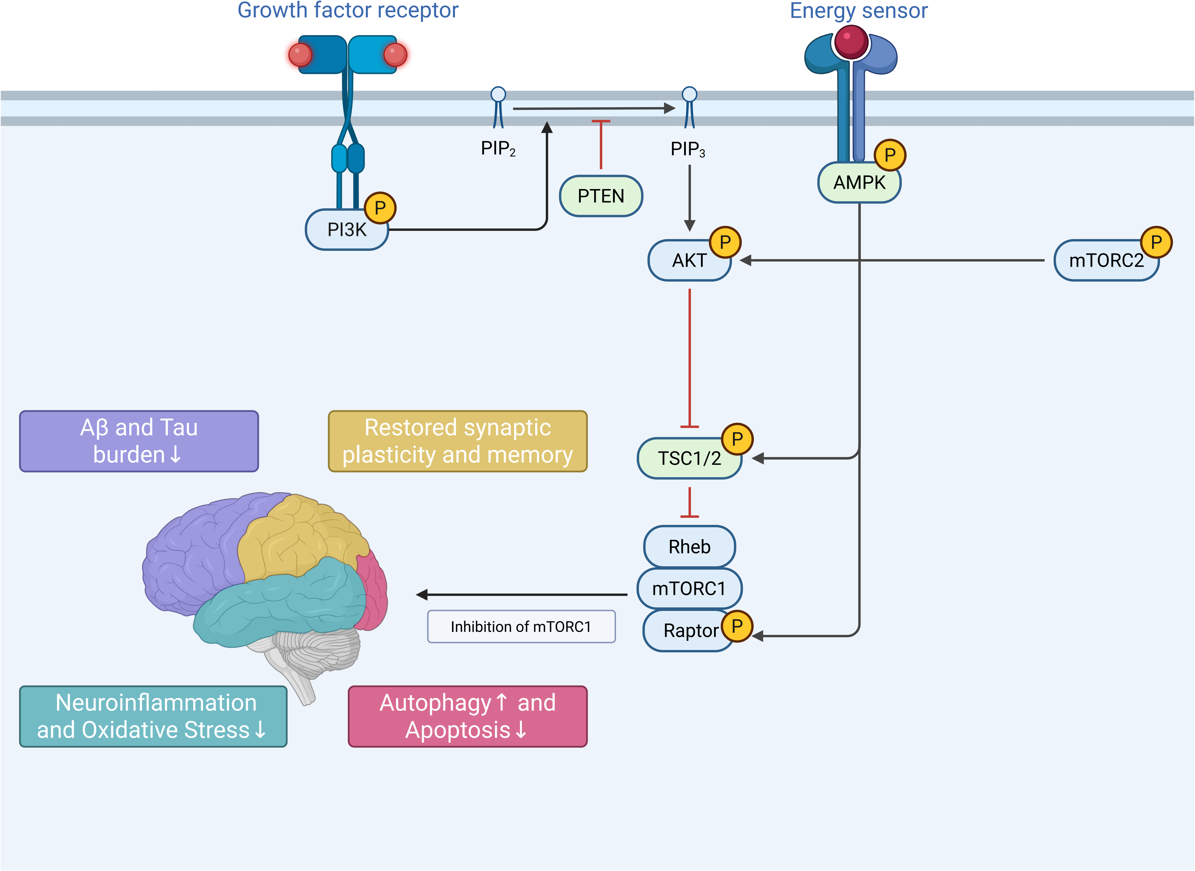
PI3K/Akt/mTOR and AMPK/mTOR signaling pathways.
AMPK/mTOR pathway
The AMPK/mTOR axis serves as a metabolic checkpoint linking cellular energy status to biosynthetic processes. AMPK, a heterotrimeric serine/threonine kinase conserved across eukaryotes, functions as a cellular energy sensor. Upon energy depletion, AMPK integrates signals from nutrient availability, mitochondrial activity, and stress responses to suppress energy-consuming biosynthesis and promote catabolic pathways.137–140 By phosphorylating TSC2 and Raptor, AMPK inhibited Rheb-mediated mTORC1 activation, thereby downregulating protein and lipid synthesis. Simultaneously, AMPK activated ULK1 to initiate autophagy, ensuring neuronal survival under energy stress.141,142 In AD and related neurodegenerative diseases, decreased AMPK activity and hyperactive mTORC1 were implicated in impaired autophagy, accumulation of Aβ and tau, and synaptic degeneration. Pharmacological modulation of this axis (e.g., rapamycin or AMPK activators) was shown to restore metabolic balance and mitigate disease progression. 45
An experimental study demonstrated that AMPK activation enhanced autophagic flux, increased LC3-II and Beclin-1, reduced p62, and promoted clearance of Aβ and damaged organelles, thereby reducing oxidative stress and neuronal toxicity. 143 In AD mouse models, AMPK activation inhibited mitochondrial dysfunction, reduced oxidative stress, and improved cognitive performance.144,145 A study reported that upregulation of phosphorylated AMPK with concomitant suppression of phosphorylated mTOR restored autophagic activity, reduced neuroinflammation (IL-1β, IL-6, TNF-α), and lowered amyloid precursor protein (APP) and phosphorylated-tau burden. 146 A combination therapy (e.g., empagliflozin with memantine) enhanced AMPK/mTOR signaling, reduced Aβ/BACE1, suppressed hippocampal inflammation, alleviated oxidative stress, and increased BDNF levels, collectively improving cognition. 147 Natural compounds such as mangiferin and magnolol similarly exerted neuroprotective effects via AMPK activation and mTOR inhibition, promoting autophagy, suppressing microglial activation, and reducing apoptosis.148,149 A detailed schematic of the AMPK/mTOR signaling pathway is presented in Figure 2.
The AMPK/mTOR axis represents a promising therapeutic target, yet its systemic modulation may disrupt metabolic processes in peripheral tissues. Furthermore, chronic AMPK activation could impair anabolic processes essential for neuronal plasticity. Future strategies should focus on CNS-specific delivery of AMPK activators or dual modulators that achieve selective regulation of autophagy and inflammation, minimizing off-target effects while maximizing therapeutic efficacy.
mTOR-targeted therapeutics for AD
Rapamycin
Rapamycin (sirolimus) is the prototypical mTOR inhibitor, a natural macrolide antibiotic first isolated from Streptomyces hygroscopicus on Rapa Nui (Easter Island) in the 1970s. 150 Rapamycin forms a complex with the intracellular immunophilin FKBP12 and binds the FRB domain of mTORC1, producing allosteric inhibition that prevents substrate access to the kinase active site.151,152 Owing to dual anchoring of 4E-BP1 to the Raptor subunit via its N-terminal RAIP motif and C-terminal TOS motif, certain 4E-BP1 phosphorylation sites could partially escape rapamycin inhibition. 139 Clinically, rapamycin is FDA-approved as an immunosuppressant to prevent transplant rejection and to treat lymphangioleiomyomatosis. 18 An early-phase clinical study suggested that rapamycin may indirectly modulate AD-related mechanisms, but its specific efficacy and optimal clinical use in AD remain to be clarified. 153
Systemic rapamycin mitigated AD-like phenotypes, including memory impairment and synaptic pathology, in a preclinical model. 103 Mechanistically, rapamycin downregulated mTORC1 to restore autophagic activity, enhance aggregate clearance, and reduce Aβ and tau burden, leading to significant cognitive improvement in AD animal models.110,154 Additional mechanisms were proposed to include modulation of insulin signaling, relief of endoplasmic reticulum stress, and TFEB-dependent enhancement of lysosomal biogenesis.13,155,156 Caution is warranted: chronic mTORC1 inhibition was reported to overload lysosomal degradative capacity, provoking “autophagic stress” and potentially worsening amyloid deposition in specific contexts. 157 Moreover, immunosuppression and limited blood–brain barrier penetration have constrained clinical utility in AD.103,153 Intermittent, low-dose regimens have therefore been considered to attenuate mTOR activity while limiting adverse effects.
Rapalogs
Rapalogs are first-generation mTOR inhibitors derived from rapamycin to overcome limitations such as low aqueous solubility, poor oral bioavailability, broad tissue distribution, and a narrow therapeutic window.152,158–161 Common rapalogs include everolimus (RAD001), temsirolimus (CCI-779), and ridaforolimus (deforolimus, AP23573). 162 Structural modifications—for example, esterification in temsirolimus—improve solubility and stability, facilitating intravenous administration compared with the parent compound.158,163 Like rapamycin, rapalogs require FKBP12 to allosterically inhibit mTORC1 and generally do not directly inhibit mTORC2.164,165 However, rapamycin/rapalogs typically show limited blood–brain barrier (BBB) penetration, posing a challenge for CNS indications. 166 Thus, optimizing BBB delivery and drug-delivery systems remains a key direction for AD therapy.
Mechanistically similar to rapamycin, rapalogs enhanced autophagy and reduced Aβ/tau accumulation. In 3 × Tg-AD mice, continuous intracerebroventricular infusion of everolimus significantly lowered human APP/Aβ and tau levels, improved learning and memory, and alleviated depression-like behaviors. 167 In 5 × FAD mice, alternate-day intraperitoneal everolimus for 8 weeks improved spatial learning and memory; via mTOR-dependent mechanisms it enhanced hippocampal mitochondrial bioenergetics, reduced oxidative stress, improved energy metabolism, and preserved neuronal survival and synaptic integrity. 168 Temsirolimus likewise promoted Aβ clearance and reduced amyloid pathology in APP/PS1 mice by activating autophagy and increasing Aβ removal, thereby lowering brain Aβ levels and plaque burden.169,170 Across recent studies, everolimus and temsirolimus reduced Aβ deposition, attenuated tau hyperphosphorylation, and improved synaptic/neuronal integrity and cognition, whereas ridaforolimus currently lacks robust AD-model evidence. Given mTOR hyperactivation in AD, further investigation of rapalogs—especially ridaforolimus—together with BBB-penetrant formulations could refine their clinical feasibility.
ATP-competitive small-molecule kinase inhibitors
Second-generation mTOR inhibitors are ATP-competitive small molecules that bind the catalytic kinase domain of mTOR, thereby inhibiting both mTORC1 and mTORC2 and fully blocking mTOR kinase activity. 171 Unlike the macrolide rapamycin, these compounds do not rely on FKBP12 but directly target the active site to prevent ATP binding. 172 Representative chemotypes include morpholine-substituted heterocycles (e.g., WAY-001, WAY-600), quinoline-based inhibitors (Torin1, Torin2), pyrazolo[3,4-d]pyrimidin-4-amine derivatives (PP242, MLN0128), and other structural classes. 173 Owing to their relatively small size and hydrophobicity, many second-generation inhibitors display improved BBB permeability. 174
Evidence in AD has been dominated by cellular and early preclinical studies. In iPSC-derived neuronal models of familial AD, Torin1 reduced tau hyperphosphorylation and aggregation, suggesting potential for tauopathy modulation. 175 In a microglial model, Torin2 reduced endocytosis/phagocytosis of Aβ oligomers by inhibiting mTOR, highlighting a role for mTOR in innate immune function and offering mechanistic insight into neuroinflammation. 176 In tauopathy cell models, PP242 promoted clearance of pathological tau via mTORC1/2 inhibition and autophagy activation, reducing the fraction of tau-aggregate–bearing neurons from 37% to 5%. 177 While pan-inhibition of mTORC1/2 can deliver stronger target engagement, it raises safety concerns (e.g., metabolic effects, impaired survival signaling). Dose, schedule, and brain-selective delivery will be pivotal to balance efficacy and tolerability in AD.
Novel mTOR inhibitors
Dual-target inhibitors simultaneously modulate mTOR and a second pathway to enhance efficacy and delay resistance. Examples include PI3K/mTOR, mTOR/DNA-PK, and HDAC/mTOR dual inhibitors.178–180 Compared with single-target drugs, dual inhibitors were reported to reduce adverse events and suppress compensatory pathway activation.181–183 In a transgenic AD mouse model, the PI3K/mTOR inhibitor NVP-BEZ235 improved cognition by enhancing autophagy–lysosome function and reducing neuroinflammation—lowering the hyperactivated lysosomal marker CD68 in microglia and restoring the anti-inflammatory cytokine IL-10. 184 By contrast, most dual-target programs remain oncology-focused, and evidence in AD is still limited.
RapaLink-1 represents another innovation that chemically links a second-generation ATP-competitive inhibitor (MLN0128) to rapamycin via a PEG linker, thereby engaging both the FRB and kinase domains of mTOR in a single molecule. 185 RapaLink-1 overcame resistance conferred by mutations in either the rapamycin-binding or ATP-binding sites. Compared with rapamycin, RapaLink-1 more potently suppressed mTORC1, inhibited proliferation, and induced autophagy. 186 Notably, RapaLink-1 exhibited CNS penetrance, enabling direct brain target engagement. 187 Relative to ATP-competitive agents alone, RapaLink-1 more effectively suppressed phosphorylation of mTORC1 substrates while sparing mTORC2 activity to some extent, potentially limiting resistance driven by kinase mutations or parallel-pathway compensation.188,189 Although AD-focused in vivo data remain limited, BBB permeability and robust mTOR suppression make RapaLink-1 a compelling candidate for future AD studies.
Traditional Chinese medicine approaches
Traditional Chinese Medicine (TCM)-based interventions—including isolated natural products, herbal formulae, and acupuncture—exert multi-target effects that converge on mTOR signaling and autophagy, thereby attenuating AD-related pathology. In APP/PS1 mice, the alkaloid berberine reduced Aβ production and deposition, lowered tau phosphorylation and neuroinflammation, and improved spatial learning and memory by suppressing mTORC1 hyperactivation and enhancing autophagy. 190 Astragalin, found in multiple medicinal herbs, downregulated phosphorylation within the PI3K/Akt–mTOR pathway while upregulating autophagy markers, ameliorating cognitive deficits, protecting hippocampal neurons, and alleviating Aβ pathology in APP/PS1 mice. 130 The herbal formula Lingguizhugan decoction augmented autophagy by downregulating mTOR signaling—increasing Beclin-1 and LC3-II and reducing p62—thereby improving learning/memory and reducing hippocampal Aβ deposition. 191 Danggui Shaoyao activated AMPK, suppressed p-mTOR, restored autophagy (LC3-II, Beclin-1 up; APP and p-Tau down), increased the synaptic protein PSD-95, and improved cognition in AD models. 146
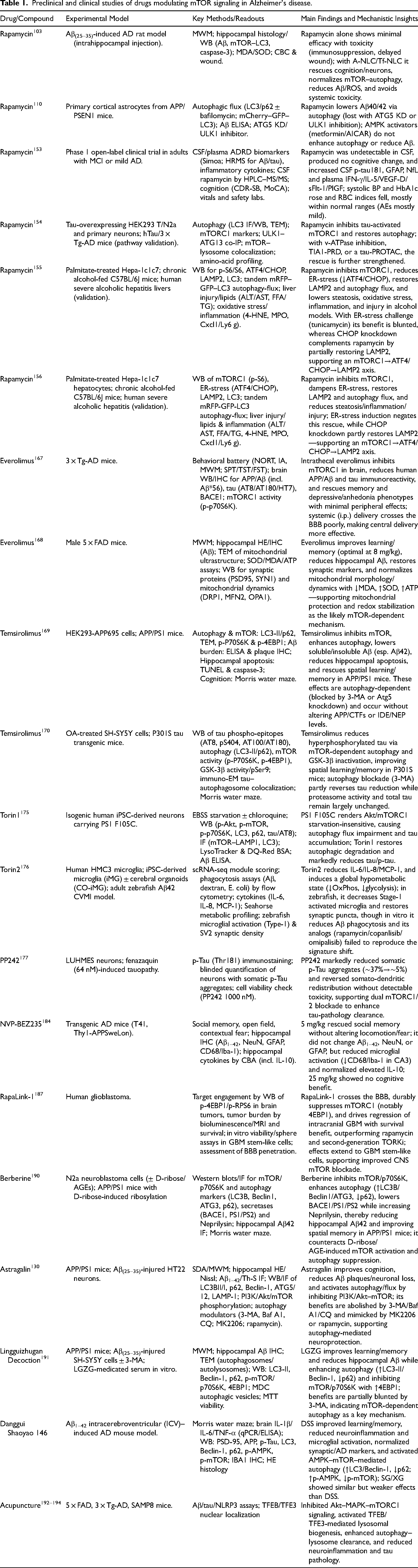
Acupuncture demonstrated convergent modulation of mTOR-related pathways. Electroacupuncture at GV24 and bilateral GB13 in 5 × FAD mice improved cognition, reduced Aβ burden and glial activation, inhibited the Akt–MAPK–mTORC1 pathway, activated TFEB, and enhanced autophagy–lysosome clearance. 192 Stimulation at ST36 in 3 × Tg-AD mice improved memory, reduced tau hyperphosphorylation and NLRP3 inflammasome activation, suppressed mTORC1, activated TFEB/TFE3, and facilitated clearance of tau and inflammatory mediators. 193 Stimulation at GV29, GV20, and GV26 in SAMP8 mice attenuated PI3K/Akt/mTOR hyperactivation, reduced tau-Ser396 phosphorylation, increased PSD-95, and improved spatial memory. 194 Collectively, these TCM interventions were reported to downregulate mTOR signaling and enhance autophagy, thereby improving Aβ/tau pathology and cognitive outcomes. Although not classical “mTOR inhibitors,” their modulatory effects position TCM as an important component of AD intervention research. A comprehensive summary of representative preclinical and clinical studies evaluating mTOR-related compounds—including experimental models, analytical methods, and mechanistic outcomes—is presented in Table 1. In addition, an integrated overview of mTOR-targeted therapeutic strategies, outlining their mechanisms of action, stages of evidence, and translational limitations, is provided in Supplemental Table 1.
Preclinical and clinical studies of drugs modulating mTOR signaling in Alzheimer's disease.
While biologically plausible and mechanistically aligned with mTOR restraint and proteostasis support, much of the current literature remains preliminary. Cross-cutting limitations include reliance on static autophagy markers without flux validation and incomplete reporting of randomization/blinding or sample-size justification. For herbal preparations, additional constraints include batch-to-batch variability and limited chemical standardization of complex extracts, uncertain brain exposure/BBB penetration, and scarce target-engagement evidence (e.g., decreased p-S6 K/p-4E-BP1, TFEB nuclear translocation) when assessed. For acupuncture, key issues include the design and masking of credible sham controls, practitioner- and site-related heterogeneity, and incomplete reporting of allocation concealment. Accordingly, causal attribution to the mTOR pathway and clinical relevance should be interpreted cautiously pending multi-site replication with preregistered protocols and pharmacodynamic confirmation. Overall, where quality criteria were met, TCM single compounds, standardized preparations/multi-herb formulas, and acupuncture were reported to modulate mTOR signaling and autophagy in AD models; however, reproducibility and CNS target engagement still need to be demonstrated to support.
Summary and prospects
This review underscores the central role of mTOR signaling in AD pathogenesis, with abnormal activity impinging on multiple core pathological processes. In preclinical systems, excessive mTOR activation was reported to increase β- and γ-secretase activities and APP processing, thereby elevating Aβ generation, while concomitant inhibition of autophagy reduced Aβ degradation and promoted plaque deposition. Aberrant mTOR signaling was tightly linked to tau pathology: pathway hyperactivation was associated with tau hyperphosphorylation—potentially via downstream kinases and protein-synthesis regulators—and autophagy impairment hindered the clearance of misfolded tau, accelerating NFT formation. Together, mTOR-mediated suppression of autophagy facilitated Aβ plaque buildup, and accumulating Aβ, in turn, drove excessive tau phosphorylation and further mTOR activation—thereby creating a self-reinforcing pathological loop.
Beyond Aβ and tau, dysregulated mTOR signaling disrupted synaptic plasticity, neuroinflammation, and neuronal survival. Under physiological conditions, moderate mTOR activity is required for long-term synaptic plasticity and memory formation by supporting local protein synthesis at synapses; by contrast, chronic hyperactivation in AD was associated with synaptic dysfunction and cognitive deficits. In the immune–metabolic domain, overactive mTOR promoted microglial activation and pro-inflammatory mediator release, while autophagy suppression heightened oxidative stress and aggravated mitochondrial damage. Collectively, these perturbations shifted the balance between neuronal survival and death by impairing proteostasis and increasing susceptibility to degeneration.
Converging evidence also implicated the AMPK–mTOR axis in AD. AMPK functions as a cellular energy sensor that restrains mTOR signaling and promotes autophagy; in preclinical models, AMPK-dependent restraint facilitated the clearance of Aβ and tau aggregates. Consistently, either direct inhibition of mTOR or indirect activation of AMPK enhanced autophagic flux and reduced aggregate burden. These observations support the concept that restoring balance to mTOR signaling—preventing its overactivation while bolstering AMPK-mediated negative regulation—may simultaneously ameliorate Aβ, tau, and synaptic pathologies. Overall, this synthesis reinforces the critical roles of the PI3K–Akt–mTOR and AMPK–mTOR axes and highlights their coordinated involvement across multiple facets of AD pathology, with implications for mTOR-targeted therapeutic strategies.
Therapeutically, first-generation allosteric mTORC1 inhibitors (rapamycin and analogues such as everolimus and temsirolimus) alleviated autophagy suppression in AD models, lowered Aβ and tau, and improved cognition; however, immunosuppressive liabilities and incomplete pathway coverage constrain clinical utility. Second-generation ATP-competitive “mTOR kinase” inhibitors (e.g., Torin-1, PP242) more comprehensively inhibited mTORC1/2 and showed cognitive benefits preclinically, but broader target engagement raised safety concerns. Third-generation agents (e.g., RapaLink-1) combined allosteric and catalytic mechanisms to deliver potent mTORC1 suppression with potentially improved tolerability in preclinical studies, although clinical data remain limited. Complementary approaches—including dual-target compounds and candidates derived from TCM—showed preclinical signals via multi-node modulation of mTOR-related pathways. Non-pharmacological modalities such as acupuncture were reported to downregulate mTOR signaling and enhance autophagy in preclinical settings; human evidence, however, is preliminary and heterogeneous. Together, these findings underscore both the therapeutic promise and the translational challenges of modulating mTOR signaling in AD.
To maintain a translational focus, this review does not attempt an exhaustive treatment of several mechanistic layers; these include TFEB/TFE3-driven lysosome biogenesis; lysosomal amino-acid sensing via the Rag GTPases–Ragulator–v-ATPase axis; S6K1-mediated inhibitory phosphorylation of IRS-1 linking mTORC1 to insulin signaling; mTORC2-dependent control of the actin cytoskeleton; and cell-type–specific programs in microglia and astrocytes. These topics are mentioned only when directly pertinent and will be elaborated in future work.
Notwithstanding the overall trend toward mTOR hyperactivation in AD, context-dependent or region-specific reductions in pathway activity were also described, particularly at late disease stages. Moreover, while rapamycin-based regimens frequently alleviated pathology in rodents, effects on cognition and lifespan varied across models—a finding that highlighted the delicate balance between enhancing proteostasis and sustaining activity-dependent translation at synapses. These inconsistencies exposed gaps in our understanding of temporal, regional, and cell-type–specific mTOR regulation and underscored the need for standardized models, harmonized outcome measures, verified target engagement, and longitudinal, mechanism-anchored human studies.
Despite the progress summarized here, several challenges remain. First, AD pathogenesis reflects interacting networks of Aβ, tau, inflammation, and oxidative stress; targeting a single pathway such as mTOR may be insufficient for disease modification. Second, much of the evidence is preclinical and may not recapitulate the complexity of human AD—especially late-stage pathology and common comorbidities. Third, mTOR has essential physiological roles in metabolism, immunity, and memory; chronic systemic inhibition therefore risks on-target toxicities. Finally, limited brain penetration for many mTOR modulators remains a practical barrier. Overcoming these hurdles—through brain-penetrant, context-aware modulators; rational combinations (e.g., with proteostasis or neuroimmune targets); biomarker-guided dosing; and rigorous clinical-trial designs—will be crucial to determine whether mTOR-targeted strategies can translate into effective, disease-modifying therapies for AD.
Supplemental Material
sj-docx-1-alz-10.1177_13872877251400667 - Supplemental material for Dysregulated mTOR signaling in Alzheimer's disease: Linking pathogenic mechanisms to emerging therapeutic strategies
Supplemental material, sj-docx-1-alz-10.1177_13872877251400667 for Dysregulated mTOR signaling in Alzheimer's disease: Linking pathogenic mechanisms to emerging therapeutic strategies by Siqi Zhang, Tianyi Wang, Guocheng Xue, Ruwen Zheng, Ning Ding, Jing Yang and Miao Zhang in Journal of Alzheimer's Disease
Footnotes
Acknowledgements

Author contribution(s)
Funding
The authors disclosed receipt of the following financial support for the research, authorship, and/or publication of this article: This work was supported by the National Natural Science Foundation of China (Grant Number:82174509); Postgraduate of Heilongjiang University of Traditional Chinese Medicine (Grant Number:2024yjscx009).
Declaration of conflicting interests
The authors declared no potential conflicts of interest with respect to the research, authorship, and/or publication of this article.
Supplemental material
Supplemental material for this article is available online.
References
Supplementary Material
Please find the following supplemental material available below.
For Open Access articles published under a Creative Commons License, all supplemental material carries the same license as the article it is associated with.
For non-Open Access articles published, all supplemental material carries a non-exclusive license, and permission requests for re-use of supplemental material or any part of supplemental material shall be sent directly to the copyright owner as specified in the copyright notice associated with the article.
