



Abstract
Background
Alzheimer's disease (AD) is a progressive neurodegenerative characterized by amyloid-β (Aβ) peptide aggregation and tangles. Protein translation deregulation and viral exposures, including SARS-CoV-2, have been implicated in increased AD risk. Genes such as YIF1A, PABPC4, and MRPS27, which are involved in mRNA translation and protein folding have been linked to neurodegeneration and host responses to SARS-CoV-2.
Objective
This study investigates the association of three genetic variants, rs7945723G>A, rs6587A>G, and rs6831A>G, respectively correlated with YIF1A, PABPC4, and MRPS27, with AD, and explored their expression in COVID-19 samples to assess potential shared pathways.
Methods
A KASP genotyping assay was performed on 127 AD patients and 250 controls. A Binary logistic regression model assessed the association between AD and the single nucleotide polymorphisms, adjusting for age, sex, body mass index, and education. Gene expression was quantified by RT-qPCR in nasopharyngeal samples from 32 COVID-19 patients and 39 controls. The Mann- Whitney test compared gene expression between groups, and logistic regression evaluated associations with COVID-19 status.
Results
Results showed that rs6587A>G in PABPC4 is significantly associated with AD risk with the GG genotype conferring increased susceptibility (OR = 4.3, p = 0.010). Gene expression analysis revealed no significant differences in PABPC4, YIF1A, or MRPS27 between COVID-19 and control groups.
Conclusions
Our findings suggest that post-transcriptional regulatory mechanisms, particularly involving PABPC4, may contribute to AD-related pathways. Larger multi-ethnic cohorts and functional studies are needed to clarify the role of PABPC4 and potential shared mechanisms between AD and COVID-19.
Introduction
Alzheimer's disease (AD) is a severe neurodegenerative disorder characterized by irreversible and progressive loss of cognitive function. 1 AD is a major public health concern, ranking among the leading causes of mortality worldwide and representing the most common subtype of dementia accounting for 60–80% of all cases.2,3
As with other neurodegenerative disorders, AD is characterized by the misfolding and aggregation of specific proteins, such as amyloid-β (Aβ) and tau, whose abnormal conformations acquire toxic properties that disrupt cellular homeostasis and contribute to neuronal disfunction. 4 The neuropathological hallmarks of AD include the extracellular accumulation of insoluble Aβ peptides, and the formation of neurofibrillary tangles composed of hyperphosphorylated tau protein.5,6 These features are associated with a series of detrimental events, including exacerbated brain inflammation and oxidative stress, leading to synaptic dysfunction and neuronal death. 7 Protein translation deregulation has also been implicated in neurodegenerative disorders and other human diseases.8,9 Translation deregulation can be induced by various factors including epigenetic modifications, genetic mutations, deregulated translation factors or regulatory proteins, alterations in mRNA stability or localization, as well as environmental and cellular stress conditions.10–12
Exposure to viruses, such as Herpes Simplex type 1 and 2, Epstein–Barr virus, human cytomegalovirus, influenza virus, hepatitis C virus and recently SARS-CoV-2, has also been associated with cognitive decline and increased risk of AD.13,14 Individuals with dementia are three-fold more likely to contract severe COVID-19 condition than those without dementia,15,16 and the mortality rate is 30% higher in patients who suffer from dementia. 17 A recent study has reported that severe COVID-19 patients display increased expression of genes implicated in AD pathology. 18
This study aims to investigate the association of three genetic variants, rs7945723G>A, rs6587A>G, and rs6831 A>G with AD risk. These variants have been associated with expression variation of YIF1A, PABPC4, and MRPS27 genes respectively. 19 YIF1A is essential for forming and restructuring the ER network, which is crucial for proper protein folding and trafficking. 20 Cytoplasmic poly(A) binding protein (PABP) is a key translational machinery component, and PABPC4, an isoform of PABP, plays an important role in enhancing translation and mRNA stability.21,22 MRPS27 facilitates mitochondrial mRNA translation, and its downregulation has been linked to AD progression. 23 In addition, these genes have been previously reported as SARS-Cov-2 human host genes, experimentally validated or bioinformatically predicted to interact with SARS-CoV-2 proteins. 19 We studied the differential expression of the YIF1A, PABPC4, and MRPS27 in nasopharyngeal samples of COVID-19 positive patients versus controls to validate these findings and explore whether genetic variation and gene expression differences in these loci could provide a molecular link between AD risk and COVID-19 susceptibility.
Methods
Participant recruitment and blood sample collection
The Institutional Review Board of the Beirut Arab University approved all the recruitment and data collection procedures for AD patients and controls (IRB code 2019H-0092-HS-R-0361). All participants or their legal guardians signed an informed written consent. Blood samples were collected from 377 Lebanese individuals, among whom 127 participants were diagnosed with AD by neurologists, based on memory and cognitive tests, functional assessment, physical and neurological exams, diagnostic tests, and brain imaging. Disease-free subjects (controls; n = 250) were 58 years or older, chosen based on the absence of personal or familial psychiatric or cognitive impairment history and a Mini-Mental State Examination (MMSE) score above 26 (Table 1).
Demographic and clinical characteristics of study participants.
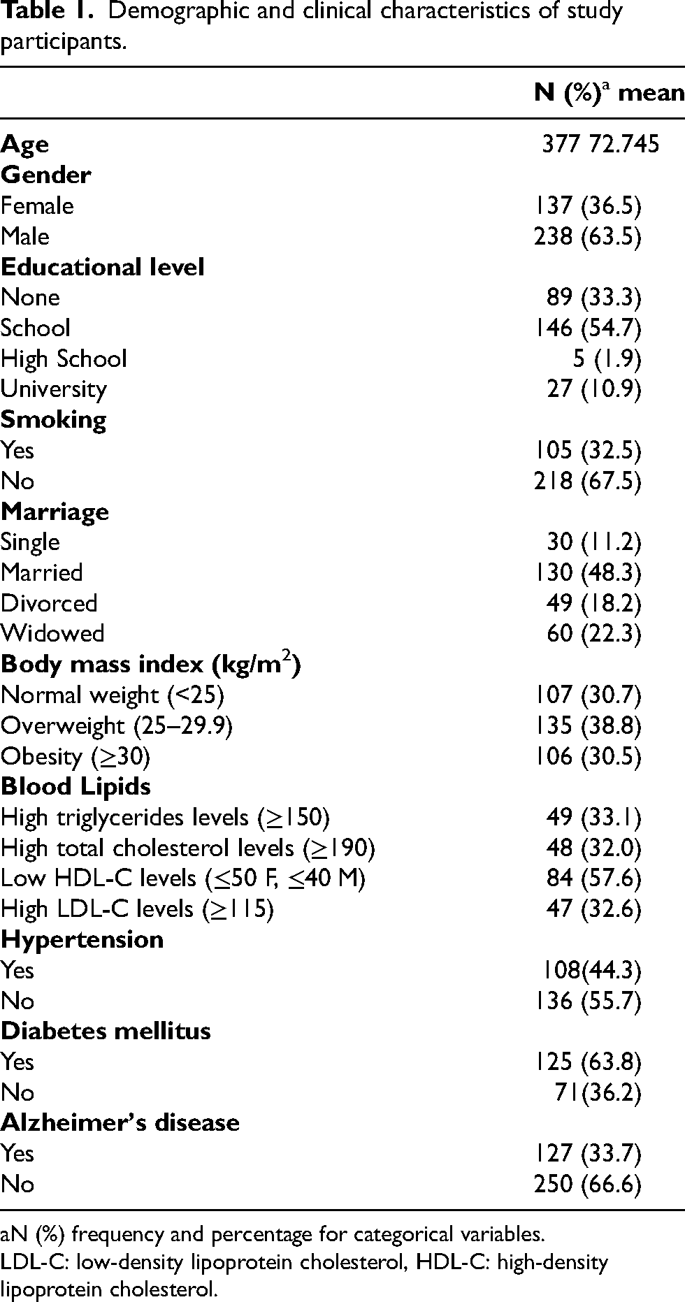
N (%) frequency and percentage for categorical variables.
LDL-C: low-density lipoprotein cholesterol, HDL-C: high-density lipoprotein cholesterol.
In a subset, 71 nasopharyngeal samples were collected for analysis of the expression of the selected genes in COVID-19-infected patients (n = 32) and non-infected controls (n = 39). Exclusion criteria included chronic autoimmune diseases, active malignancy, uncontrolled diabetes mellitus, chronic renal disease, and acute infections other than COVID-19. These samples were collected in full accordance with ethical guidelines and approved by the Institutional Review Board (IRB) of Hospital Libanais Geitaoui under protocol code 2024-IRB-010. Written informed consent was obtained from the study participants.
Single nucleotide polymorphism (SNP) selection and KASP genotyping protocol
The selected SNPs, loci, and respective eGenes are listed in Table 2. Genomic DNA was extracted from peripheral blood leukocytes of AD patients and controls using FlexiGene® DNA kit (QIAGEN) according to the manufacturer's instructions. Genotyping was conducted by the LGC group (Berlin, Germany) using the KASP genotyping assay. KASP is a homogeneous, fluorescence resonance energy transfer-based assay that enables accurate biallelic discrimination of known genetic variations, including SNPs and insertions/deletions, as described previously. 24
The loci, minor allele frequencies, population frequencies (GnomAD), and respective eGenes of the SNPs analyzed in this study.

SNP: single nucleotide polymorphism; MAF: minor allele frequency.
RNA extraction and cDNA synthesis
Nasopharyngeal swab samples from a subset of 71 COVID-19 affected patients and controls were processed for RNA extraction using the ANDiS Viral RNA Auto Extraction & Purification Kit in conjunction with the ANDiS 350 Automated Nucleic Acids Extraction System. In this automated protocol, viral particles were first lysed to release RNA, which was then captured on magnetic beads. Following washes to remove impurities, the RNA was eluted into a clean solution for further analysis. RNA concentration and purity were assessed using a NanoDrop spectrophotometer (Thermo Fisher Scientific), ensuring A260/A280 ratios between 1.8 and 2.0. The extracted RNA was quantified, stored at −80°C, and subsequently reverse-transcribed into complementary DNA (cDNA) using a commercial reverse transcription kit, following the manufacturer's instructions.
Quantitative real-time PCR for gene expression
RT-qPCR was performed to quantify the expression PABPC4, YIF1A, and MRPS27 genes in nasopharyngeal tissue samples from COVID-19 infected and non-infected samples. Gene-specific primers were designed and validated for efficiency and specificity. Each 20 µL reaction mixture contained SYBR Green Supermix, optimized concentrations of forward and reverse primers, and 70 ng of cDNA template, with GAPDH serving as the housekeeping gene for normalization. The thermal cycling protocol commenced with an initial enzyme activation and denaturation step at 95°C for 3 min, followed by 40 cycles of denaturation at 95°C for 15 s and a combined annealing/extension step at 60°C for 30 s. All reactions were executed in duplicate to ensure reproducibility and accuracy.
Statistical analysis
All analysis was conducted using SPSS software version 24 (SPSS, Inc, Chicago, IL, USA). All continuous variables are expressed as the mean ± standard deviation, and categorical variables are presented as frequencies and percentages. Normality was tested using Shapiro-Wilk test.
Independent samples t-tests and Chi-square tests were used to compare continuous and categorical variables, respectively, between AD patients and controls.
A binary multiple logistic regression model was employed to investigate the association between the presence of AD (dependent variable) and the minor allele frequency and the genotypes of the three SNPs while adjusting for potential confounders. Covariates, including age, gender, body mass index, and educational level, were selected based on their established connections with AD and their potential to introduce confounding effects into the SNP-disease association analysis. The significance threshold was established at p ≤ 0.05. Model diagnostics were performed to assess assumptions of normality, variance homogeneity, and multicollinearity, and goodness-of-fit was evaluated using residual plots and standard tests. To control for type I error across the three prespecified genes, p-values were adjusted using Bonferroni correction (α = 0.05/3 ≈ 0.0167).
The AD sample size was primarily determined based on statistical power considerations and informed by recruitment feasibility. Assuming an additive genetic model, an odds ratio of approximately 2.0 for the association between the rs6587 variant and AD, and a minor allele frequency of 0.42 as reported in Middle Eastern populations, power calculations were performed using a two-sided significance level of α = 0.05 and 80% power. Under these assumptions, the required sample size was estimated to be approximately 120 AD cases and 230 cognitively normal controls. The study was primarily powered for the detection of moderate effects for the main variant of interest, acknowledging that smaller effect sizes may not be detectable with adequate statistical power.
The sample size in the expression analysis of YIF1A, PABPC4, and MRPS27 (71 participants) provides adequate power to detect moderate-to-large differences (Cohen's d ≈0.6), but smaller effects may remain underpowered. For each gene, a median value was calculated and further used as a cut-off to classify the gene expression as high or low. The Mann-Whitney test was employed to compare gene expression between COVID-19 infected and non-infected individuals. Binary logistic regression models, adjusted for age, sex, and BMI, were used to evaluate the association between the normalized expression of selected genes and COVID-19 status.
Results
Demographic and clinical characteristics of study participants
The demographic characteristics of the study participants, including age, gender, education, marital status, and smoking status were shown in Table 1. The mean age was 72.745, with 36.5% of participants being females. Educational levels varied as 33.3% with no formal education, 54.7% had attended some school, 1.9% had completed high school, and 10.9% had pursued university. Additionally, 32.5% were smokers. The clinical characteristics and comorbidities were also recorded. Regarding BMI, 30.7% of the participants were classified as normal, 38.8% were overweight, and 30.5% were obese. Of 148 participants, 33.1% had high triglyceride and 32.0% had high total cholesterol levels. Out of 244 participants, 44.3% had hypertension and 63.8% of 196 patients had diabetes mellitus.
AD-SNP analysis
The minor allele frequencies in our data ranged from 0.21 to 0.42, approximately similar to those found in Middle Eastern populations (GnomAD) and to those reported by Chen et al., 2020. The observed genotype frequencies of rs7945723G>A, rs6587A>G and rs6831A>G did not deviate significantly from the Hardy-Weinberg equilibrium (HWE). The minor allele frequency of rs7945723G>A, rs6587A>G and rs6831A>G was 0.21, 0.42 and 0.32, respectively, suggesting that these alleles were relatively common in the studied population (Table 2).
To explore the associations with AD risk, we used a binary logistic regression model. Age was positively associated with AD risk, with individuals over 80 years showing an odds ratio (OR = 9.6, p = 0.000). BMI was inversely associated, as those with BMI ≥30 had an (OR = 0.14, p = 0.000). Educational level and gender were not significantly associated with AD (p > 0.05). Among the three genetic variants analyzed, a significant association with AD was observed for the rs6587A>G SNP; the GG genotype showed an odds ratio of 4.32 (p = 0.010) (Table 3).
Binary logistic regression Analysis of demographic risk factors and SNP genotypes within AD.

OR: odds ratio; CI confidence interval.
Quantitative RT-PCR (qRT-PCR): gene expression analysis
We examined the differential expression of YIF1A, PABPC4, and MRPS27, hypothesizing that these genes may be associated with the human host responses to SARS-CoV-2. The comparison of gene expression based on COVID-19 disease status revealed that the normalized expression levels of YIF1A, PABPC4, and MRPS27 did not reach statistical significance (p > 0.05). This indicates that the gene expression profiles for these genes were comparable between COVID-19 positive participants and controls (Figure 1). Moreover, we conducted a binary logistic regression analysis that included the three genes, while adjusting for confounding factors such as BMI, age, and gender, according to COVID-19 disease status. None of the genes was found to significantly increase the risk of the disease (p > 0.05) (Table 4).

(A) Normalized expression levels of YIF1A mRNA in COVID-19 negative versus COVID-19 positive participants. (B) Normalized expression levels of PABPC4 mRNA in COVID-19 negative versus COVID-19 positive participants. (C) Normalized expression levels of MRPS27 mRNA in COVID-19 negative versus COVID-19 positive participants. Statistical significance is indicated (*p < 0.05).
Binary logistic regression analysis with COVID-19 disease predictors: gene expression data.

Gene expression levels were classified as low or high based on their median value.
OR: odds ratio; CI: confidence interval.
Discussion
The greatest risk factor for AD is advanced age. 25 In our study, higher age was strongly associated with higher risk of AD, with those over 80 having significantly elevated odds (OR = 9.6, p = 0.00) (Table 3). In contrast, higher BMI was associated with decreased AD risk (OR = 0.14, p = 0.00). This inverse relationship between late-life BMI and AD risk has been reported in recent meta-analyses. 26 In contrast to the increased AD risk linked to higher BMI in midlife. 27 One plausible explanation for this might be the fat-derived hormone leptin. 28
Our study found that the rs6587, which influences the expression of PABPC4, is significantly associated with AD. The GG genotype conferred a significantly increased risk (OR = 4.32, p = 0.01). PABPC4 has been shown to work, alongside translation factors like PABPC1 and RPS6, in regulating Tar DNA-binding protein 43 (TDP-43) in mRNA translation. TDP-43 proteinopathy disrupts local translation at synapses and mitochondrial functions, contributing to neurodegenerative diseases. 24 Additionally, as PABPC4 is part of MID1 protein complex, implicates a possible role in the regulation of amyloid precursor protein (APP) mRNA through mTOR signaling pathway. 29 Emerging evidence indicates that PABPC4 influences mitochondrial oxidative metabolism under metabolic stress, a pathway known to be disrupted in AD.30–32 A computational study, analyzing regulatory perturbations underlying the molecular etiology of AD using gene expression data, inferred associations between AD pathology and PABPC4. The study also revealed that an upstream variant of the PABPC4 gene is associated with a longer lifespan among AD patients. 33 Together, these findings demonstrate the impact of post-transcriptional regulatory proteins, including PABPC4, on brain cell function and suggest its possible association with AD risk. Notably, PABPC4 has not been identified as a genome-wide significant locus in large AD GWAS, predominantly conducted in European ancestry cohorts. Our study population, drawn from a Middle Eastern cohort, may capture population-specific allele frequencies or linkage disequilibrium patterns that differ from those in larger datasets. Moreover, regulatory variants such as rs6587 often exert modest, tissue-specific effects that may not reach genome-wide significance thresholds in large heterogeneous cohorts.
On a different note, PABPC4 is involved in various cellular process. It is essential for erythroid mRNA expression, telomerase regulation and cell growth. 34 It is also upregulated during T cell activation and is known as activated platelet protein-1.21,35–37 PABPC4 serves as a general viral target, as viruses modify PABP activity to inhibit host translation and evade immune responses. 34 Coronavirus N proteins have shown to bind PABP, negatively regulating translation of coronaviral RNA and host mRNA both in vitro and in cells. 38 Recent studies have also implicated PABPC4 in COVID-19-related coagulation disorders. 39 PABPC4 has been reported to inhibit CoV replication by targeting and degrading the CoV N protein through selective autophagy via the autophagy receptor NDP52. 36 A similar mechanism was observed with swine acute diarrhea syndrome coronavirus (SADS-CoV), where PABPC4 recruits MARCHF8 to ubiquitinate the N protein for autophagic degradation. 40 Our results did not show a variation in PABPC4 expression in COVID-19 patients versus controls. This may be attributed to the complex molecular regulation of its expression. The downregulation of transcription factor SP1 during coronavirus infection has been shown to stabilize PABPC4 levels despite viral presence. 36
Our findings indicate that the rs7945723 variant, influencing the expression of YIF1A, is not linked to an increased risk of AD (p > 0.05). Yip1- interacting factor homolog A (YIF1A) is essential for forming and restructuring the ER network, which is crucial for proper protein folding and trafficking. 21 YIF1A, FAM8A1, and members of the SLC30 family, expressed in the Golgi apparatus or the ER-Golgi intermediate compartment (ERGIC) and are involved in vesicular transport, cellular zinc homeostasis, and membrane trafficking. 41 Mutations in genes coding for trafficking proteins have been shown to cause severe neurodevelopmental delay, white matter defects and intellectual disability in humans. 42 A study shows that YIF1A interaction with vesicle-associated membrane protein-associated protein B (VAPB), and the mitochondrial membrane protein, protein tyrosine phosphatase interacting protein-51 (PTPIP51), is involved in signaling between the ER and mitochondria, with dysfunction linked to neurodegenerative diseases. On the other hand, YIF1A has been implicated in cellular response to SARS-CoV-2 infection. During SARS-CoV-2 infection, the virus hijacks host cellular machinery, including components like YIF1A, to facilitate its replication and assembly. 43 SARS-CoV-2 proteins can interact with various host proteins related to the Golgi apparatus. 44 SARS-CoV-2 interaction with YIF1A enhances the ability to manage ER morphology and maintains the protein production and transport balance, thereby limiting the induction of ER stress and preventing apoptosis. 43 Additionally, YIF1A, FASTKD5, and AASS, are linked to the SARS-CoV2 host protein-protein interaction network. These genes have significant regulatory roles in mitochondria metabolism.45,46
Despite the involvement of YIF1A in SARS-CoV2 response, our expression analysis revealed no significant difference in YIF1A expression levels between COVID-19 positive and negative subjects (p > 0.05). The expression of YIF1A may be influenced by interactions with other proteins, such as Yip1p homologs (YIPF4, YIPF5, YIPF6), which, when knocked down, lead to a significant reduction in YIF1A levels. This suggests that while YIF1A is implicated in viral processes, its overall expression may be maintained through complex regulatory networks that do not solely depend on the presence of SARS-CoV-2. 47
Our study investigated the association of rs6831, which influences the expression of mitochondrial ribosomal protein S27 gene (MRPS27), with AD. Mitochondria play a crucial role in energy production and metabolic regulation in neurons, and their dysfunction is linked to AD through oxidative damage and impaired synaptic plasticity. 48 A recent study identified novel biomarkers related to mitochondrial dysfunction in AD by combining bioinformatics and machine learning methodologies. MRPS27, which is essential for synthesizing electron transport chain complexes, was one of the six key mitochondrial dysfunction-related genes with strong diagnostic potential for AD. 49 MRPS27 facilitates mitochondrial mRNA translation, and its downregulation is linked to AD progression. 23 Moreover, SARS-CoV-2 proteins interact with mitochondrial proteins (NDUFAF1, NDUFAF2, NDUFB9, MRPS2, MRPS5, MRPS25, MRPS27) that play crucial roles in the mitochondrial metabolic pathways, supporting viral establishment and replication. 45 Our findings did not indicate a link between rs6831 variant of MRPS27 and AD (p > 0.05) and did not reveal differential expression of MRPS27 in COVID-19 patients. Further studies may elucidate the potential role of this gene in AD pathogenesis and response to sars-cov2 infection.
Conclusion
This study examined the association of the genetic variants rs7945723G > A, rs6587A>G, and rs6831A>G, which influence the expression of YIF1A, PABPC4, and MRPS27, with AD, and explored their expression in COVID-19 samples to assess potential shared molecular pathways. Among the three variants, only rs6587A>G in the PABPC4 gene was significantly associated with increased AD risk, suggesting that post-transcriptional regulation may contribute to mechanisms implicated in AD. These findings provide preliminary evidence and lay the groundwork for future investigations in larger, more diverse populations to clarify the role of PABPC4 in AD-related molecular processes. Although PABPC4 expression was not significantly altered in COVID-19 patients in this cohort, replication in expanded multi-ethnic datasets with greater statistical power may help resolve subtle differences. Multi-tissue transcriptomic approaches and functional studies, will determine whether common molecular mechanisms link COVID-19 susceptibility and AD.
Study limitations
The restricted genotyping panel in our study, which included three SNPs does not provide the genomic depth necessary to rule out spurious associations. However, the observed link between rs6587 in PABPC4 and AD risk is hypothesis-generating. This variant lies in a non-coding region, possibly within a promoter or enhancer, and its functional role remains uncertain. Larger studies employing genome-wide approaches and functional validation will help explore the potential causal role of PABPC4 or other variants in AD.
Our gene expression analysis, done on nasopharyngeal swabs, may not capture brain-relevant regulation. In addition, the analysis is underpowered to detect small effect sizes. With this sample size, the minimal detectable difference is approximately Cohen's, and power is further influenced by expression variance, covariate adjustment, and any multiple-testing correction. Subgroup analyses within the 32 cases are particularly limited by event sparsity.
Footnotes
Acknowledgements
We would like to express our sincere gratitude to all geriatric care facilities and hospitals including Dar-Al Ajaza Al-Islamia Hospital in Beirut, the social services association in North Lebanon, Bayt Al Shaikhookha in Tripoli, Bayt Al Raha Ozanam in Batroun, and Dar Al Inaya in Jbeil for their support in sample collection and consent approval. We also like to extend our appreciation to the study participants and to the neurologists Dr Nabil Mohsen and Dr Nabil Naja, as well as to Dr Nadia Ramadan, for their expertise and invaluable assistance in diagnosing participants with Alzheimer's disease.

Ethical considerations
The Institutional Review Board of the Beirut Arab University approved all the recruitment and data collection procedures for Alzheimer's Disease patients and controls (IRB code 2019H-0092-HS-R-0361).
The samples from COVID-19 patients and controls were collected in full accordance with ethical guidelines and approved by the Institutional Review Board (IRB) of Hospital Libanais Geitaoui under protocol code 2024-IRB-010.
Consent to participate
All participants, or their legal guardians, signed an informed written consent
Consent for publication
Not applicable
Author contribution(s)
Funding
The authors received no financial support for the research, authorship, and/or publication of this article.
Declaration of conflicting interests
The authors declared no potential conflicts of interest with respect to the research, authorship, and/or publication of this article.
Data availability statement
The data supporting the findings of this study are available within the article. De-identified genotype and expression datasets are available from the corresponding author upon reasonable request.