



Abstract
Significance:
Oxidative mechanisms contribute to both vascular function and pathogenesis of many diseases, but their role in the microvasculature remains poorly understood.
Recent Advances:
The role of reactive oxygen and reactive nitrogen species (ROS/RNS) in the vasculature has been well-established for years. Our knowledge of microvascular responses to ROS/RNS has relied on extrapolation of studies performed in large vessels or cultured endothelial cells from large vessels. In healthy tissue, ROS/RNS are implicated in microvascular cell survival and death, angiogenesis, vasodilation, and barrier function, and, in disease, they contribute to increased permeability, leukocyte extravasation, and inflammation. Redox-mediated microvascular dysfunction underlies a multitude of conditions, including cardiovascular diseases, autoimmune diseases, infectious diseases, hemoglobinopathies, inflammatory diseases, vasculitides, and metabolic diseases.
Critical Issues:
New single-cell RNA sequencing studies reveal that endothelial cells from different vascular beds have unique gene signatures. Moreover, microvessels respond differently than large vessels, yet findings are frequently extrapolated across vascular beds. Technical challenges have limited our ability to reliably link alterations in ROS/RNS levels to microvascular outcomes. Moreover, successful therapeutics targeting redox signaling in general and in the microvasculature in particular are lacking. While numerous associations exist between common diseases and the microvasculature, the precise contribution of redox-mediated microvascular dysfunction to disease pathogenesis has been challenging.
Future Directions:
Additional research in organ-specific microvasculature focusing on the redox mechanisms underlying microvascular function and dysfunction is needed, as well as the development of new targeted therapeutics that can be locally delivered. Comparison of redox responses between different diseases may uncover general mechanisms to exploit therapeutically. Antioxid. Redox Signal. 43, 566–621.
Introduction
For much of human history, the function of the human circulatory system was unclear. Several millennia ago, it was recognized that humans must somehow distribute nutrition from the digestive tract to the rest of the body. In ancient Egypt, the Ebers Papyrus described the cardiac vessels and observations likely representing congestive heart failure (Katz, 1957; Saba et al., 2006). The ancient Greeks distinguished arteries from veins, and Indian Ayurveda physicians described the heart acting as a pump and hinted at the cardiovascular system being a closed circuit (Fig. 1) (Patwardhan, 2012). Then, in the second century, Galen described the movement of blood, albeit as a one-way system. In the 8th to 10th centuries, two Iranian physicians, Razi and Ahwazi, described arteries, veins, and the circulation of blood (Azizi et al., 2008). In 1628, William Harvey published De Motu Cordis, stating that blood “moves around in a circle continuously” (Fig. 1) (Aird, 2011).
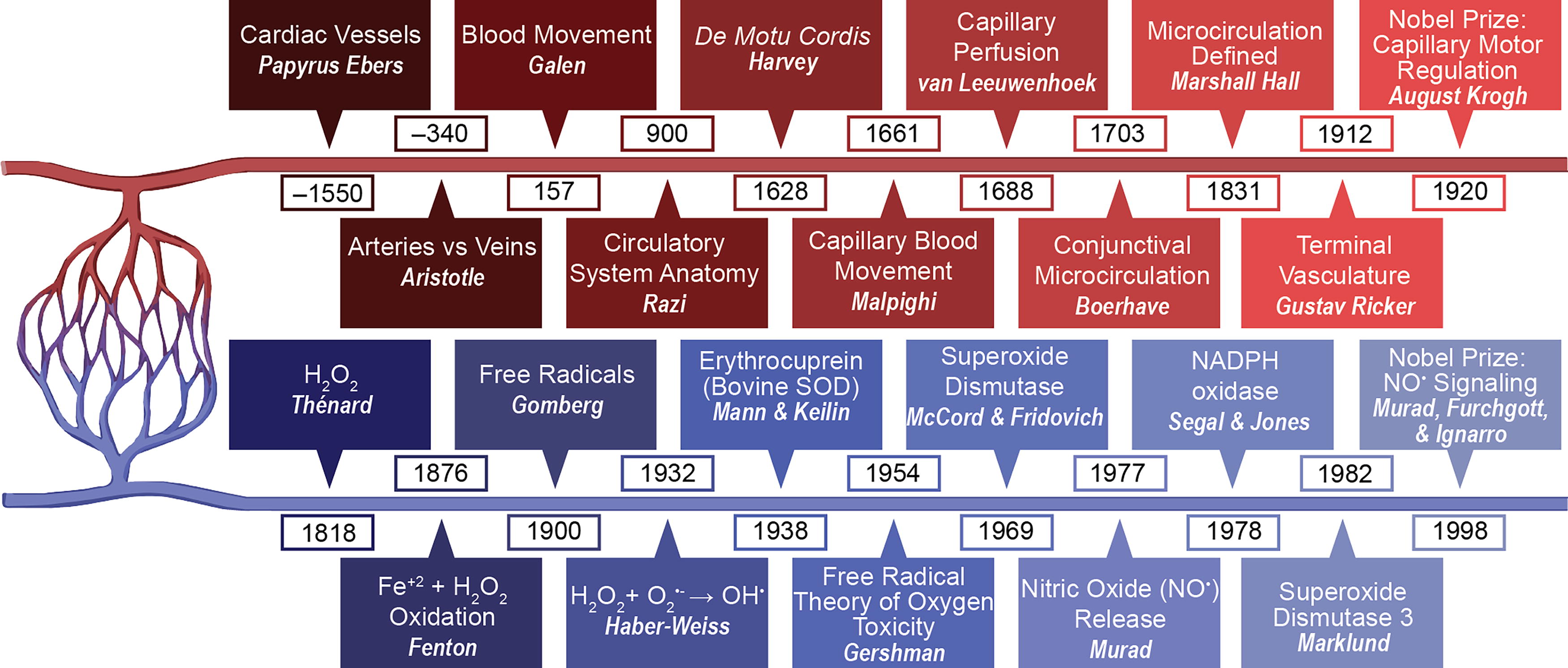
While capillaries were discovered as early as the 1500s when Leonardo da Vinci injected blood vessels with wax, the role of capillaries in bridging arterioles and venules was not recognized until 1661, when Marcello Malpighi used microscopy to identify that blood moved through capillaries, although he thought the particles seen were clots rather than blood cells, which were not described until Antoni van Leeuwenhoek observed them in glass eel caudal fin capillaries (Pearce, 2007). Then, in 1703, Hermann Boerhaave described the microcirculation in the bulbar conjunctiva of patients, although the definition of microcirculation was only clearly established by Marshal Hall in 1831 (Fig. 1). In 1912, the smallest vessels were described and termed by Gustav Ricker as the terminal vasculature, which now refers to the microvasculature. The significance of the microvasculature system was recognized with the 1920 Nobel Prize in Physiology or Medicine, awarded to August Krogh for his discovery of the capillary motor regulating mechanism. Over the past century, much progress has been made in our understanding of the microvascular system, which includes arterioles, venules, and capillaries (Fig. 1) (Jung, 2024).
Chemically reactive species have been increasingly recognized as contributing to numerous functions of the microvasculature (Fig. 1). While Fenton discovered the oxidizing properties of ferrous ions and hydrogen peroxide (H2O2) in 1876, and H2O2 itself was first discovered in 1818 by Louis Jacques Thénard (1819), free radicals were not identified until 1900 by Moses Gomberg (1900). Then, in 1954, Gershman suggested a connection between oxygen’s toxicity and free radicals (Gerschman et al., 1954). One of the main defenses against free radicals, superoxide dismutase (SOD), was first discovered in 1938 by Mann and Keilin as bovine erythrocuprein, which was later identified in humans as SOD1 by McCord and Fridovich (Mann and Keilin, 1938; McCord and Fridovich, 1969). SOD3 was then discovered by Marklund in 1982 (Fig. 1) (Marklund et al., 1982). In 1978, NADPH oxidase (Nox), one of the major producers of reactive oxygen species (ROS), was discovered (Fig. 1) (Segal and Jones, 1978). Then, in 1986, discoveries concerning the role of nitric oxide (NO•) signaling in the cardiovascular system led to the 1998 Nobel Prize in Physiology or Medicine (Fig. 1) (Moncada and Higgs, 2006). Since then, ROS and reactive nitrogen species (RNS) have been found to not only contribute to toxicity but also play an integral role in the physiology and pathophysiology of the microvasculature (Kadlec and Gutterman, 2019; Moncada and Higgs, 2006). In this article, we focus on the role of ROS in the normal microvasculature and in a wide range of diseases associated with microvascular dysfunction.
Terminology
Before delving into the various pathways involved, an overview of the terminology and key players in the microvasculature is warranted. ROS specifically refers to chemically reactive species containing oxygen, while RNS specifically refers to chemically reactive species containing nitrogen. ROS is often used to include RNS that have reactive oxygen. Since 1989 (Halliwell, 1989), the term ROS has been used to refer to both free radical and non-free radical oxygen derivatives. Free radicals include ROS/RNS containing one unpaired reactive electron in the outer orbit. Non-free radicals, conversely, include ROS/RNS without an unpaired electron (Table 1). Oxygen has several unique properties that contribute to its reactivity and tendency to form free radicals. It has the largest free energy change per electron transferred (except for chlorine and fluorine) while still being stable enough to be an excellent terminal electron acceptor, allowing for its central role in the electron transport chain and cellular respiration (Zhang et al., 2019). Several properties of free radicals differentiate them and contribute to their various roles. Key differentiating properties include lifetime, diffusion distance, solubility, propensity for oxidation and reduction, and the types of molecules with which they tend to react. Examples of free radicals include NO•, superoxide anion (O2 •−), hydroxyl radical (•OH), carbonate radical anion (CO3 •−), nitrogen dioxide (•NO2), alkoxy peroxyl (R′OO•), and alkyl peroxyl (ROO•). Non-free radical ROS and RNS are typically more stable than free radicals and can act as two-electron oxidants. Examples of non-free radicals include H2O2, peroxynitrite (OONO−), peroxynitrous acid (HNO3), and hypochlorous acid (HOCl) (Kadlec and Gutterman, 2019). ROS/RNS are produced both intracellularly and extracellularly through a variety of mechanisms, as reviewed by Forrester et al. (2018).
Radicals and Their Corresponding Antioxidant Defense
Summary of the free radicals and non-free radicals discussed in this article. We included the corresponding antioxidant defense in each case.
NOS, nitric oxide synthases.
General Experimental Strategies to Study Oxidative Stress
This table summarizes experimental strategies used to study how redox states contribute to microvascular disease.
ROS, reactive oxygen species; Nox, NADPH oxidase; iNOS, inducible/calcium-independent NOS2; PPARγ, peroxisome proliferator- activated receptor gamma; SOD, superoxide dismutase; GPx, glutathione peroxidase; Nrf2, nuclear factor erythroid 2-related factor 2; PAD, peripheral arterial disease; MV, microvascular; LPS, lipopolysaccharide; TNFα, tumor necrosis factor α.
Nitric oxide
While it has been known for over a thousand years that compounds later identified to contain nitrates can relieve anginal pain, it was not until recently that this effect was found to occur via the known poison and air pollutant NO•. NO• was first identified as a simple inorganic gas by the pneumatic chemist Joseph Priestly in 1772. It is one of the smallest known molecules, consisting of a single oxygen atom paired with a single nitrogen atom. This combination leaves a single free electron in the highest occupied electron orbital, accounting for both the molecule’s simultaneous inert and highly reactive properties. In the absence of molecules with unpaired electrons, NO• is less reactive, although it still can react with some molecules with paired electrons such as ozone. In the presence of other free radicals and transition metals, it is highly reactive and can lead to the production of RNS. These properties contribute to its role in cell signaling, which is key in numerous aspects of vascular endothelial (VE) physiology, as recognized by the 1998 Nobel Prize in Physiology or Medicine (Fig. 1) (Furchgott, 1983; Ignarro et al., 1987; Moncada and Higgs, 2006; Moncada et al., 1986; Murad et al., 1978).
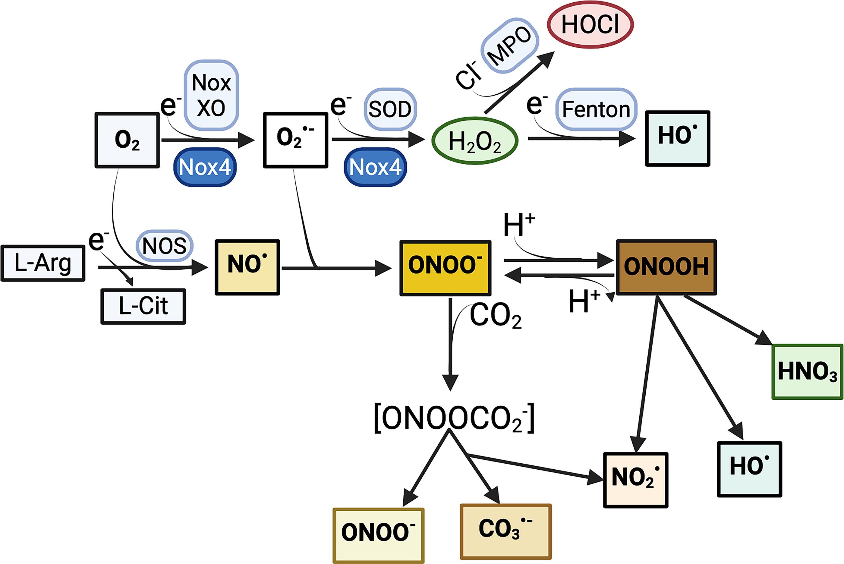
Some of the many reactions regulated by NO• include termination of lipid peroxidation, generation of OONO− by interaction with O2 •−, generation of protein radicals via nitrosylation of proteins, and formation of nitrosyl-metal complexes (Fig. 2). These reactions are involved in numerous physiological pathways, most notably cyclic guanosine monophosphate (cGMP)-mediated vascular relaxation, which will be discussed in more detail later. OONO− is a particularly important metabolite of NO• in the microvasculature because it can react with carbon dioxide, thiols, and various metals to form CO3 •− and •NO2, resulting in further oxidative and nitrative effects (Fig. 2). Compared with many other free radicals, NO• has a longer life, is highly soluble, and is a weak antioxidant.
NO• synthesis is highly regulated and can occur both enzymatically and nonenzymatically (Lancaster, 2020). It is enzymatically produced by nitric oxide synthases (NOS). Additionally, nitrite can be reduced to NO• enzymatically by reductase enzymes such as xanthine oxidoreductase, cytochrome c oxidase, aldehyde dehydrogenase 2, cytochrome P450 reductase, and cytochrome P450 (Zweier et al., 1995). In contrast, NO• can be produced nonenzymatically in tissues as a function of pH or reducing equivalents, or from S-nitrosothiol via transition metal ions in combination with thiol-reducing agents. NOS are encoded by three separate genes: “neuronal” NOS1 (nNOS), “inducible/calcium-independent” NOS2 (iNOS), and “endothelial” NOS3 (eNOS). NOS produce NO• by converting l-arginine into l-citrulline. The vascular endothelium primarily expresses eNOS, although iNOS can be induced by inflammation and nNOS can be found to a lesser degree. eNOS functions as a homodimer and contains two main domains linked by a calmodulin-binding sequence. The C-terminal reductase domain includes flavin mononucleotide (FMN), flavin adenine dinucleotide (FAD), and nicotinamide adenine dinucleotide phosphate (NADPH) binding sites, while the N-terminal oxygenase domain includes l-arginine, tetrahydrobiopterin (BH4) cofactor, heme, and zinc-binding sites. eNOS is located in caveolae at baseline and is released and activated as intracellular calcium levels increase (Vanhoutte et al., 2016).
Superoxide anion radical
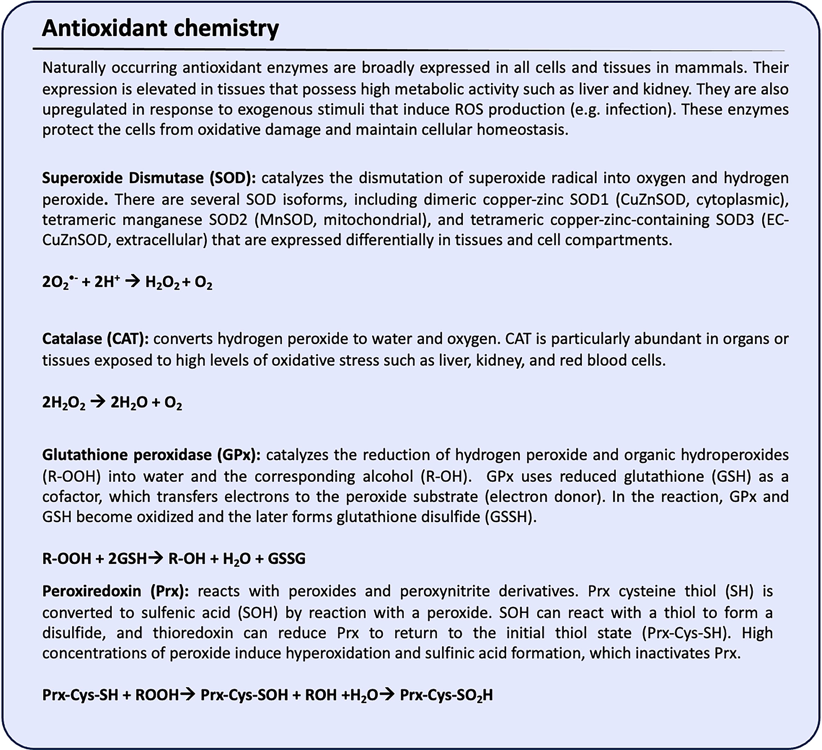
O2 •− is a weak radical, partly due to a charge limiting access to other electron-rich molecules. It is generated during cellular respiration, by NADPH oxidases or xanthine oxidase (XO) (Fig. 2), or via eNOS uncoupling, aldehyde oxidase, cyclooxygenase (COX), and others (Piacenza et al., 2022). Even though it is not highly reactive itself, it can form other highly reactive species, such as OONO− when reacting with NO•. As such, O2 •− is a major biologically significant free radical, and the identification by McCord and Fridovich in 1969 of the enzyme copper-zinc-containing superoxide dismutase (SOD1), which accelerates the conversion of O2 •− into H2O2 (Fig. 2 and Fig. 3), was a landmark finding in the history of free radical biology (McCord and Fridovich, 1969) (Fig. 1). There are three main types of SODs: dimeric copper-zinc-containing SOD1 (cytoplasmic), tetrameric manganese-containing SOD2 (mitochondrial), and tetrameric copper-zinc-containing SOD3 (extracellular) (Wang et al., 2018).
Of note, cytoglobin, which shares structural similarity with myoglobin and hemoglobin, has recently been identified as a fourth type of molecule with SOD activity (Zweier et al., 2021). SODs are highly expressed, and part of the reason that O2 •− is so short-lived is that SODs are close to where it is produced. It should be noted, however, that the formation of OONO− by NO• and O2 •− is much faster than the removal of O2 •− by SODs, and the highly reactive nature of OONO− can thus contribute to tissue injury.
Nitrogen dioxide
•NO2 is a moderately reactive radical produced primarily through electron transfer during OONO− decomposition in the presence of carbon dioxide (Fig. 2) (Martínez and Andriantsitohaina, 2009). It is also a common air pollutant due to emissions from motorized vehicles, industry, and fires (Mirowsky et al., 2016). It can oxidize thiols, resulting in downstream production of nitrosothiol in combination with NO•. •NO2 and carbonate produced from OONO− homolysis can then result in the generation of the radical forms of tyrosine and cysteine, as well as the oxidation of nucleotides (Szabó et al., 2007).
Hydrogen peroxide
H2O2 plays a key role in the microvasculature as a substrate for peroxidases, but more importantly, it is a redox signaling molecule. It is ubiquitous and has a longer half-life than all of the above molecules. Its small size and lack of polarity allow it to passively cross cellular membranes. Additionally, water channels such as aquaporin-3, along with other aquaglyceroporins and unorthodox aquaporins, can also facilitate H2O2 uptake across the plasma membrane of mammalian cells (Miller et al., 2010). In the vascular endothelium, H2O2 has been found to regulate a variety of protein signaling pathways and ion channels, which affect functions including vasodilation, cytoskeletal remodeling, cellular growth and proliferation, and response to inflammation (Cai, 2005).
Antioxidants
As noted above, ROS and RNS can be damaging if left unchecked. The human body counteracts free radicals’ harmful effects to maintain redox homeostasis by producing both enzymatic and nonenzymatic antioxidants. Enzymatic antioxidants, such as SOD, catalase, glutathione peroxidase (GPx), and peroxiredoxin, directly catalyze the conversion of ROS into less reactive molecules (Fig. 3). Nonenzymatic antioxidants, such as vitamin C and E, melatonin, alpha-lipoic acid, glutathione (GSH), and coenzyme Q10 (endogenous), or ascorbate, tocopherols, carotenoids, and flavonoids (exogenous), work primarily by scavenging ROS through electron donation. Antioxidants usually act synergistically, such that the oxidized form of one antioxidant can be reduced back to its active form by another, thus forming an integrated network that maintains cellular redox balance. Intracellular antioxidant systems are also tightly regulated via localization, expression, and substrate availability (Gutterman et al., 2016).
In the cytoplasm and mitochondria, GPx replenishes other cellular antioxidants (thioredoxins, peroxiredoxins) by maintaining GSH stores (Fig. 3). GSH can interact with NO• to form S-nitrosoglutathione (Baldelli et al., 2019), and NO• itself can function as both an antioxidant and an oxidant. Catalase, in the presence of metal ion cofactors, converts H2O2 into water in peroxisomes, mitochondria, and the nucleus (Fig. 3). SODs can function as balancing enzymes or mediators, which in the presence of metal ion cofactors, can neutralize O2 •− in mitochondria, cytosol, and peroxisomes (Fig. 3) (Andrés et al., 2023; Fukai and Ushio-Fukai, 2011). In Table 1, we summarize the most common antioxidants for the corresponding ROS/RNS free and non-free radicals discussed in this article. We will explore various antioxidant pathways and potential antioxidant therapeutics later in this article.
Experimental models for studying redox signaling in the microvasculature
The methods for studying ROS/RNS have been extensively described elsewhere (Griendling et al., 2016). More recently, a group of experts in redox biology have published guidelines for measuring ROS and oxidative damage in cells and in vivo, emphasizing the importance of accurate and reproducible methodologies (Murphy et al., 2022). Throughout this review, we explore various experimental approaches to study the effects of ROS/RNS in the microvasculature. To enhance comprehension, we have summarized key experimental strategies and animal models in two tables. Table 2 outlines general experimental strategies for studying oxidative stress, while Table 3 highlights commonly used animal models for investigating oxidative stress in the microvasculature. These tables provide a structured overview of the strategies and models that have advanced our understanding of oxidative stress-related mechanisms and potential therapeutic interventions.
Experimental Animal Models Used to Study Oxidative Stress in Microvascular Pathologies
Summary of various animal models in which altered ROS levels impact the microvasculature.
KO, knockout; Tg, transgenic; EC, endothelial cell; SMC, smooth muscle cell; NO•, nitric oxide; MMP-9, matrix metalloproteinase-9; MV, microvascular; INOCA, ischemic and no obstructive coronary artery disease; ROS, reactive oxygen species.
Clinical Trials Evaluating Antioxidant Drugs Targeting Microvascular-Associated Pathologies
T1D, type 1 diabetes; T2D, type 2 diabetes; SOD, superoxide dismutase; GSH, glutathione; Nrf2, nuclear factor erythroid 2-related factor 2; MV, microvascular; PAD, peripheral artery disease; CKD, chronic kidney disease; NA, not applicable.
Source: www.clinicaltrials.gov
Cellular and Molecular Basis of Redox Regulation in the Microvasculature
Both ROS and RNS are important physiological regulators of endothelial function. NO• is not only paramount in the regulation of vasodilation, but it also affects mitochondrial dynamics (Borniquel et al., 2006), limits ROS production from mitochondria and NADPH oxidases (Beltrán et al., 2000; Jiang et al., 2006; Selemidis et al., 2007), and inhibits apoptosis (Kang-Decker et al., 2007). Similar to NO•, H2O2 induces vasodilation of microvessels (Liu et al., 2011; Matoba et al., 2000), but it also increases endothelial permeability (Lee et al., 2004) and controls contraction/relaxation of pericytes (Kerkar et al., 2001). Overall, ROS finely modulate kinase and phosphatase activity to regulate cell signaling (reviewed in Truong and Carroll, 2013 and Parsons and Gates, 2013). Intracellular physiological concentrations of ROS (e.g., 10–100 nM H2O2) are crucial for cell signaling, mitochondrial function, gene expression, and cell homeostasis (Sies, 2017). However, when ROS levels exceed this normal range (>100 nM H2O2), oxidative stress can occur, leading to the irreversible oxidation of proteins, lipids, and DNA/RNA, which ultimately results in cell death. In this section, we will discuss how ROS regulate normal physiological processes in the microvasculature, considering not only the toxic effects of excess ROS but also signaling roles essential to microvascular function and the intersection of ROS/RNS signaling. Many of the published studies regarding the role of ROS in the vasculature have been conducted in cells cultured from large arteries. Where the results have been confirmed in microvascular cells, there is general concordance in outcomes, although sometimes responses vary in different vascular beds (Table 3). In the following sections, we summarize the fundamental work that establishes the role of ROS in vascular function, including references to the microvasculature where appropriate.
Redox regulation of endothelial permeability
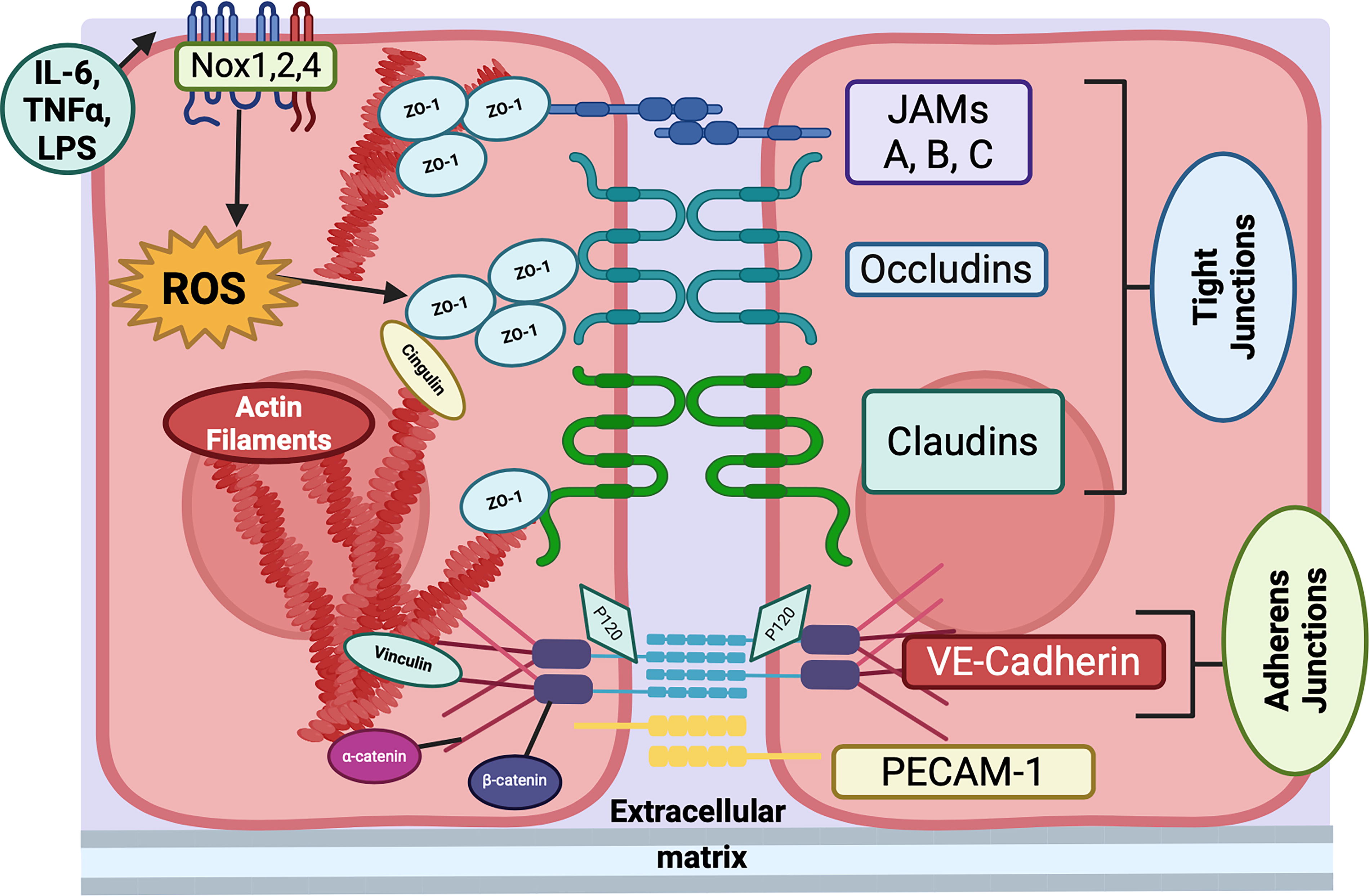
Increased endothelial permeability is a hallmark of a variety of vascular diseases, including acute lung injury, sepsis, ischemic stroke, hemorrhagic fevers, cancer, diabetic retinopathy, and atherosclerosis. Normally, the endothelium maintains close apposition between adjacent cells to create a tight barrier that allows only selective permeability to fluid and solutes. Disruption of the barrier by inflammation or pathogens, for example, leads to excessive flux of plasma and transmigration of inflammatory cells into the surrounding tissue. The occlusive junctions between endothelial cells are classified as adherens junctions, formed by interaction of VE-cadherins expressed on neighboring cells, and tight junctions composed of occludins, claudins, and/or junctional adhesion molecule A (JAM-A) (Fig. 4) (Duong and Vestweber, 2020).
In general, adherens junctions are important in the microvasculature of the heart and lungs (Corada et al., 1999), while tight junctions regulate the blood–brain barrier (BBB) (Nitta et al., 2003; Yang et al., 2007), although each type of junction can also be found in other endothelial beds. In both junctional types, a close relationship with the actin cytoskeleton is pivotal to dynamic regulation of permeability. VE-cadherin is linked to actin by β-catenin and plakoglobulin, while p120-catenin stabilizes VE-cadherin at the junction by preventing endocytosis (Fig. 4). Tyrosine phosphorylation of VE-cadherin disrupts the junctions. Tight junctions interact with the actin cytoskeleton through zonula occludens 1 (ZO-1) and cingulin proteins and are disrupted by matrix metalloproteinase-mediated degradation (Fig. 4) (Yang et al., 2007). Platelet/endothelial cell adhesion molecule-1 (PECAM-1) also bridges adjacent endothelial cells but does not regulate permeability; rather, it facilitates leukocyte diapedesis (Muller, 2016).
Work in animal models provides strong evidence for a role of ROS in diseases accompanied by edema, as summarized here for septic lung injury. EUK-8, a synthetic inhibitor of SOD and catalase, inhibits edema in a pig model of acute respiratory distress syndrome (ARDS) (Gonzalez et al., 1995). The mitochondrial-targeted redox modulator with antioxidant activity, MitoQ, protects mice against lipopolysaccharide (LPS)-induced lung injury by reducing oxidative stress and attenuating the degradation of the antioxidant transcription factor Nrf2, leading to inhibition of endothelial permeability (Cen et al., 2021). Moreover, pneumolysin, a toxin that is produced by Streptococcus and causes endothelial permeability, increases ROS production (Martner et al., 2008), an effect that is inhibited by the mitochondrial chaperone Hsp70, which in turn protects the endothelial barrier (Li et al., 2018b). Finally, induction of sepsis in rats via cecal ligation and puncture leads to pulmonary edema, which is mitigated by co-treatment with antibiotics and antioxidants, including N-acetyl-cysteine (NAC) (Galvão et al., 2014). Similar outcomes were observed in rats treated with oleic acid to induce acute lung injury, where NAC mitigated lung injury and improved survival, but in contrast had no effect on lung edema as measured by histology (Kumar et al., 2022). These studies showcase the pathological aspects and negative impact of ROS production. It should be noted, however, that ROS are also critical to the initial inflammatory response, and complete inhibition of ROS may paradoxically worsen edema by blocking anti-inflammatory pathways, such as induction of IL-10 (Deng et al., 2012). In a meta-analysis of patients with ARDS, NAC treatment led to a shorter stay in the Intensive Care Unit (ICU) but had no benefit to mortality; however, the number of studies and patients was small, and edema was not assessed, indicating a need for more robust and larger studies (Zhang et al., 2017). Because oxidative stress is closely associated with infection by pathogens such as SARS-CoV-2 (Zarkovic et al., 2022), additional trials with other, more targeted antioxidants are warranted.
Drilling down to a molecular level, ROS production is induced by various stimuli associated with barrier dysfunction, including growth factors (e.g., vascular endothelial growth factor [VEGF]), cytokines, and chemokines (e.g., tumor necrosis factor α [TNFα], IL-10, CCL8) that regulate Rho GTPases (e.g., Rac), mitochondria, and NADPH oxidases (Fig. 5). Subsequent increases in ROS regulate a diverse set of molecules, such as matrix metalloproteinases (MMPs), Ca2+ channels, kinases, phosphatases, and deacetylases (Fig. 5). Through these interactions, ROS exert direct effects on endothelial permeability and serve as signaling molecules to regulate the dynamic changes necessary for proper barrier function (Fig. 5). For example, simply transfecting human umbilical vein endothelial cells with a constitutively active Rac causes a ROS-dependent loss of VE-cadherin-mediated cell–cell adhesion (van Wetering et al., 2002).
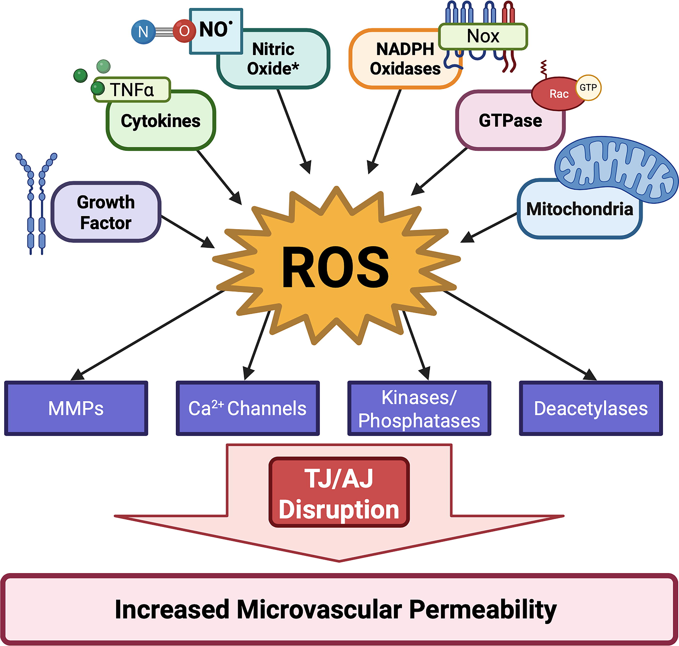
In pulmonary microvascular endothelial cells, H2O2 induces its pro-permeability effects via activation of TRPV4-mediated calcium influx, Fyn kinase, and protein kinase C (Siflinger-Birnboim et al., 1992; Suresh et al., 2015). Conversely, interfering with VE-cadherin junctions by incubation with an antibody causes activation of Rac and production of ROS, leading to redox-dependent activation of Pyk2, which is then recruited to adhesions. Inhibiting any one of these signals prevents the loss of junctional integrity, suggesting that these pathways are important regulators of adherens junctions (van Buul et al., 2005). Furthermore, knockdown of polymerase delta interacting protein-2 (Poldip2), which inhibits Nox4 activation and mitochondrial ROS production, prevents TNFα-induced disruption of VE-cadherin-containing junctions in pulmonary microvascular endothelial cells (Dolmatova et al., 2022; Forrester et al., 2018).
In other studies, treatment of lung microvascular endothelial cells with mitoTempo attenuated LPS-induced increases in ROS and permeability (Fu et al., 2021), while treatment of tail arteries from aging rats with manganese(III) tetrakis (1-methyl-4-pyridyl)porphyrin (MnTMPyP) or Tempol to break down O2 •− increased the intensity and width of VE-cadherin staining at endothelial junctions, suggesting these junctions were stabilized (Chang et al., 2018). It should be noted that MnTMPyP and Tempol not only reduce O2 •− levels but also increase H2O2; thus, this latter study is inconsistent with previous reports of H2O2 causing permeability. However, some SOD mimetics with dual (SOD/GPx or SOD/Catalase) antioxidant properties (e.g., MnTMPyP) can also decrease H2O2 levels, which may have been the case in this study. Finally, an indirect effect of sustained ROS generation may be the inactivation of NO•, which normally inhibits permeability (Kubes and Granger, 1992).
A specific target of ROS in junctional turnover is protein tyrosine phosphatase-1B (PTP1B), which associates with the adherens junctional proteins β-catenin, p120-catenin, and VE-cadherin and is inhibited by oxidation. In an LPS model of acute lung injury, oxidation of PTP1B inhibited its interactions with β-catenin and enhanced β-catenin tyrosine phosphorylation. Prevention of oxidation blocked these effects, leading to reduced permeability and lung edema (Grinnell et al., 2012). Similarly, in microvascular endothelial cells treated with VEGF, Rac1-mediated ROS production was increased, and treatment with NAC blocked the resulting tyrosine phosphorylation of VE-cadherin and β-catenin and the subsequent permeability increase, ultimately restoring adherens junction integrity (Monaghan-Benson and Burridge, 2009).
Another mechanism by which ROS regulate barrier function is through sirtuins, nicotinamide adenine dinucleotide (NAD+)-dependent deacetylases that regulate transcription factors, histones, and enzymes. The sirtuin family consists of seven members (SIRT1-7) that are critical for endothelial redox homeostasis and barrier function. In particular, SIRT1 reduces ROS via FOXO and NFκB transcriptional regulation, NADPH oxidases, and eNOS pathways, while its suppression amplifies ROS generation (reviewed in Man et al., 2019). SIRT1 also inhibits SIRT3 through AMPK-PGC1α, increasing mitochondrial ROS and decreasing BBB permeability (Chen et al., 2018). SIRT7 silencing reduces LPS-induced inflammation and endothelial-mesenchymal transition (EndoMT), decreasing VE-cadherin and increasing permeability (Wyman et al., 2020). Similarly, SIRT5 increases BBB permeability by promoting occludin degradation in microvascular cells exposed to hypoxia/reoxygenation. Consequently, SIRT5 knockdown enhances tight junction integrity via the PI3K/Akt pathway (Diaz-Cañestro et al., 2018). Thus, sirtuins represent an interesting new potential therapeutic target for increased endothelial permeability.
In the brain, tight junctions are mainly responsible for the integrity of the BBB, with adherens junctions supporting them. Treatment of human brain microvascular endothelial cells with TNFα or IL-6 increases permeability in concert with increased ROS and decreased expression of VE-cadherin, occludin, and claudin-5. These effects can be reversed by SOD, catalase, or NAC, as well as siRNA against Nox2 (Rochfort et al., 2014). In cultured bovine brain microvascular endothelial cells, H2O2 itself increases permeability, which is correlated with changes such as small alterations in the expression of occludin, actin, and ZO-1, rearrangement of occludin and ZO-1 at tight junctions, actin stress fiber formation (Lee et al., 2004), and activation of focal adhesion kinase (Usatyuk and Natarajan, 2005).
The increased permeability in response to treatment of human brain microvascular endothelial cells with either TNFα or IL-6 is dependent upon redox-mediated decreases in ZO-1 expression and movement away from the junctions (Rochfort and Cummins, 2015). Increased permeability of the BBB is also seen after ischemia/reperfusion injury. Rat brain microvascular endothelial cells subjected to oxygen-glucose deprivation exhibit disruption of ZO-1-containing junctions and increased permeability that can be inhibited by treatment with the antioxidant l-ascorbic acid (Alluri et al., 2014). Similarly, exposure of brain endothelial cells to moderate hypoxia/reoxygenation leads to disruption of ZO-1 and claudin-5, increased permeability, and increased production of cytosolic ROS, all of which are inhibited by the flavin oxidase inhibitor, diphenylene iodonium (DPI) (Zehendner et al., 2013). These studies underline the importance of ROS signaling in dynamic regulation of the BBB.
Numerous papers have implicated NADPH oxidases in endothelial barrier function, usually in the setting of response to infection. An early study showed that treatment of pulmonary microvascular endothelial monolayers with TNFα induces permeability, and this effect is blocked by antisense oligonucleotides to p22phox (Gertzberg et al., 2004). In Pseudomonas aeruginosa-infected mice, Nox2 and Nox4 expression in the lung is increased, and ROS are elevated in bronchoalveolar lavage fluid, but only knockdown of Nox4 attenuates increased endothelial permeability and apoptosis. Nox2 is more related to inflammatory endpoints (Fu et al., 2013). Similar results were found in a cecal-ligation puncture model of sepsis (Jiang et al., 2020). In cultured human pulmonary artery endothelial cells, pharmacological inhibition of Nox4 reduces LPS induction of endothelial permeability (Palumbo et al., 2017); however, in human lung microvascular endothelial cells exposed to LPS, permeability is dependent on ROS derived from eNOS that has become uncoupled due to Nox2-dependent S-glutathionylation (Wu et al., 2014). Similarly, in endothelial cells exposed to CCL8, a pro-inflammatory agent, knockdown of Nox2 prevented the increase in permeability (Xue et al., 2021). Finally, in human umbilical vein endothelial cells, amyloid-β1 impairs endothelial barrier function by activating Nox1 (Tarafdar et al., 2022). Thus, while it is clear that increased ROS from NADPH oxidases increase endothelial permeability, the identity of the oxidase appears to be stimulus- and cell-type dependent.
Redox regulation of endothelial glycocalyx maintenance
The endothelium is coated with glycoproteins, glycolipids, and proteoglycans that extend their carbohydrate side chains, creating a polysaccharide-rich matrix. This carbohydrate mesh is mainly comprised of glycosaminoglycans (GAGs) (e.g., heparan sulfate, chondroitin sulfate, dermatan sulfate, hyaluronan, and keratin sulfates) that attach to core proteins to form proteoglycans (e.g., syndecans and glypicans). Notably, heparan sulfate GAG proteoglycans account for 50%–90% of the proteoglycans expressed in the endothelium. GAGs are essential for the structure and function of the glycocalyx, playing a key role in regulating endothelial function, vascular permeability, and cell signaling.
GAG sulfation and/or acetylation allows glycocalyx structural rearrangements, and their negative charge facilitates electrostatic interactions with growth factors, ECM proteins, adhesion molecules, inflammatory factors, and other soluble molecules present in the blood. The glycocalyx also plays an important role in regulating barrier function and leukocyte trafficking, as well as anticoagulation and vascular tone (Fig. 6) (Sieve et al., 2018).

The maintenance of the glycocalyx relies on a tightly regulated balance between GAG biosynthesis and turnover. GAG biosynthesis begins in the cytoplasm with the synthesis of five uridine diphosphate-activated sugars, which are transported to the Golgi apparatus for sulfation. There, glycosyltransferases catalyze the covalent linkage of sulfated GAGs to proteoglycans. A noteworthy exception is hyaluronan, which does not undergo sulfation modifications and is transported directly from the cytoplasm to the membrane (Ghiselli, 2017). Conversely, glycocalyx remodeling is mediated by enzymatic degradation, primarily through heparanase, hyaluronidases, and matrix metalloproteinases, which regulate GAG shedding in response to physiological and pathological stimuli. This turnover is crucial for adapting to shear stress, modulating endothelial permeability, and responding to inflammatory signals (reviewed in Ricard-Blum et al., 2024). However, excessive degradation or shedding, as seen in conditions such as diabetes and sepsis, leads to glycocalyx dysfunction and vascular dysfunction. Restoring GAG homeostasis through upregulation of biosynthetic pathways and inhibition of enzymatic breakdown represents a potential therapeutic strategy for preserving endothelial function in disease states.
The glycocalyx plays a role in maintaining redox homeostasis in the endothelium. Heparin and heparan sulfate, the latter of which is part of the glycocalyx, bind extracellular SOD3 (Karlsson et al., 1988), suggesting that the glycocalyx has the capacity to regulate the redox state of the endothelium (Fig. 6). In fact, SOD3 protects heparin/heparan sulfate from oxidative fragmentation in the setting of high ROS or lung inflammation (Kliment et al., 2008). Alternatively, the glycocalyx has been shown to protect the microvascular endothelium from ischemia-reperfusion injury mediated by XO, likely in part due to the antioxidant properties of hyaluronan (Rubio-Gayosso et al., 2006) in addition to SOD3, and enhances shear stress-induced generation of NO• in a hyaluronic acid-dependent manner (Fig. 6) (Kumagai et al., 2009).
In arterial endothelial cells, both sialic acid and heparan sulfate protect against shear-induced O2 •− production (Kumagai et al., 2009), such that overall, the glycocalyx supports vasodilation. This may be in part due to the fact that unidirectional shear stress-mediated induction of Nrf2-dependent antioxidant gene expression is dependent upon sialic acids in the glycocalyx (Psefteli et al., 2021). Conversely, ROS and RNS can degrade glycocalyx components (Sieve et al., 2018), leading to increased adhesion of inflammatory cells, coagulation, and constriction (Fig. 6) (van Golen et al., 2012). Treatment of glomerular endothelial cells with exogenous H2O2 leads to loss of wheat germ agglutinine (WGA) lectin and causes shedding of heparan sulfate on the cell surface, which contributes to increased permeability (Singh et al., 2013).
Together, these studies attest to the fact that redox regulation of and by the glycocalyx is central to many fundamental roles of the endothelium, but as most of these studies were performed in cells from large arteries, more work needs to be done specifically in the microvasculature.
Redox regulation of endothelial-leukocyte trafficking
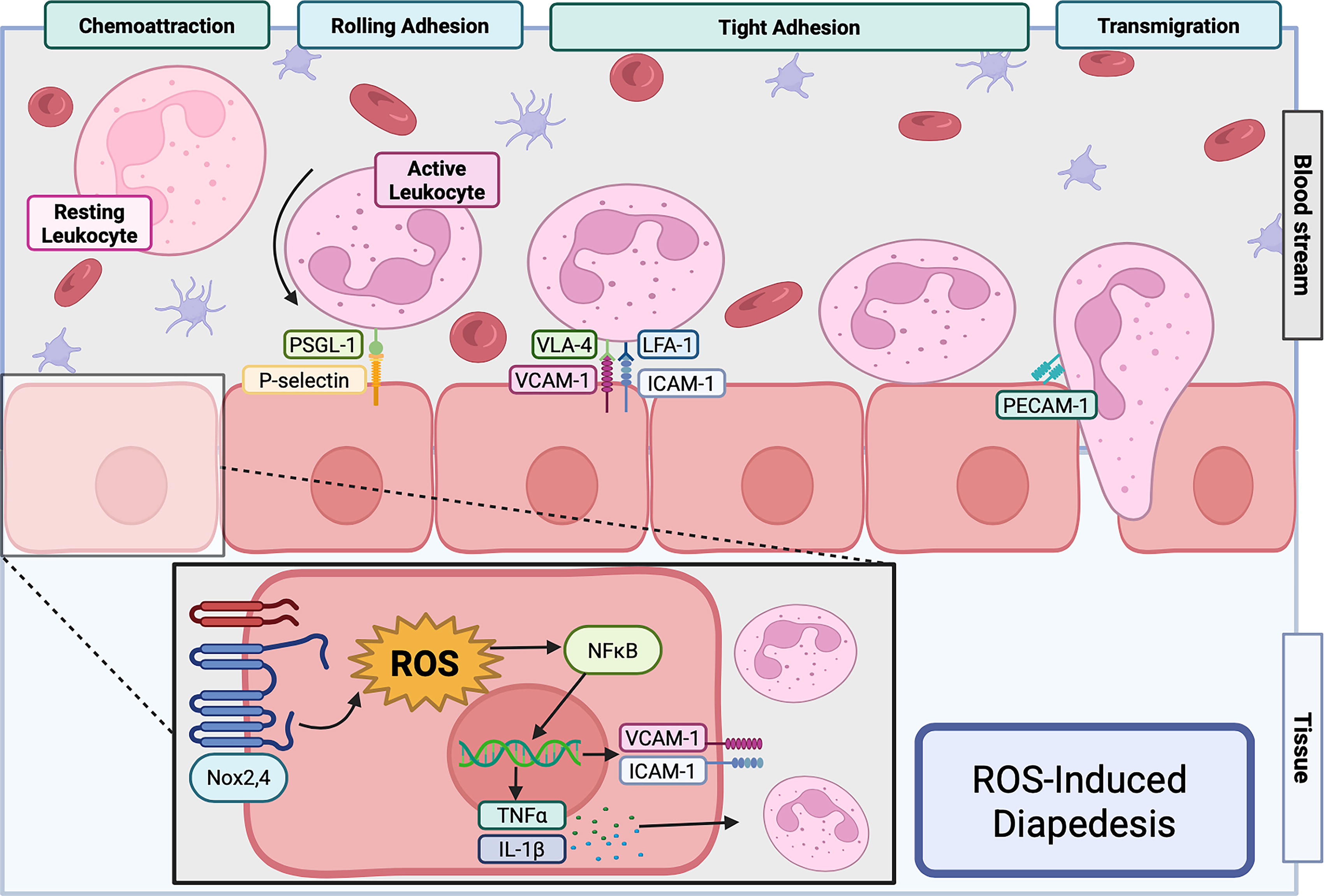
Increased endothelial permeability and disruption of the glycocalyx allow the efflux of leukocytes into surrounding tissue (diapedesis), but extravasation is a carefully coordinated event. Diapedesis occurs mainly in postcapillary venules and comprises four steps: chemoattraction, rolling adhesion, tight adhesion, and transmigration (Filippi, 2016). The most common migrating cells are neutrophils, T lymphocytes, monocytes, and natural killer cells. Upon tissue injury or infection, tissue-resident macrophages release cytokines (e.g., TNFα, IL-1, IL-6) and chemokines (e.g., CCL2, CCL5, MCP-1, IL-8), which stimulate endothelial cells to express cell adhesion molecules, such as vascular cell adhesion molecule-1 (VCAM-1), intercellular adhesion molecule-1 (ICAM-1), P-selectin, and E-selectin. Low-affinity binding of ligands on circulating leukocytes to selectins causes them to slow down and start to roll. Leukocytes are activated by chemokines or factors released from endothelial cells to switch their integrins to an activated state, inducing tight adhesion to the endothelium via counter-ligands. Leukocytes then extend pseudopodia into the gaps between adjacent endothelial cells created by exposure to cytokines and assisted by PECAM-1 molecules, and begin the process of diapedesis (Fig. 7).
Leukocyte trafficking across the microvascular endothelium is a precisely regulated process, with ROS playing a key role in modulating endothelial permeability as well as leukocyte chemoattraction, adhesion, and trafficking. For example, ischemia-reperfusion and the resulting ROS release can directly increase endothelial permeability, leading to neutrophil extravasation and hemorrhage (Granger and Kvietys, 2017). Similarly, in vitro assays using HUVEC cells have shown that ROS regulate cell junction integrity by activating proline-rich tyrosine kinase (Pyk2), which induces VE-cadherin phosphorylation, causing local junction disassembly and increased endothelial permeability (van Buul et al., 2005).
Activated neutrophils also release ROS, enhancing microvascular permeability (Zhu and He, 2006). In phagocytic cells, Nox2 is the primary source of ROS, generating O•− that is subsequently converted into H2O2 (Paclet et al., 2022). H2O2 modulates signaling pathways involved in cytokine production, although the precise mechanisms have not been fully identified. Notably, NFκB, a key transcription factor for inflammatory mediators, has been shown to be subject to redox regulation (Fig. 7) (reviewed in Bode et al., 2023 and Kabe et al., 2005).
Of interest, the inflammatory profile induced by ROS seems to be elevated in endothelium from veins as compared with arterial endothelium. Comparing arterial and venous endothelial responses to an oxidative insult in culture, Shrestha et al. (2018) found that venous cells respond with a much larger decrease in GSH/GSSG (glutathione disulfide) than do arterial cells, supporting an elevated response to oxidants and greater macrophage adhesion. ROS can also modulate neutrophil chemotaxis, as NADPH inhibition with DPI or knockdown of p22phox led to defective directional migration toward N-formyl-methionyl-leucyl-phenylalanine (fMLP), leukotriene B4 (LTB4), and IL-8-mediated chemotaxis (Hattori et al., 2010).
ROS also regulate leukocyte adhesion and rolling (Fig. 7). Indeed, direct infusion of H2O2 into the mesenteric microcirculation leads to an increase in rolling and adherent leukocytes (Scalia and Lefer, 1998). ROS affect these processes by stimulating NFκB-mediated expression of adhesion molecules such as LFA-1, VLA-4, ICAM-1, P-selectin, E-selectin, or VCAM-1 on the cell surface, which in turn dictates which cells adhere and transmigrate (Ichikawa et al., 1997; Kokura et al., 2000). Just as Fraticelli et al. (1996) demonstrated, PMNs treated with H2O2, iron chelate Fe3+ nitrilotriacetate (1:2) to generate OH−, or XO and its substrate xanthine to generate O2 •− exhibited increased expression of Mac-1 (CD11b [integrin αM] and CD18 [integrin β2]), along with L-selectin shedding, an effect that was inhibited by catalase.
Conversely, blocking ROS impairs adhesion of leukocytes to the endothelium. For example, leukocyte adhesion and transmigration induced by ischemia-reperfusion are inhibited by ROS scavengers or by overexpression of antioxidant enzymes in transgenic mice (reviewed in Yu et al., 2019). Similarly, ischemia-reperfusion-induced expression of E-selectin and subsequent adhesion of neutrophils to the intestinal microvasculature is attenuated in mice that overexpress SOD1 (Russell et al., 2000). Concordant results were found in mice fed a high-cholesterol diet, where leukocyte-endothelial cell adhesion in cremasteric postcapillary venules was blocked by transgenic overexpression of SOD1 and attenuated by knockout of the Nox subunit p47phox (Stokes et al., 2001). Because NO• is typically anti-adhesive (Binion et al., 1998), inactivation of NO• by increased ROS may also contribute to enhanced leukocyte extravasation (Kurose et al., 1994).
Redox regulation in inflammation
Inflammation is a hallmark of many diseases, regardless of their etiology. Multiple cell types in the microcirculation, including endothelial cells (O’Carroll et al., 2015), pericytes (Kaushik et al., 2021), and leukocytes (Riaz and Sohn, 2023), release cytokines and chemokines, which in turn stimulate ROS production. ROS activate NFκB, which regulates expression of additional inflammatory cytokines, chemokines, matrix metalloproteinases, and adhesion molecules. Moreover, neutrophil adhesion to TNFα-stimulated endothelial cells induces ROS production in the endothelial cells (Wang and Doerschuk, 2000), further amplifying the response. Thus, ROS are both a cause and a consequence of inflammation.
Inflammation can also be initiated by simple ischemia-reperfusion. For example, in colonic venules exposed to ischemia-reperfusion, ROS induce CCL3 (also known as MIP-1α) and CXCL-1 (also known as GRO-α or MGSA), which mediate leukocyte rolling and adhesion (Riaz et al., 2003). In another study, hypoxia-reoxygenation increased ICAM-1 expression in rat coronary microvascular endothelial cells via redox-sensitive NFκB activation, which was inhibited by NO• (Kupatt et al., 1997). In general, in the setting of ischemia-reperfusion, ROS induce complement activation, cytokine expression and release, neutrophil and platelet adhesion, and thrombosis, at times occluding flow (termed “no re-flow”) and eventually causing tissue death (Widgerow, 2014). Dead or dying cells release damage-associated molecular pattern molecules (DAMPs), which trigger inflammatory responses in cells. Intriguingly, some DAMPs highly expressed in neutrophils, namely S100A8 and S100A9, which are chemotactic and promote adhesion to the extracellular matrix, can be oxidized, serving to protect against oxidative stress (Lim et al., 2011). Such modifications may then naturally limit the inflammatory response.
One potential difference in inflammatory signaling in endothelial cells from large arteries versus those from the microcirculation lies in the response to extracellularly administered H2O2. LPS stimulation of human dermal microvascular endothelial cells leads to ROS-dependent activation of NFκB, but treatment of these cells with H2O2 does not (Chan and Murphy, 2003). A separate study suggested that H2O2 can in fact activate NFκB in these cells but is not sufficient to induce ICAM-1 expression (True et al., 2000). This is in stark contrast to the effect of H2O2 on endothelial cells from large arteries, where ROS induction of inflammatory cytokines and adhesion molecules is well documented (Lin et al., 2004; Lo et al., 1993). Of note, some studies in the microvasculature do report an effect of H2O2 on inflammatory gene expression (Jiang et al., 2017; Willam et al., 1999), suggesting that not only is the vascular bed important in determining the outcome but also that the concentration of H2O2 is critical.
Infectious agents and obesity are two other triggering factors of inflammation. Several studies in animals and humans suggest that the antioxidant NAC can blunt the inflammatory response to viruses (Fraternale et al., 2021; Ungheri et al., 2000), and more recently, can potentially reduce the cytokine storm that occurs in response to SARS-CoV2 (Ibrahim et al., 2020). In vivo, infection with P. aeruginosa not only leads to lung inflammation but also induces neuroinflammation as a consequence of increased cytokine expression (Villalba et al., 2023). However, a potential role of ROS in this response remains to be determined. In animal models and cultured cells, infection is often mimicked by administration of the bacterial agent LPS or its downstream effector TNFα. Such models have been used to probe the redox sensitivity of inflammatory responses. Expression of ICAM-1, VCAM-1, and E-selectin in human dermal microvascular endothelial cells or human pulmonary microvascular endothelial cells is induced by oxidants and TNFα, and attenuated by antioxidants or NO• donors, in part by inhibiting NFκB (Jiang et al., 2005; Jiang et al., 2004; True et al., 2000). In one study, the increase in ROS induced by TNFα was shown to be a result of Nox2 activation (Li et al., 2002). Administration of LPS also increases ICAM-1 expression in rat pulmonary microvascular endothelial cells in a ROS-dependent manner (Zhang et al., 2021), while co-administration of MitoQ and LPS in mice reduces ICAM-1 and VCAM-1 induction in the lung (Cen et al., 2021).
A link between obesity and systemic inflammation has now been well established. In particular, perivascular adipose tissue secretes TNFα and IL-6, which in turn release ROS (Virdis et al., 2019). Elevated TNFα is considered to be the driving factor behind endothelial dysfunction in obesity, with ROS-mediated inactivation of NO• serving as a major impediment to vasorelaxation (Virdis et al., 2019). In addition, these cytokines and others create a low level of inflammation, causing circulating mononuclear cells and inflammatory cells to adopt a pro-inflammatory phenotype (Ghanim et al., 2004). In a human study of obese, postmenopausal women subjected to 8 weeks of dietary supplementation with fruit and vegetable juice powder, the reduction in blood oxidants and inflammatory markers was associated with improved capillary blood flow (Lamprecht et al., 2013). The prevailing literature on obesity thus supports a link between inflammation, ROS, and impaired vasodilation, but careful analysis of redox responses in the microvasculature is currently lacking.
Inflammation is inextricably linked to endothelial permeability, the glycocalyx, and diapedesis of inflammatory cells. Thus, it is not surprising that ROS play such an integral role in inflammatory responses. Strikingly, however, inflammation also affects other aspects of endothelial function, including vasomotion as suggested above and outlined in the next section.
Redox regulation of vascular tone
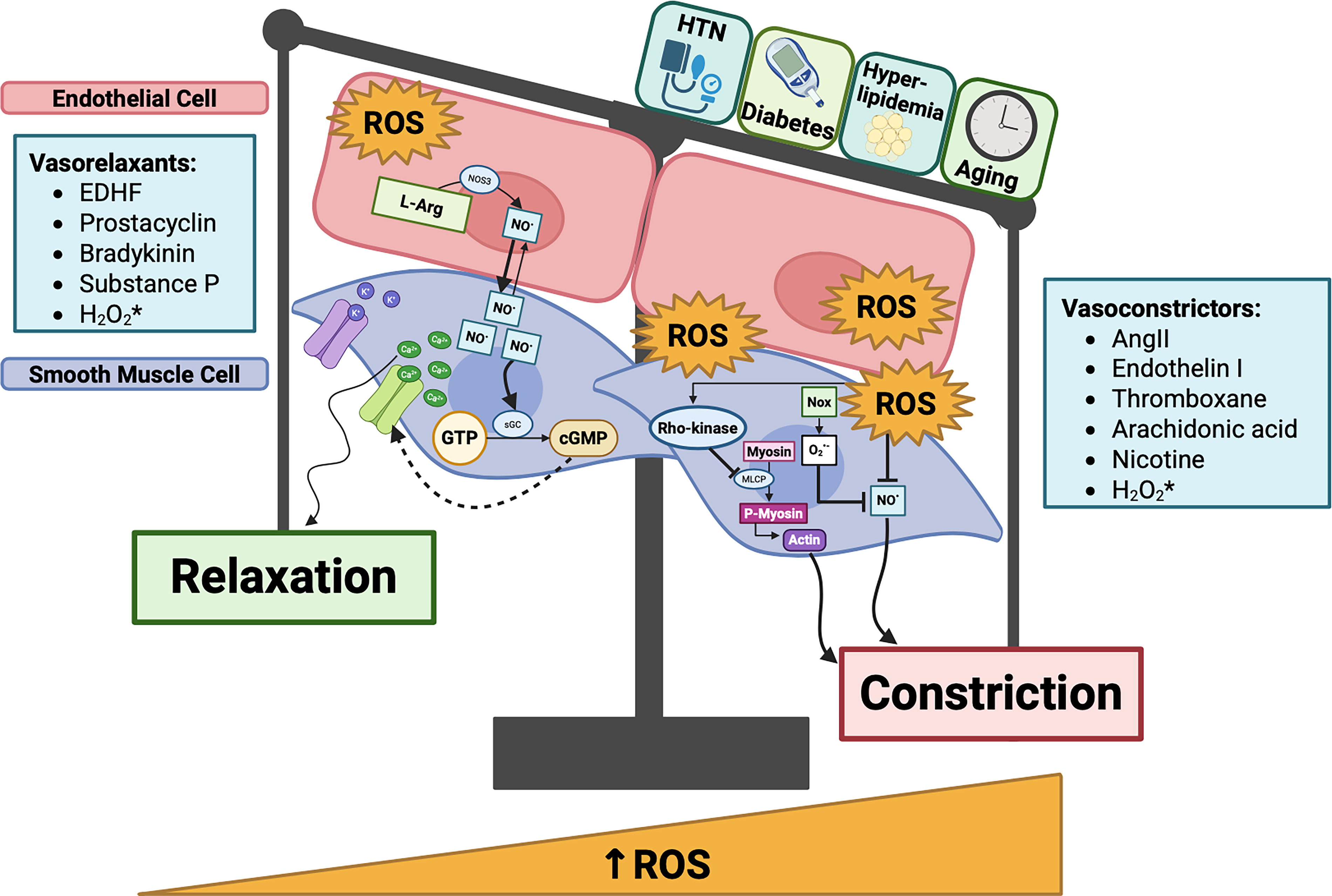
The microcirculation plays a critical role in auto-regulating organ blood flow based on oxygen and metabolic demands. For example, approximately 80% of myocardial flow is determined by resistance vessels of the coronary microcirculation (Camici and Crea, 2007). Structural alterations related to arteriolar remodeling or extravascular compressive forces, as well as functional pathobiological alterations, impact the ability of the microvasculature to autoregulate and lead to impaired flow reserve and demand-perfusion mismatch. Under normal physiological conditions, low levels of ROS modulate normal vasomotion. However, in the setting of cardiovascular risk factors that trigger excessive production of ROS, bioavailable NO• is depleted, leading to impaired microvascular vasodilation and/or enhanced vasoconstriction (Fig. 8). ROS overabundance has been shown to inhibit the function of eNOS in the vasculature, compounding NO• depletion (Forstermann et al., 2017). Disrupted vascular antioxidant defense systems (e.g., SOD, catalase, GPx) also contribute to ROS imbalance and thereby can lead to endothelial dysfunction (Fukai and Ushio-Fukai, 2011).
Excessive RNS also have deleterious effects in the vasculature, and NO dioxygenases (NOD) play a critical role in regulating RNS levels by catalyzing the conversion of NO• to nitrite (NO2 −) or other metabolites. In mammals, heme-containing NOD-like proteins help to regulate RNS levels in tissues. Liu et al. (2017) found that the NOD protein, cytoglobin (Cygb), is highly expressed in vascular smooth muscle cells (VSMCs) and regulates 70%–75% of NO metabolism in the presence of cellular reducing systems (cytochrome b5 reductase/cytochrome b5/NADH). Interestingly, in hypoxic conditions, the NOD function of Cygb declines, indicating that this mechanism of NO• degradation is highly oxygen dependent. In Cygb knockout animals, increased endothelium-dependent and endothelium-independent vasodilation was observed, along with lower systemic vascular resistance and blood pressure due to prolonged NO• decay (Liu et al. 2017).
Abnormal endothelial function is a precursor in the pathogenesis of many conditions, such as hypertension, chronic kidney disease (CKD), and atherosclerotic cardiovascular disease. In addition to vasodilators such as NO• and prostacyclin, the function of vasoconstrictors such as Angiotensin II is well known to be redox-sensitive (Garrido and Griendling, 2009). More recently, endothelin-1 (ET-1)-mediated vasoconstriction in the vasculature has also been found to be enhanced by ROS (Tsai et al., 2017). In the coronary arterioles, ROS-propagated Rho kinase (ROCK) activation can inhibit myosin light chain phosphatase, sustaining vasoconstriction, and ET-1-enhanced microvascular constriction is associated with elevated oxidative stress and overexpression of ROCK (Fig. 8) (Tsai et al., 2017).
Increased ROS production adversely impacts both endothelium-dependent and endothelium-independent vascular function (Amanso and Griendling, 2012). Lavi et al. (2008) showed that in humans with coronary endothelial dysfunction (detected by abnormal acetylcholine response), basal NO• production did not decrease, but local oxidative stress was enhanced, as measured by products of lipid peroxidation, F2-isoprostanes. There is heterogeneity in ROS-mediated responses depending on the type of ROS species, the concentration of ROS, the vascular bed interrogated, and also changes in local conditions (Lee and Griendling, 2008; Lucchesi et al., 2005). For example, NADPH oxidase-derived ROS such as H2O2 modulate protein kinase C and mitogen-activated protein kinase (MAPK) signaling, which controls vascular smooth muscle contraction and relaxation (Oeckler et al., 2003). Potassium channels also play an important role in vasodilation. In addition to inactivating NO•, O2 •− has been shown to inhibit the opening of various potassium channels (voltage-sensitive [Kv], Ca2+-sensitive [Kca], and ATP-sensitive K channels) on VSMCs, which impacts hyperpolarization and enhances contractility (Gutterman et al., 2016; Gutterman et al., 2005). H2O2 can directly induce vasodilation by activating Kv channels through reversible oxidation of thiol groups. This mechanism may play a significant role in microvascular dysfunction observed in coronary artery disease (CAD), Takotsubo cardiomyopathy, and other microvascular disorders (Appiah et al., 2012; Dong et al., 2023; Nishijima et al., 2017).
After ischemia-reperfusion injury, excessive ROS-mediated detrimental effects occur in the arterioles (leading to impaired flow reserve), in the capillaries (with no-reflow as well as leaky capillaries), and in the venules (impacting resorption and triggering platelet plugging) (Yu et al., 2019). Disrupted redox balance also impacts pathways involved in sympathetic regulation of vascular tone, but further work is needed to understand the role of ROS in triggering autonomic imbalance and abnormal microvascular reactivity (Zhao et al., 2006).
While there are several redox-sensitive mechanisms involved in maintaining vascular tone (Fig. 8), our understanding of the differential mechanisms involved in ROS-mediated vascular control of larger vessels versus microvessels is limited. For example, while shear stress is an important mechanism that influences epicardial vascular tone, microvascular tone is regulated not only by intravascular pressure but also by products of metabolism such as carbon dioxide, lactate, hydrogen ions, etc., which adds to the complexity. Regarding this latter mechanism, Saitoh et al. (2007) showed that coronary vasodilation that occurs with increased myocardial oxygen demand is redox sensitive and also partially dependent on thiol oxidation. H2O2 released from metabolically active cardiomyocytes isolated from Wistar rats caused VSMC relaxation and arteriolar dilation. This vasodilatory response to H2O2 and to cardiac metabolic products was attenuated by thiol reduction. Further, while NADPH oxidases are the main sources of ROS in the larger vessels, there are some data that mitochondrial ROS may trigger pressure-induced vasoconstriction in the microvasculature, a response that is inhibited by ROS scavenging in rat cerebral arteries (Gebremedhin et al., 2013).
While NO• is a well-established vasodilator, in the microcirculation, endothelium-derived hyperpolarizing factor and H2O2 may play a key regulatory role in vasorelaxation (Chabowski and Gutterman, 2015), especially in the coronary microvasculature, but further work is needed to understand the relationships of shared and differential ROS-dependent mechanisms controlling macro- versus microvascular tone.
Redox regulation of hemostasis
Hemostasis is the physiological response to injury that stops bleeding at the site of damage. During primary hemostasis, platelets adhere to the exposed collagen fibers of the damaged blood vessel wall and become activated, releasing various chemical signals and recruiting more platelets to the injury site. This process forms a temporary platelet plug that helps to seal the damaged blood vessel and prevent further bleeding. The coagulation cascade is activated during secondary hemostasis. This cascade is a series of enzymatic reactions culminating with soluble fibrinogen conversion into insoluble fibrin strands (Fig. 9). Fibrin strands interweave with the platelet plug to form a stable blood clot, effectively sealing the wound and preventing excessive blood loss. Coagulation can be initiated in response to tissue damage (extrinsic pathways) or by internal damage of the endothelium (intrinsic pathway). Both pathways converge on the common pathway to form a blood clot (Fig. 9).
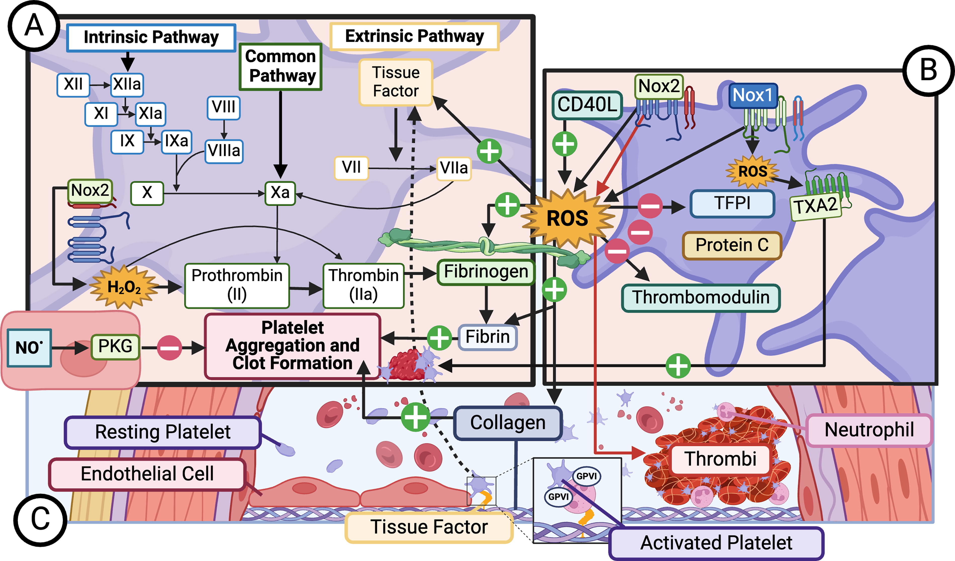
Thrombosis is pathological clot formation inside the blood vessels in the absence of vascular injury. These clots can obstruct blood flow, leading to heart attack, stroke, pulmonary embolism, deep vein thrombosis, and in severe cases, death. Thrombi are initiated in an injured vessel when platelets roll and adhere to the collagen-containing subendothelial matrix through glycoprotein receptors such as glycoprotein VI (GPVI) (Fig. 9). GPVI can also regulate the formation of platelet–leukocyte complexes. Venous thrombi are composed of platelets, leukocytes, and fibrin surrounding an erythrocyte mass, while arterial thrombi tend to be platelet-rich (Gutmann et al., 2020). Platelet activation and aggregation are increased by ROS, such as those released upon hypoxia/reoxygenation, and decreased in the presence of antioxidants (Dong et al., 2010; Handin et al., 1977; Jang et al., 2015; Leo et al., 1997). Using a microfluidic device coated with collagen followed by in vivo studies in mice, Bresette et al. (2024) showed that NAC reduced arterial thrombi formation. High-dose NAC prevented formation of an occlusive clot, while low-dose destabilized clots.
Platelet-activating agonists, such as soluble CD40L (Chakrabarti et al., 2005), also increase ROS production, although the source of ROS is not clear (Fig. 9). A fairly definitive study revealed no role for platelet Nox2 in thrombin-induced platelet activation/aggregation or arterial thrombosis in large vessels (Sonkar et al., 2019), a conclusion supported by a study in human platelets from patients with a gp91phox hereditary deficiency, which showed normal aggregation but impaired recruitment of platelets (Pignatelli et al., 2011). However, Vara et al. (2019) showed that in isolated platelets from Nox1−/y and Nox2−/− knockout mice, intracellular ROS, specifically O2 •− for collagen and H2O2 for thrombin, are required for platelet activation and aggregation, but the sources differ: Nox1 mediates collagen-induced responses, while Nox2 is required for activation by thrombin. Conversely, an earlier study by Delaney et al. (2016) using platelets from these same Nox1−/y and Nox2−/− mice, concluded that Nox1 mediates thrombin- and thromboxane A2-, but not collagen-related peptide-, induced platelet aggregation, while Nox2 was responsible for collagen-stimulated platelet aggregation. Only platelets from Nox2−/− mice showed impaired arterial thrombosis (Fig. 9). These authors concluded that Nox2 is important for thrombosis in vivo, but not hemostasis. An important difference between these studies was the sex of the animals used to isolate platelets, a variable that deserves additional attention.
Another investigation targeted the Rac-p67phox interaction to show that a small-molecule inhibitor of this complex attenuated collagen-related peptide ROS production and platelet activation, concluding that Nox2 was responsible (Akbar et al., 2018). However, since p67phox and Rac are also capable of activating Nox1, these data are not consistent with those of either Vara et al. (2019) or Delaney et al. (2016). Finally, Del Principe et al. (1991) showed that collagen-induced platelet aggregation is dependent on H2O2, but thrombin stimulation is not. Given these conflicting reports from high-quality studies, further work is needed to reconcile these paradoxical findings.
Under normal conditions, NO• released by endothelial cells inhibits platelet activation through protein kinase G (PKG) activation (Fig. 9) (Gkaliagkousi et al., 2007; Ignarro, 1990), and loss of NO•, for example by interaction with O2 •− to form OONO−, leads to platelet activation (Meng et al., 1995). Activated platelets release procoagulant factors, causing thrombin generation from endothelial cells and fibrin formation. With respect to coagulation, ROS can increase tissue factor (TF) expression in vascular cells both directly and in response to thrombin (Golino et al., 1996; Herkert et al., 2002). Both NADPH oxidases and mitochondrial ROS have been implicated in this step (Banfi et al., 2009; Jacobi et al., 2005). TF pathway inhibitor (TFPI) and other anticoagulant proteins such as Protein C and thrombomodulin are inhibited by ROS, thus enhancing coagulation (Cimmino et al., 2015; Glaser et al., 1992; Nalian and Iakhiaev, 2008; Ohkura et al., 2004). ROS also oxidize fibrinogen, augmenting its conversion to fibrin (Fig. 9) (Upchurch et al., 1998). However, while oxidation of Factor VIIa by H2O2 does not affect its activity, it does reduce its affinity for TF and slightly reduces its ability to activate Factor X (Kornfelt et al., 1999), which would tend to be antithrombotic. Moreover, activated phagocytes produce HOCl, which acts as an anticoagulant through the oxidizing action of singlet oxygen on factors I, V, VIII, and X, resulting in a local zone of anticoagulation (Stief et al., 2000). Thus, while the overall effect of oxidants tends to be procoagulant, the balance between oxidant and antioxidant signaling is critical.
ROS have been implicated in diseases associated with coagulation. In sepsis, overactivation of the coagulation system can result in disseminated intravascular coagulation (DIC) or the formation of microthrombi that then occlude small vessels. DIC results not only from activation of the clotting cascade but also impaired fibrinolysis. An early study showed that Tempol inhibits the formation of thrombin–antithrombin complexes (a marker of coagulation) and attenuates expression of PAI-1 (inhibitor of fibrinolysis) induced by P. aeruginosa infusion in pigs, potentially contributing to protection from circulatory failure (Matejovic et al., 2005). This protective effect of Tempol was amplified by co-administration of l-N6-[1-iminoethyl]-lysine, an inducible NOS inhibitor (Matejovic et al., 2007). IL-1-mediated induction of PAI-1 in cardiac microvascular endothelial cells (CMECs) was also shown to be dependent on ROS (Okada et al., 1998). The cytokine storm that accompanies sepsis can activate coagulation by increasing TF expression (Kinasewitz et al., 2004; Levi and van der Poll, 2017), which is dependent upon the redox-sensitive transcription factor NFκB (Mussbacher et al., 2019). In mice, exosomes carrying SOD2 derived from neutrophils reduce endothelial-derived ROS, thus inhibiting DIC in LPS-induced sepsis (Bao et al., 2022). Moreover, a correlation between elevated oxidation markers and impairment of fibrinolysis has been reported in diabetes (Lados-Krupa et al., 2015).
As with most redox-sensitive processes within cells, the overall effect of oxidative stress on hemostasis and coagulation depends upon the balance between the environmental susceptibility to pro- and anticoagulant factors as well as susceptibility to ROS, which in turn depends on the location and source of ROS and RNS, as well as the identity of the ROS and RNS produced.
Redox regulation in angiogenesis
Angiogenesis is defined as the growth of new blood vessels from the existing vasculature (Adair and Montani, 2010) and occurs in response to metabolic changes in tissue, especially hypoxia. Physiologically, it is critical to embryonic development, skeletal remodeling, regeneration of the endometrium during the menstrual cycle, and wound healing. In the adult, sprouting angiogenesis begins with secretion of MMPs by endothelial cells and is followed by protrusion of specialized tip endothelial cells toward the stimulus and proliferation of stalk endothelial cells to elongate the new vessel. When tip cells meet, they anastomose to form a new perfused vessel. The vessel then stabilizes and matures with the return of endothelial cells to quiescence and the formation of mature junctions (phalanx cells) (Eelen et al., 2020). The bulk of our understanding of angiogenesis comes from studies in cultured endothelial cells from large arteries, although the microvasculature is where angiogenesis actually occurs, opening opportunities for further research.
ROS can directly influence many of these processes; indeed, H2O2 can promote angiogenesis by activating the transcription factor ETS-1 (Yasuda et al., 1999). In tip cells, hypoxia induces mitochondrial ROS-mediated activation of mammalian sterile 20-like kinase 1 (MST1), which induces the nuclear import of FOXO1 and leads to transcription of polarity- and migration-associated genes (Fig. 10) (Kim et al., 2019). Cooperation between Notch signaling and VEGF drives the differentiation of tip and stalk cells (Naito et al., 2020). VEGF is not only induced by H2O2 (González-Pacheco et al., 2006) but also signals through H2O2 to disrupt endothelial junctions and stimulate cell migration, proliferation, and capillary tube formation. VEGF, acting through the VEGF receptor-2 (VEGFR2), activates Nox2, Nox4, and mitochondria to produce H2O2, which in turn stimulates a cascade of intracellular signals. Although the mechanism of this activation remains unknown in endothelial cells, in other cells phosphoinositide 3 kinase (PI3K) or Ras activation downstream of VEGFR2 can activate Rac to drive Nox1- and Nox2-induced ROS. VEGF triggers tyrosine phosphorylation of VEGFR2 and phosphorylation of MAPKs, PI3K, protein kinase B (Akt), and JAK-STAT. PTP1B, SH2-containing protein tyrosine phosphatase 2 (SHP2), and phosphatase and tensin homolog (PTEN) are inhibited by ROS, and together these signaling pathways induce gene transcription dependent upon HIF1α, NFκB, and AP-1 (Fig. 10) (Fukai and Ushio-Fukai, 2020; Ushio-Fukai, 2007). Similarly, PTP1B and SHP2 can dephosphorylate VEGFR2 (Corti and Simons, 2017); thus, the redox status of these phosphatases could regulate the activation of VEGFR2 (Fig. 10). A direct target of particular importance is the activation of protein kinase A (PKA) by Nox4-induced formation of an intermolecular disulfide bond between regulatory RIα subunits of PKA, leading to activation of MAPKs (Burgoyne et al., 2015).

Different intracellular sources and subcellular localizations of ROS also affect different aspects of VEGF-stimulated angiogenesis. For example, Nox2-derived ROS are involved in VEGF-mediated lamellipodial formation, an early step in endothelial migration, by oxidizing IQGAP1 and localizing to the leading edge of the migrating cell (Ikeda et al., 2005). Nox4 can enhance angiogenesis by inducing endothelial migration and proliferation and inhibiting apoptosis (Datla et al., 2007). In addition, H2O2 derived from SOD3 activation inhibits PTP1B in caveolae, which enhances VEGF signaling (Oshikawa et al., 2010). Mitochondrial-derived ROS stabilize HIF1α, leading to transcription of angiogenic genes such as VEGF itself (Reichard and Asosingh, 2019). These studies are good examples of how different ROS produced in specific subcellular compartments work together to modulate the final physiological response.
There are several other important angiogenic factors that signal through ROS, including angiopoietin-1 (Harel et al., 2017), PDGF-BB, and basic fibroblast growth factor (bFGF). Angiopoietin-1 binds to the receptor tyrosine kinase Tie-2 to increase ROS derived from mitochondria, Nox2, and Nox4, which regulates Akt, ERK1/2, and SAPK (JNK/p38) activation. Intriguingly, Nox2 is required for survival, while Nox4 regulates migration and tube formation in response to angiopoietin-1 (Harel et al., 2017; Kim et al., 2006). In mouse embryonic stem cells, PDGF-BB induces vascular sprouting via ROS- and calcium-mediated activation of ERK1/2 and JNK signaling (Lange et al., 2009). In addition, bFGF can also be induced by oxidative stress to promote angiogenesis (Yamamoto et al., 2020), but more in the context of disease than vascular homeostasis, although it is involved in wound healing as well (Oladipupo et al., 2014).
In addition to ROS regulation of angiogenesis, the presence of NO• is critical to the angiogenic response. Specifically in the microvasculature, NO• derived from NOS3 (eNOS) is required along with VEGF to produce mature arterioles (Fig. 10) (Benest et al., 2008). Even in the absence of a hypoxic stimulus, NO• can induce angiogenesis in microvascular endothelial cells (Natarajan et al., 2003). NO• acts by S-nitrosylation of β-catenin, facilitating its dissociation from VE-cadherin and disrupting junctions (Fraisl, 2013). NO• also inhibits HIF1α degradation, leading to its accumulation and stimulating pro-angiogenic gene transcription (Metzen et al., 2003). It should be noted that there is considerable crosstalk between oxygen and NO• as they relate to angiogenesis (reviewed in Fraisl, 2013).
Finally, the transcription factor Nrf2, which promotes the transcription of antioxidant genes, is critically important in angiogenesis. Global deletion of Nrf2 impairs vascular sprouting by increasing delta-like ligand 4 (Dll4) expression and Notch activity (Wei et al., 2013). In primary brain microvascular endothelial cells isolated from Nrf2−/− mice, migration and tube formation in response to VEGF are inhibited (Li et al., 2016). Nrf2 is also involved in the angiogenic switch during tumor angiogenesis (Kim and Byzova, 2014). An excellent recent review of the redox regulation of tumor angiogenesis for additional information (Manuelli et al., 2022).
Redox regulation of cell death and survival
For many years, the prevailing assumption was that free radicals were toxic byproducts of metabolism and responsible for DNA damage, lipid oxidation, and cell death. While this is true when certain ROS are produced in excess, ROS serve as critical signaling molecules for many normal physiological functions.
Cell survival requires a nontrivial amount of ROS production of a specific duration at particular subcellular locations in response to external signals. Deviation of any one of these parameters can result in cell death (Fig. 11). Indeed, activated neutrophils produce H2O2, which, when converted to •OH, is cytotoxic for human dermal microvascular endothelial cells (Varani and Dame, 1995). In separate studies, H2O2 itself enhanced TNFα toxicity in rat brain capillary endothelial cells (Ginis et al., 2000) and caused cell death in renal preglomerular arterioles (Peng and Arendshorst, 2008). Conversely, induction of heme oxygenase and iNOS protects microvascular endothelial cells from H2O2-induced cell death (Nakao et al., 2008). It is noteworthy that activation of PKG by cGMP leads to an increase in antioxidant genes that attenuate H2O2-induced death of mouse lung microvascular endothelial cells (Stephens et al., 2010).

Along these lines, it is also clear that low doses of H2O2 (10–100 nM) can promote growth and survival (Deshpande et al., 2002). As noted above, H2O2 induces VEGF expression (González-Pacheco et al., 2006), which in turn promotes endothelial viability. Medium-dose H2O2 (5 µM) also promotes differentiation of bone marrow cells to endothelial cells, promoting survival upon incorporation into microvessels (Kubo et al., 2007). In an animal model of skin flap neovascularization, treatment of exosomes derived from adipose mesenchymal stem cells or human umbilical vein endothelial cells with a moderate concentration of H2O2 (50 or 100 µM) enhanced their ability to promote microvascular growth (Bai et al., 2018; Guo et al., 2022). Interestingly, treatment of bovine lung microvascular endothelial cells with moderate amounts of vitamin C (10 mM), usually considered an antioxidant, impaired redox-dependent cell viability by inducing excess intracellular ROS and depleting GSH (Varadharaj et al., 2005), emphasizing the significance of the level of ROS to which cells are exposed.
Apoptosis is the most well-studied redox-sensitive mechanism of programmed cell death. Excess ROS can indirectly trigger apoptosis by damaging DNA, proteins, and lipids, or directly by regulating pro-apoptotic signaling through SAPK/p38MAPK (Fig. 11) (Frey et al., 2009; Kumar et al., 2004; Li et al., 2018a). In macrovascular endothelial cells, apoptosis is activated by pro-inflammatory agents and inhibited by NO•, at least in part due to S-nitrosylation of caspases (Dimmeler and Zeiher, 1999). Additional work in these cells showed that oxidized LDL promotes apoptosis, which is inhibited by NAC but not the SOD inhibitor diethyl-dithio-carbamate, implicating H2O2 in this response (Dimmeler et al., 1997; Du et al., 1998; Galle et al., 1999). In contrast, apoptosis induced by nutrient deprivation in human dermal microvascular endothelial cells or coronary microvascular endothelial cells depends on p21cip1 and p53 cell cycle arrest, which is regulated by Nox2 (Li et al., 2007). The bottom line is that redox-dependent apoptotic signaling is complex and depends upon the identity, source, and amount of ROS produced (Yuan and Ofengeim, 2024).
Anoikis is a form of apoptosis that occurs upon detachment of adherent cells. Endothelial cells rely on anchorage to the ECM for survival, and detachment is accompanied by a rapid increase in intracellular ROS, presumably O2 •− derived from mitochondria, which activates caspases and JNK. Treatment with NAC blocks the rise in ROS and subsequent anoikis (Li et al., 1999). Cell attachment to the ECM is a function of engagement of integrins, transmembrane receptors that form focal adhesions linking the ECM to the cytoskeleton. FAK is a key protein in the focal adhesion, regulating cell migration, signaling, and survival through integrin activation. Both integrins and FAK contain redox-sensitive cysteine residues that are crucial for their function (Ben Mahdi et al., 2000; Yan and Smith, 2000). ROS also control FAK phosphorylation status and activity by indirectly regulating redox-sensitive kinases and phosphatases (Lu and Rounds, 2012; Yan and Smith, 2000).
Chronic microvascular disease in diabetes is associated with the disruption of ECM interactions with adherent endothelial cells, compromising cell survival and impairing vascular integrity. Hyperglycemia promotes dicarbonyl metabolism, increasing the formation of methylglyoxal, a reactive dicarbonyl metabolite that modifies vascular basement membrane type IV collagen at integrin-binding sites. These modifications disrupt cell-ECM adhesion, leading to endothelial cell detachment and anoikis in human microvascular endothelial cells (HMEC-1) (Dobler et al., 2006). Infection can also induce anoikis in the microvasculature, as described for human cardiac microendothelial cells exposed to Ebola virus glycoprotein, through a mechanism that involves a decrease in the levels of integrin expression (Ray et al., 2004). However, endothelial cells can also display resistance to anoikis when exposed to Kaposi’s sarcoma herpesvirus latent oncoprotein v-FLIP, a fas associated death domain (FADD)-like interferon-converting enzyme or caspase 8 inhibitor. v-FLIP promotes anoikis resistance by upregulating the basal expression of COX-2 and prostaglandin E2 (PGE2). Inhibition of the COX-2/PGE2 pathway also reduces the levels of the mitochondrial antioxidant enzyme manganese superoxide dismutase (MnSOD), implying that endothelial cells resistant to anoikis may possess increased antioxidant capacity and a lower susceptibility to ROS-induced cytotoxicity (Sharma-Walia et al., 2012). ROS generated by v-FLIP were observed to directly oxidize the tyrosine kinase Src, initiating the MAPK and PI3K/Akt signaling cascades, which further drove the phosphorylation and degradation of the pro-apoptotic protein Bim, thereby enhancing cell survival (Sharma-Walia et al., 2012). Protein kinase B/Akt has also been implicated as a key determinant of anoikis resistance in human microvascular endothelial cells, with cells that constitutively activate Akt signaling showing increased resistance to anoikis (Venetsanakos et al., 2002). Moreover, increased endothelial NOS3 (eNOS) expression and NO• production have also been correlated with anoikis resistance (López-Farré et al., 1998).
Ferroptosis is another form of regulated cell death characterized by iron-dependent lipid peroxidation, which damages the cell membrane and leads to cell death. It can be initiated by either the extrinsic or intrinsic pathway. The extrinsic pathway involves the regulation of transporters, such as activation of iron transporters (e.g., transferrin and lactotransferrin) or the inhibition of the cystine/glutamate antiporter Xc-, which controls intracellular GSH levels. The intrinsic pathway is triggered by the inhibition of antioxidant enzymes, particularly GPx4 (Tang and Kroemer, 2020).
The occurrence of ferroptotic cell death has also been investigated for its close association with the progression of microvascular damage and tissue injury in cardiac pathological conditions. Hyperhomocysteinemia, a condition characterized by elevated levels of homocysteine in the blood, is a risk factor for cardiovascular disease and induces ferroptosis and oxidative stress (Li et al., 2024; Shi et al., 2023; Zhang et al., 2020). Elevated homocysteine levels disrupt cysteine biosynthesis, leading to GSH depletion, and decrease GPx4 activity, resulting in ferroptosis (Friedmann Angeli et al., 2014). Clinical studies have correlated serum homocysteine levels with coronary microvascular dysfunction (CMD) (Ahmad et al., 2020, Li et al., 2024), while in vitro experiments demonstrated that homocysteine treatment induces ferroptosis in EA.hy926 endothelial cells (Shi et al., 2023). Additionally, acyl-CoA synthetase long-chain family member 4 (ACSL4) plays a critical role in ferroptosis by catalyzing the esterification of polyunsaturated fatty acids (PUFAs) into acyl-CoA. ACSL4 preferentially targets long-chain PUFAs, such as arachidonic acid and adrenic acid, thereby promoting lipid peroxidation (Ding et al., 2023). Consequently, ACSL4 inhibition with Nicorandil or Isorhapontigenin protects human CMECs from ferroptosis in a diabetes model (Chen et al., 2023b; Chen et al., 2024). In this context, microvascular cells exposed to high glucose and free fatty acids exhibited impaired mitophagy, a process tightly regulated by redox-dependent mechanisms involving mitochondrial ROS. ACSL4 was shown to regulate the AMPK/Pink/Parkin pathway, linking mitophagy and ferroptosis during cardiac microvascular injury (Chen et al., 2024). Concurrently, overexpression of peroxiredoxin 2 (Prdx2), an antioxidant enzyme that targets mitofusin 2 (MFN2), mitigates iron overload and mitochondrial lipid peroxidation accumulation (Chen et al., 2023b). These protective effects are abolished when the expression of mitochondrial ACSL4 is enhanced, suggesting that the Prdx2-MFN2 pathway protects the cardiac microvasculature from ferroptosis by suppressing mitochondrial ACSL4-induced pathological dysfunction (Chen et al., 2023b).
Ectonucleotide pyrophosphatase/phosphodiesterase 2 (ENPP2) plays a critical role in catalyzing the production of lysophosphatidic acid (LPA). Recent studies have demonstrated that ENPP2 overexpression protects human CMECs under hypoxia/reoxygenation conditions from ferroptosis, suggesting a potential protective mechanism against oxidative stress-induced endothelial injury (Fang et al., 2023). Despite these findings, the precise molecular mechanisms and signaling pathways downstream of LPA that mediate ferroptosis resistance in microvascular endothelial cells remain unclear. Further investigation into these pathways is crucial to fully understand the protective role of ENPP2 and LPA in ferroptosis prevention and their potential therapeutic targets. Similarly, growth differentiation factor 11 (GDF11) overexpression has been shown to inhibit ferroptosis in LPS-treated human pulmonary microvascular endothelial cells by activating sirtuin 1 (SIRT1) signaling. In this case, GDF11-mediated upregulation of SIRT1 reduces Nox4 expression while restoring GPx4 levels, effectively counteracting LPS-induced lipid peroxidation (Wu et al., 2024).
Emerging evidence suggests that glucose-related metabolic pathways, including glycolysis, the pentose phosphate pathway, oxidative phosphorylation, and the tricarboxylic acid cycle, regulate ferroptosis by modulating cellular redox balance (Fang et al., 2024b; He et al., 2022; Liu et al., 2025). While the role of these pathways in microvascular endothelial ferroptosis remains relatively underexplored, their involvement in redox regulation highlights their potential significance in ferroptosis-related vascular dysfunction.
Pyroptosis is a form of programmed cell death driven by the activation of caspase-1 or caspase-4/5/11, leading to the cleavage of gasdermin D (GSDMD) and the formation of GSDMD pores in the cell membrane. This process results in cell swelling, membrane rupture, and the release of pro-inflammatory cytokines such as IL-1β and IL-18 (Tian et al., 2023; Yao et al., 2024). Pyroptosis is typically triggered by pathogen-associated molecular patterns (PAMPs) or DAMPs sensed by inflammasomes. Inflammasomes are cytosolic multiprotein complexes composed of a pattern recognition receptor, such as NOD-like receptors (NLRs) or AIM2-like receptors (ALRs), adaptor protein, and caspases. The most well-characterized inflammasomes include NLRP3, NLRP1, AIM2, and NLRC4 (Bulté et al., 2023). ROS play a key role in inflammasome activation by modulating NLRP3 activation and promoting pyroptosis (Bai et al., 2021; Tian et al., 2023; Toldo and Abbate, 2024).
Pyroptosis has been extensively studied in the context of neurodegeneration, cardiovascular diseases, and metabolic disorders. For instance, traumatic brain injury was found to induce NLRs (NLRP1, NLRP3, and NLRC4) and AIM2 inflammasome-mediated pyroptosis in brain microvascular endothelial cells (Ge et al., 2018). Similarly, in diabetic kidney disease, glomerular endothelial cells also undergo pyroptosis, contributing to the progression of glomerulopathy. This process is closely linked to the formation of neutrophil extracellular traps (NETs), which are web-like structures composed of DNA, histones, and antimicrobial proteins released by neutrophils (Zheng et al., 2022). A key enzyme in NET formation, peptidylarginine deiminase 4 (PAD4), is activated by NADPH oxidase-derived ROS and H2O2 (Li et al., 2010; Neeli et al., 2009; Papayannopoulos, 2018). PAD4 activation exacerbates endothelial injury by promoting NETosis, amplifying inflammation and cellular damage in the glomerular microvasculature (reviewed in Papayannopoulos, 2018).
Moreover, persistent hyperglycemia also damages microvascular endothelial cells by inducing pyroptosis. High glucose levels activate the thioredoxin 1 (Trx1)/thioredoxin-interacting protein (Txnip)-NLRP3 inflammasome axis, leading to pyroptosis in glomerular microvascular endothelial cells (Wu et al., 2023) and CMECs (Liu et al., 2014). Trx1 antioxidant activity is inhibited by Txnip binding; therefore, their interaction has been linked with oxidative damage observed in pathologies such as autoimmune disease, cancer, and diabetes (Yoshihara et al., 2014).
Myocardial ischemia-reperfusion (MI/R) injury is another condition associated with pyroptosis (Sun et al., 2019). Overexpression of Beclin1 has been shown to suppress caspase-4 activation in both in vivo and in vitro models, highlighting its role in inhibiting pyroptosis while promoting autophagy. This dual effect contributed to increased animal survival and reduced myocardial infarct size (Sun et al., 2021). Moreover, MI/R injury elevates lactate dehydrogenase (LDHA) levels, and silencing LDHA ameliorates myocardial damage. In fact, LDHA promotes myocardial damage by enhancing NLRP3 acetylation and triggering pyroptosis during MI/R injury (Fang et al., 2024a).
Together, these observations demonstrate the importance of considering which ROS are produced, in what amounts, and by what source when investigating the role of ROS in cell survival, and offer insight into why broad antioxidant therapy fails to be beneficial in many diseases. While antioxidants may scavenge injurious ROS, they also scavenge physiological ROS, thus impairing signaling pathways crucial for cell survival. More targeted approaches with respect to oxidant source, dose, timing, and vascular bed are warranted.
Role of Exercise in Acute ROS Production and Microvascular Function
Physical exercise induces a transient increase in ROS, which modulates microvascular function, including angiogenesis, capillary function, and tissue perfusion. Although excessive ROS induced by unaccustomed and/or exhaustive exercise can be detrimental, the adaptive upregulation of antioxidant defenses in response to regular exercise ensures that ROS act as beneficial signaling molecules supporting overall cardiovascular health. Conversely, cardiovascular diseases are often driven by mitochondrial dysfunction, redox imbalance, and excessive oxidative stress, while multiple studies confirm that exercise delays cellular aging and prevents endothelial dysfunction, which are closely associated with disease development and progression (He et al., 2016).
Regular physical activity has been shown to enhance antioxidant capacity and promote the upregulation of peroxisome proliferator-activated receptor gamma coactivator-1α (PGC-1α), thus delaying aging-related endothelial dysfunction (Scarfò et al., 2023). PGC-1α is integral to mitochondrial biogenesis and regulation, balancing ROS production and facilitating mitochondrial adaptation through redox signaling. Exercise has also been shown to improve microvascular remodeling and mitigate dysfunction induced by hypertension. In hypertensive humans, exercise reduces the wall-to-lumen ratio of retinal arterioles and venules (Streese et al., 2023) and upregulates antioxidant enzymes such as SOD and GPx within the microvasculature, mitigating harmful ROS and reversing hypertension (Hansen et al., 2022). Similarly, a study using spontaneously hypertensive rats revealed that low-intensity training significantly lowered blood pressure while normalizing arteriole morphology and improving skeletal muscle microcirculation resistance (Amaral et al., 2000).
In general, exercise improves antioxidant capacity, thereby increasing NO• bioavailability and restoring vascular tone (Scarfò et al., 2023). Exercise-induced H2O2 vasodilation is mediated by PKA signaling and involves the coupling of large-conductance calcium-activated potassium (BKCa) channels and 4-aminopyridine (4AP)-Kv channels, which restore impaired vasodilation sensitivity of coronary arterioles (Johnson et al., 2023). Moreover, the release of extracellular vesicles (EVs), which contain microRNAs, proteins, and lipids, is stimulated under hypertensive and hypoxic conditions, and these EVs counteract oxidative stress in microvascular endothelial cells by modulating the angiotensin II and Nox2 signaling pathways (Sigdel et al., 2025). The renin–angiotensin system is an important regulator of oxidative stress, especially in cardiovascular and metabolic diseases, where angiotensin-converting enzyme 2 (ACE2) protects endothelial cells from angiotensin II-mediated oxidative stress through an angiotensin-(1–7)-dependent mechanism (Hu et al., 2024). Exercise has been shown to reduce the plasma and cardiac Ang II/Ang-(1–7) ratio, thereby decreasing oxidative stress and inflammation (Hu et al., 2024; Sigdel et al., 2025).
Reactive Oxygen and Nitrogen Species in Microvascular Disease
Microvascular dysfunction is associated with many disease processes and, depending on the severity of dysfunction and the organ(s) involved, has major adverse consequences, such as myocardial infarction, heart failure (systolic and diastolic), stroke, and death. While certain diseases are self-limited, with localized microvascular dysfunction limiting blood flow in a small area with no immediate major consequence, other pathophysiologic processes involve systemic microvascular dysfunction and multiorgan failure. Our understanding of pathophysiologic mechanisms that contribute to microvascular dysfunction is evolving, but conditions that trigger oxidative stress and inflammation (acute or chronic) are implicated. These conditions can be due to (1) common risk factors such as aging, hypertension, insulin resistance and diabetes, smoking, obesity, and CKD; (2) chronic autoimmune and inflammatory disorders; and (3) infectious etiologies. Major mechanisms leading to reduced microvascular blood flow include impaired endothelial dilation, arteriolar remodeling, impaired angiogenesis and capillary rarefaction, microvascular neutrophilic infiltration, and microvascular thrombi/emboli. While it is not possible to review the whole spectrum of diseases where oxidative stress and microvascular disruption have been implicated, in this section we focus on some common diseases to convey that microvascular disruption is implicated in a broad range of diseases and impacts all organ systems (Fig. 12) (also reviewed in Chiurchiù and Maccarrone, 2011).

Role of ROS
Underlying endothelial dysfunction is increasingly thought of as a generalized, systemic process that impacts microvascular beds in many organs, including the brain, eyes, heart, and kidneys. The microvasculature is influenced by many conditions, such as aging, cardiometabolic factors, autoimmune dysfunction and inflammation (Dorge et al., 2000; Marroquin et al., 2005; Recio-Mayoral et al., 2009; Sagris et al., 2021; Taqueti and Ridker, 2013; Tona et al., 2014; Zanatta et al., 2019), autonomic dysfunction (Chen et al., 2021a; Mehta et al., 2018), and hormonal axis dysregulation (Kim et al., 2014; Lamas et al., 2015; Lee et al., 2001; Tunc et al., 2020; Virdis et al., 2000; Yung et al., 2011). In general, ROS contribute to microvascular disease via their actions on arterial remodeling, impaired angiogenesis, increased thrombosis, impaired endothelial dilation, and neutrophilic infiltration.
Endocrine disease
Diabetes
Type 2 diabetes mellitus is a chronic metabolic disorder characterized by elevated blood sugar levels due to insulin resistance and impaired insulin secretion. Chronic hyperglycemia triggers the formation of advanced glycation end-products (AGEs) and ROS, damaging endothelial cells, impairing their ability to regulate vascular tone and permeability, and promoting inflammation. Microvascular oxidative stress has been proposed to be implicated in the pathogenesis of many diabetic complications (Yang et al., 2024). Microvascular endotheliopathy results in diabetic retinopathy, diabetic nephropathy, CMD, peripheral microvascular dysfunction, and stroke. In particular, the effects of diabetes on angiogenesis and neovascularization can result in excessive growth of abnormal blood vessels in diabetic retinopathy, while reduced angiogenesis in the peripheral circulation contributes to ischemia and poor wound healing in peripheral arterial disease (PAD) (Cheng and Ma, 2015). Given the relevance of ROS in diabetic microvascular dysfunction, many of the most effective diabetes therapeutics not only control blood sugar levels but also display an antioxidant effect.
Cardiovascular disease
Coronary microvascular dysfunction
Approximately 50% of patients who are suspected of having myocardial ischemia as a cause of their chest pain symptoms have no significant obstructive epicardial CAD on left heart catheterization. This condition of ischemia and no obstructive CAD (INOCA) is more prevalent in women compared with men (Bairey Merz et al., 2017). Due to advances in imaging techniques, mounting evidence over the past 30 years has shown that a vast majority of patients with INOCA have CMD, triggered by endothelium-dependent and/or endothelium-independent mechanisms. CMD occurs in both men and women, but women have more symptoms and report more disability with CMD (Shimokawa et al., 2021). Coronary vasospasms due to impaired endothelial function and smooth muscle hyperactivity are another important cause of INOCA, and some patients have both coronary vasospasm and CMD. Importantly, even in the absence of obstructive CAD, these vascular dysfunction abnormalities are associated with an increased risk of major adverse cardiovascular events, including myocardial infarction, stroke, and heart failure (Bairey Merz et al., 2017).
Patients with CMD have impaired microcirculatory flow in response to vasodilators such as adenosine, which occurs due to heterogeneous mechanisms including oxidative stress (Fig. 13). In general, risk factors and conditions that adversely shift the homeostatic balance toward more ROS production or decreased antioxidant capacity within the vasculature trigger endothelial dysfunction and lead to CMD. A study of resistance arterioles from gluteal biopsies in patients with INOCA (diagnosed as having microvascular or vasospastic angina) demonstrated attenuated acetylcholine-mediated vasodilation compared with controls, while endothelium-independent relaxation tested with sodium nitroprusside was not different (Ford et al., 2018). Peripheral microvascular function, determined by reactive hyperemia index (RHI) digital tonometry, is abnormal in patients with CMD, which points to microvascular dysfunction as a systemic process; of note, an impaired RHI response is also prognostic of adverse events (Godo et al., 2021). It is important to recognize that patients with CMD often have underlying diffuse atherosclerosis and associated vulnerable plaque characteristics (Godo et al., 2020).
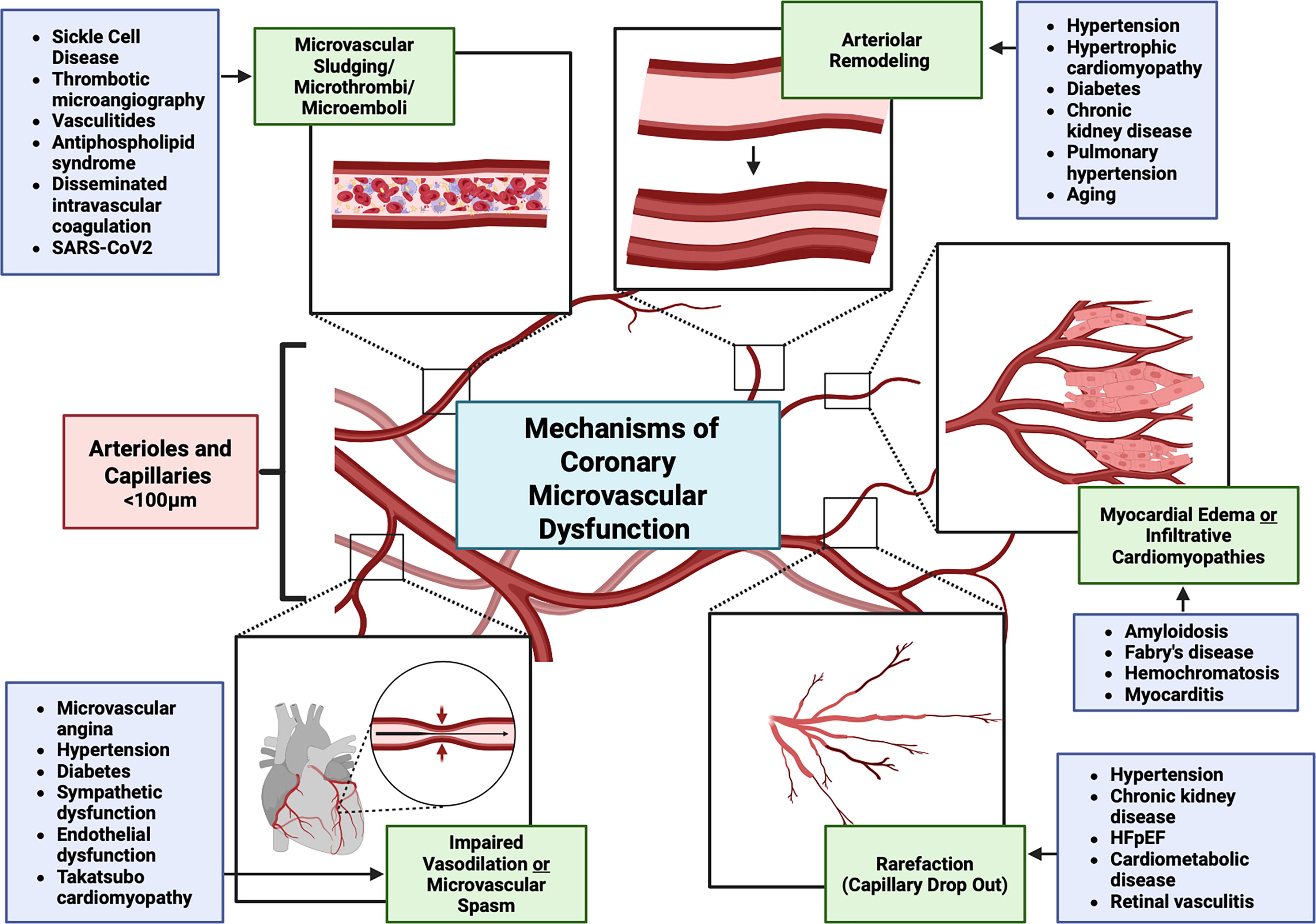
While the relationship between oxidative stress and CAD is established, a limited number of human studies have investigated oxidative stress in patients with CMD. Oxidative stress, measured by elevated levels of plasma cystine (an aminothiol), was found to be associated with left ventricular diastolic dysfunction in 75 midlife women with INOCA, even after adjusting for risk factors for diastolic dysfunction, although GSH levels were not associated with diastolic dysfunction (Raad et al., 2020). In another study, high levels of basal O2 •− production from circulating mononuclear cells, thought to contribute to intravascular oxidative stress, were prognostic of cardiovascular events in patients with INOCA (Leu et al., 2006). Indeed, therapies that influence ROS and improve endothelial function (such as angiotensin-converting enzyme inhibitors and statins) are used to manage this complex condition, but improved therapies are needed (Burke et al., 2000; Chade et al., 2006; Corban et al., 2020; Ford et al., 2020; Guddeti et al., 2016).
Heart failure
Heart failure is a syndrome of left ventricular dysfunction in the presence of symptoms such as shortness of breath, fatigue, and volume overload. Prevalence is rising, and it is a leading cause of morbidity and mortality. Heart failure with preserved ejection fraction (HFpEF) has been associated with microvascular dysfunction, with up to 75% of hospitalized patients with HFpEF noted to have CMD (Mehta et al., 2022). Furthermore, HFpEF prognosis is worse in the presence of systemic inflammation, which is defined as an increase in cytokines, including TNFα, IFN-γ, and IL-1β. However, anti-inflammatory therapies have not had a significant therapeutic effect in patients with HFpEF (Shirazi et al., 2017). Heart failure (with both reduced and preserved EF) has been associated with increased oxidative stress in the setting of decreased NO•, resulting in dysfunction of the cardiac microvasculature, impaired cardiac conduction, and impaired ventricular remodeling, particularly in HFpEF (Mehta et al., 2022). It is unclear whether the association is related to shared risk factors discussed above, such as age, hypertension, diabetes, smoking, elevated cholesterol, and obesity, or if there is a direct causal relationship.
Stroke
Stroke poses a significant global health challenge, ranking as the second leading cause of death worldwide according to the World Health Organization, and is projected to claim even more lives in the coming decades. Microvascular dysfunction is a critical component in the pathogenesis of stroke, particularly in the context of ischemic strokes, such as lacunar strokes, which constitute a substantial portion of cases. At the cerebral microvasculature level, stroke sets off inflammatory and thrombotic cascades, leading to progressive neuronal demise and infarct expansion (De Meyer et al., 2016). Although recanalization of major arteries remains a primary therapeutic goal, the microvascular perfusion sustaining the surrounding tissue often remains compromised, exacerbated by ischemia–reperfusion injury-induced oxidative stress and inflammation (Kalogeris et al., 2016). ROS generated during reperfusion directly assail endothelial cells, precipitating vascular dysfunction. Inflammatory mediators released poststroke can directly damage the microvasculature, disrupt endothelial integrity, and perpetuate chronic neuroinflammation, exacerbating microvascular dysfunction and prolonging secondary brain injury (Candelario-Jalil et al., 2022).
Ischemic stroke-induced BBB disruption triggers vasogenic edema, amplifying tissue hypoxia and neuronal injury. Edema formation, stemming from compromised BBB integrity, further compromises cerebral perfusion, exacerbating ischemic damage. The interplay of oxidative stress, inflammation, and endothelial dysfunction underscores the multifaceted pathophysiology of stroke (Chen et al., 2021b). Key enzymatic sources of ROS, including Nox2, Nox4, XO, and COX-2, along with NOS, significantly contribute to microvascular dysfunction during stroke (Duan et al., 2021; Jiang and Yu, 2021; Maciejczyk et al., 2022). Consequently, the inhibition of reperfusion-induced ROS from the parenchyma restores microvascular perfusion after recanalization and provides neuroprotection (Taskiran-Sag et al., 2018). Neutrophil influx poststroke worsens oxidative stress via myeloperoxidase (MPO) activity, compromising BBB integrity (Roy et al., 2023). Activated microglia further elevate ROS production through Nox2 and Nox4, exacerbating inflammation and BBB dysfunction (Qiu et al., 2021). Notably, inhibition of Poldip2, a regulator of Nox4, presents a promising avenue for attenuating immune cell recruitment under the inflammatory milieu, offering a potential therapeutic intervention for barrier dysfunction-related pathologies (Eidson et al., 2021; Forrester et al., 2019).
Chronic autoimmune and inflammatory conditions
Systemic lupus erythematosus
Systemic lupus erythematosus (SLE) is an autoimmune disease characterized by multiorgan damage in the setting of circulating immune complexes. SLE has been associated with an increased risk of CAD, heart failure, hypertension, and CMD, particularly in women (Prasada et al., 2020). SLE is also associated with pulmonary vasculitis and pulmonary hypertension, especially when anti-endothelial cell antibodies are present (Hassoun, 2009). Similar to other chronic autoimmune diseases, SLE autoantibodies result in immune-mediated vascular damage associated with increased oxidative stress. Oxidative stress contributes to the damage to various organs, including the kidney, heart, and skin seen in SLE, and the level of oxidative stress has been correlated with disease severity (Perl, 2013). Of interest, NAC treatment leads to improvement in central nervous systetem complications, endothelial function, and decreased oxidative stress in mild SLE (López-Pedrera et al., 2016).
Rheumatoid arthritis
Rheumatoid arthritis (RA) is a systemic disease characterized by inflammatory arthritis and has been associated with increased oxidative stress (Mateen et al., 2016). It often occurs in a milieu of pro-inflammatory cytokines, complement, and immune complexes (Gravallese and Firestein, 2023). RA has increasingly been recognized as an independent risk factor for cardiovascular diseases, particularly atherogenesis. Interestingly, RA has been associated with both macrovascular and microvascular endothelial dysfunction. As the disease progresses, reduced endothelial vasodilation is observed independent of increased levels of C-reactive protein or erythrocyte sedimentation rate, both markers for monitoring chronic inflammatory conditions. Patients with RA have giant capillaries, neoangiogenesis, hemorrhages, and avascular areas (Anghel et al., 2023). However, it is unclear how much microvascular disease and ROS contribute to overall RA disease activity or if they share an underlying disease process, such as chronic systemic inflammation. RA is commonly treated with methotrexate and anti-TNFα agents, and while treatment with these agents has been associated with improvement in macrovascular disease, there have been inconsistencies regarding the effect on the microvasculature (Bordy et al., 2018). Other clinical trials have noted an increase in soluble ICAM-1 (sICAM-1) levels in patients with RA with clinical signs of systemic vasculitis, which has been associated with capillary abnormalities (Gorska et al., 2008). The mechanisms of RA are still not fully understood, highlighting the need for further research into this area, especially on the potential role of ROS.
Juvenile idiopathic arthritis
Juvenile idiopathic arthritis (JIA) is the most common rheumatic disease in children. These patients have increased capillary loop size, increased frequency of abnormal capillaries, and wide subpapillary venular plexus associated with increased sICAM-1 and VEGF (Gorska et al., 2008). Excessive oxidative stress has also been implicated in the pathogenesis of JIA, with evidence for increased oxidative downstream products and decreased total antioxidant plasma capacity (Lipińska et al., 2015). Another study found that patients with JIA also have lower SOD and catalase levels and higher malondialdehyde (arachidonic acid oxygenation product) levels, suggesting reduced antioxidant capacity (Guney et al., 2009).
Psoriasis
Psoriasis is a common chronic inflammatory skin disease that often results in raised erythematous plaques. These plaques are thought to be a result of hyperproliferative basal epidermis in combination with dermal inflammatory infiltrates. One of the earliest signs of psoriasis lesions is the development of dilated and tortuous blood vessels within dermal papillae (Micali et al., 2010). The redness of these lesions is associated with increased amounts and tortuosity of capillaries, as well as thinning of the overlying epithelium (Nestle et al., 2009). Psoriasis has an established association with microvascular dysfunction related to increased levels of pro-angiogenic cytokines such as VEGF, endothelial cell-stimulating angiogenesis factor, TNFα, transforming growth factor (TGF)-α, and platelet-derived growth factor (PDGF). The pathogenesis of psoriasis is also associated with increased oxidative stress and decreased antioxidant capacity (Pleńkowska et al., 2020). Because skin is exposed to a higher level of ROS given the exposure to UV light (Magenta et al., 2016), damage to the stratum corneum by ROS is thought to be part of the pathogenesis of psoriasis (Shilov and Sergienko, 2000). On the other hand, excess ROS can be beneficial in psoriasis as they can inhibit cellular proliferation. In fact, phototherapy with UV light is a common component of psoriasis treatment (Hu et al., 2022). While phototherapy is associated with increased oxidative stress, it also enhances total antioxidant status. These findings suggest that phototherapy may contribute to redox modulation by altering ROS concentration, localization, or their chemical nature; however, the exact mechanisms behind this effect remain unclear (Dobrică et al., 2022).
Atopic dermatitis
Atopic dermatitis (AD), like psoriasis, is a common chronic inflammatory skin disease associated with dermal and epidermal inflammatory infiltrates. The vascular endothelium is thought to play a central role in regulating acute and chronic inflammation during AD through regulation of inflammatory cell adhesion, leukocyte extravasation, and vascular permeability (Steinhoff et al., 2006). In an IL-4 transgenic mouse model of AD, researchers found a reduction in tight junction protein expression with changes in interendothelial junctional complexes and increased vascular permeability (Agha-Majzoub et al., 2005). Chronic inflammation can limit oxygen diffusion into the tissues, resulting in a relatively hypoxic environment, which can trigger angiogenesis through VEGF upregulation by hypoxia-inducible factors. Both VEGF and angiopoietic factors were found to be increased in these mice, leading to increased dermal angiogenesis with significantly more capillary beds. Children with AD have increased oxidative stress markers such as 8-hydroxy-2′-deoxyguanosine, acrolein-lysine adducts, and bilirubin oxidative metabolites (Steinhoff et al., 2006). Conversely, iNOS is increased in the upper dermal microvasculature of inflamed skin lesions from these patients (Steinhoff et al., 2006), and NO• plays a key role in the regulation of the dermal microvasculature vasodilation in AD. These alterations in ROS and RNS, vascular permeability, and chronic systemic inflammation generate endothelial dysfunction that likely contributes to AD.
Systemic sclerosis
Systemic sclerosis (SSc) is a heterogeneous progressive disease characterized by immune dysfunction, microvascular disease, and multiorgan fibrosis. Microvascular injury and endothelial cell dysfunction occur in the early stages of pathogenesis, along with the transition of endothelial cells to myofibroblasts (Piera-Velazquez and Jimenez, 2021). Local immune and inflammatory reactions progressively destroy microvessels in skin and visceral organs, which are replaced by fibrosis, resulting in irreversible damage. Interestingly, high levels of ROS have been implicated in the pathogenesis of SSc as evidenced by increased indicators of cellular and molecular oxidative damage and elevated production of ROS by cells involved in the fibrotic process (Piera-Velazquez and Jimenez, 2021). Of note, SSc dermal fibroblasts have constitutively increased Nox4 expression, and administration of antioxidants or Nox4 inhibitors can reduce the expression of genes implicated in fibrosis (Piera-Velazquez and Jimenez, 2021). Nox4-stimulated ROS production has also been implicated in the generation of SSc-specific autoantibodies (Piera-Velazquez and Jimenez, 2021).
Dermatomyositis
Dermatomyositis is an inflammatory muscle disease characterized by microvascular injury, perivascular inflammation, and loss of capillaries. Nailfold videocapillaroscopy (NVC) shows a variety of changes in the microvasculature, including giant capillaries, microhemorrhages, severe capillary loss, and angiogenesis (Cutolo and Smith, 2021). Dermatomyositis is associated with enhanced type 1 interferon (IFN) signaling in skeletal muscle, which is accompanied by increased ROS production in myocytes. Microvascular depletion of capillaries in the setting of increased local ROS production was seen in muscles from patients with DM (Gitiaux et al., 2013). Transcriptomics showed downregulation of mitochondrial genes, and in situ experiments showed mitochondrial abnormalities, including decreased respiration and increased ROS (Meyer et al., 2017). IFN-β is thought to contribute to these mitochondrial malfunctions, potentially via ROS, since NAC prevents mitochondrial dysfunction in a mouse model of dermatomyositis (Meyer et al., 2017).
Antiphospholipid syndrome and antisynthetase syndrome
Antiphospholipid syndrome (APS) is an autoimmune disorder with antiphospholipid antibodies (aPL) characterized by the presence of both venous and arterial thrombi. These autoantibodies are associated with increased ROS as well as reduced antioxidants, leading to vascular damage following immune cell activation (López-Pedrera et al., 2016). Human umbilical vein endothelial cells treated with IgG from patients with APS show increased oxidative stress and VCAM-1 expression, which is prevented by NAC and vitamin C (Simoncini et al., 2005). Lipid peroxidation has also been identified as an independent predictor of endothelial dysfunction in patients with APS (Nocella et al., 2021; Stanisavljevic et al., 2016).
Antisynthetase syndrome (ASS) is a heterogeneous autoimmune disease also associated with autoantibodies. This disorder is characterized by arthritis, myositis, and interstitial lung disease, as well as symptoms such as Raynaud phenomenon, altered microcirculation, fever, and hyperkeratosis of hands. The most prevalent NVC finding is the presence of angiogenesis (Cutolo and Smith, 2021). ASS autoantibodies target tRNA-synthetases, most commonly HisRS, which is responsible for synthesizing histidyl-tRNA. Histidine is a precursor for carnosine biosynthesis, which is an antioxidant with SOD-like activity. Thus, HisRS may have antioxidant and anti-inflammatory roles such that anti-HisRS antibodies result in an increase in oxidative stress (Gallay et al., 2018). One study noted increased ROS response gene expression in patients with ASS (Zhu et al., 2022). Given the altered microcirculation and increased oxidative stress observed in this disease, it is plausible that redox imbalances contribute to its pathogenesis.
Hidradenitis suppurativa
Hidradenitis suppurativa (HS) is a chronic inflammatory disease associated with painful skin boils in areas with a significant number of apocrine glands (Jemec, 2012). Optical coherence tomography of HS nodules shows increased blood flow in the dermis and increased skin microvascularization (Manfredini et al., 2022). Smoking and obesity, both associated with increased oxidative stress, significantly increase the risk of HS (Scala et al., 2021). The pathogenesis is still not well understood, although it does involve loss of the sebaceous glands, infiltration by lymphocytes, and hyperkeratosis of the pilosebaceous unit with eventual destruction of the hair follicle and granuloma formation, which can lead to sinus tract formation and scarring in later stages. The IL-1β, IL-12, IL-23, and TNFα pathways have been found to be upregulated in patients with HS, and chronic systemic inflammation is thought to be involved in its pathogenesis (Witte-Händel et al., 2019). The immune response to the damage and inflammation in HS then results in increased free radicals, leading to oxidative stress and nucleotide nitration (Preda-Naumescu et al., 2021), which in turn increases the risk of mutations that can result in the conversion of HS into squamous cell carcinoma (Chapman et al., 2018).
Ankylosing spondylitis
Ankylosing spondylitis (AS) is characterized by inflammation of entheses, synovia, and bone. In later stages, chronic inflammation can result in the fusion of sacroiliac, vertebral, and apophyseal joints. AS is also associated with anterior uveitis, inflammatory bowel disease (IBD), and psoriasis (Taurog et al., 2016). It is predominantly a genetic disease, with about 90% of the susceptibility attributable to genetic factors. Interestingly, AS is also associated with increased oxidative stress (Ye et al., 2020). Patients with AS have impaired endothelium-dependent vasodilation and capillary recruitment and show improvement in microvascular function following treatment with anti-TNFα therapy (van Eijk et al., 2009). Increased IL-23/IL-17 pathway activation is also crucial in the pathogenesis of AS, and treatment with anti-IL-17 monoclonal antibody has been efficacious (Ranganathan et al., 2017). Thus, the exacerbated inflammation and oxidative stress may potentially explain the endothelial dysfunction observed in AS.
Behçet’s disease
Behçet’s disease (BD) is an inflammatory disorder with relapsing oral ulcers and skin lesions associated with intermittent acute inflammatory attacks. Repeated uveitis from BD can also lead to blindness. While rare, involvement of the lower gastrointestinal and central nervous systems can be life-threatening (Sakane et al., 1999). BD can result in multisystemic vasculitis of both large and small vessels, in part by induction of adhesion molecule expression and activation of neutrophil-derived ROS due to circulating TNFα, IL-1β, and IL-8 (Sakane et al., 1999). Of importance, increases in oxidative stress and decreases in antioxidants correlate with disease severity in patients with BD (Najim et al., 2007; Onur et al., 2011).
Inflammatory bowel disease
Inflammatory bowel disease (IBD), which includes Crohn’s disease and ulcerative colitis, presents as a multifaceted systemic condition characterized by persistent gastrointestinal inflammation. Recent studies have shed light on the heightened risk of microvascular complications in individuals with IBD, particularly emphasizing the significance of endothelial dysfunction. Perturbations in microcirculatory dynamics, such as alterations in vessel caliber, capillary density, and blood flow velocity, contribute significantly to the microvascular dysfunction observed in IBD. Patients with IBD exhibit diminished coronary microvascular flow velocity reserve and impaired endothelium-dependent flow-mediated vasodilation (Kakuta et al., 2021). Dysregulated angiogenesis and vascular remodeling further exacerbate microvascular architectural and functional anomalies in IBD, as evidenced by increased microvascular density correlating with disease severity in human IBD and various murine colitis models (Alkim et al., 2015; Chidlow et al., 2006; Danese et al., 2006).
The intricate mechanisms underlying microvascular dysfunction in IBD represent a convergence of factors, including inflammatory cytokines, oxidative stress, and a multifactorial interplay of genetic predisposition and environmental influences. These factors modulate endothelial cell function, culminating in impaired vascular reactivity, heightened leukocyte adhesion, augmented endothelial permeability, and a prothrombotic milieu. In IBD, endothelial cells produce less NO• due to diminished expression of both eNOS and iNOS, leading to impaired NO•-mediated vasodilation and an elevation in ROS production within the microvasculature of affected intestinal regions (Gravina et al., 2018; Hatoum et al., 2003). Localized microvascular endothelial dysfunction may progress to systemic vascular barrier impairment. Robust evidence links IBD with conditions such as atherosclerosis, coronary dysfunction, and an increased risk of cardiovascular morbidity and mortality. Serum levels of von Willebrand factor (VWF) in individuals with IBD reliably indicate systemic inflammation associated with the disease.
The presence of ROS within inflamed regions promotes the accumulation of ultra-large VWF multimers, which play a pivotal role in platelet adhesion and aggregation, ultimately contributing to the pathogenesis of microvascular thrombosis (Lancellotti et al., 2010). Thrombi formation within mucosal tissues exacerbates ischemic inflammation within the intestinal microvasculature, thereby amplifying tissue injury. Given the proximity of the intestinal microvasculature to the epithelial barrier, which represents a secondary defense against commensal and microbial translocation, compromised epithelial integrity during IBD exacerbates microvascular perturbations. Subsequent exposure of mucosal microvascular endothelial cells to bacterial antigens elicits an inflammatory cascade, mediated in part by toll-like receptor engagement, thereby perpetuating microvascular endothelial dysfunction amid the inflammatory milieu (Bush et al., 1998; Cornet et al., 2001).
Vasculitides
Kawasaki disease
Kawasaki disease (KD) is a pediatric vasculitis primarily affecting medium-sized vessels and is the most common cause of acquired heart disease in children. In addition to macrovascular inflammation, the microvasculature is also involved, as seen on retinal microvasculature imaging. Patients with KD have significantly larger retinal venules than healthy subjects, which is thought to reflect chronic inflammation and endothelial dysfunction (Chen et al., 2017). Indeed, KD has been associated with increased oxidative stress and vascular inflammation (Yahata and Hamaoka, 2017). The acute stage of KD includes inflammatory cell infiltration into the arterial wall and increased inflammatory cytokines. The inflammatory infiltrates are likely responsible for the increased oxidative stress, which is evident in elevated urine 8-isoprostane, an acute oxidative stress marker, and reduced metabolic products of NO• produced in endothelial cells (Yahata and Hamaoka, 2017). Moreover, patients with KD were found to have chronically elevated reactive oxygen metabolites (Yahata and Hamaoka, 2017). However, it is unclear if oxidative stress contributes to the pathogenesis of KD or if it is a side effect of the inflammatory infiltrates that worsen KD.
Takayasu arteritis
Takayasu arteritis (TAK) is a chronic vasculitis primarily affecting large-sized vessels such as the aorta, its main branches, and pulmonary arteries. TAK is characterized by obstructive and aneurysmal vascular lesions originated by the loss of elastic fibers in the vessel walls. Like KD, TAK also affects the microvasculature even in the absence of upstream arterial insufficiency (Svensson et al., 2022). NVC in patients with TAK shows significantly fewer nailfold capillaries as well as other capillary abnormalities (Wang et al., 2023). Interestingly, TAK was first described in 1908, with microvasculature changes noted in the form of wreath-like retinal arteriovenous anastomoses (Numano and Kakuta, 1996). While much of the pathophysiology of TAK remains unknown, oxidative stress has been identified as a possible contributor, with patients having increased levels of 8-iso-prostaglandin F2α, which may contribute to increased matrix metalloproteinase-mediated destruction of elastic fibers (Mahajan et al., 2010).
Thromboangiitis obliterans
Thromboangiitis obliterans (TAO) is a nonatherosclerotic segmental inflammatory disease affecting peripheral small and medium arteries, veins, and nerves. Patients with TAO also have impaired endothelium-dependent vasorelaxation in the peripheral vasculature (Olin, 2000). Unlike other forms of vasculitis, TAO often leads to highly cellular and inflammatory thrombi, affecting both arteries and veins with a relative sparing of the blood vessel wall and without an increase in circulating acute-phase reactants. TAO is associated with increased TNFα, IL-6, and IL-10, as well as malondialdehyde from lipid peroxidation (Chen et al., 2023a). In fact, oxidative stress has been identified as a key player in TAO, and the main risk factor for developing TAO, smoking tobacco, has also been associated with increased oxidative stress and reduced antioxidant protection, which then contributes to microvascular endothelial dysfunction (Chen et al., 2023a).
Hemoglobinopathies
Sickle cell disease (SCD), thalassemias, and other hemoglobinopathies are genetic disorders that affect the structure or production of hemoglobin. Thalassemias, the most common hemoglobinopathy worldwide, result in defects in protein synthesis with preserved hemoglobin structure. SCD is caused by mutations in the hemoglobin gene and has been associated with endothelial dysfunction, which has been partially attributed to sickled erythrocytes blocking vessels, hemolysis, and direct damage to the microvascular endothelium (Mannino et al., 2012). Thalassemia is associated with cardiovascular endothelial dysfunction in the setting of hemolysis and increased oxidative stress with increased macrophage activation, loss of NO•, oxidation of LDL, and increased microthrombi (Gullu et al., 2013). Many patients with hemoglobinopathies receive treatment with frequent blood transfusions and often develop iron overload. As noted earlier, increased iron load is associated with elevated oxidative stress, given iron’s role in forming ROS/RNS (Fig. 14) (Güvenç et al., 2023).

Neuroinflammatory diseases
Multiple sclerosis
ROS-induced microvascular damage and neuroinflammation have been associated with neurodegenerative diseases such as Alzheimer’s and Parkinson’s, which are major causes of morbidity in our aging population. Here we highlight multiple sclerosis (MS), another neuroinflammatory condition, typically diagnosed in young adults (ages 20–40 years). It involves chronic inflammation of the central nervous system leading to demyelination. In this setting, the neuronal microvasculature is exposed to significant oxidative stress, given its high metabolic consumption of glucose, increased ROS/RNS production inherent in neuronal signaling, and increased metal ion accumulation during aging (Beal, 1992). MS is also associated with reduced cerebral perfusion. While vascular endothelial inflammation was once thought to be the cause of reduced perfusion, the loss of blood flow is more diffuse than would be expected from inflammation alone and may also be related to increased metabolic demands in the setting of demyelination (Chiurchiù and Maccarrone, 2011). Interestingly, mice with experimental autoimmune encephalomyelitis, an animal model for MS, have increased TNFα, iNOS, and ICAM/VCAM-1 expression. In these animals, the neurological deficits are improved following treatment with iNOS inhibitor (tricyclodecan-9-xyl-xanthogenate), NO• scavenger (2-phenyl-4,4,5,5-tetramethylimidazoline-1-oxyl-3-oxide), or peroxynitrite scavenger (uric acid) (Hooper et al., 1997), suggesting that NO•-derived RNS may contribute to disease progression.
Infectious disease
Chronic infections and inflammation
Longstanding infections can lead to chronic inflammation and affect the microvasculature. A few examples of infectious diseases that can contribute to oxidative stress in the microvasculature include chronic hepatitis C virus and human immunodeficiency virus infections, which have both been associated with increased cardiovascular risk in the setting of chronic systemic inflammation (Moran et al., 2022). Even localized infections such as Helicobacter pylori have been associated with increased chronic inflammation and oxidative stress (Han et al., 2022; Suzuki et al., 2009), which may be related to increased infiltration of macrophages and neutrophils that express large amounts of Nox2 and iNOS (Fu et al., 1999), and increased paracellular permeability of human microvascular endothelial cells (de Jesus Souza et al., 2019).
Sepsis
Sepsis comprises a life-threatening organ dysfunction caused by microvascular system failure, which is related to a massive and dysregulated host immune response (Singer et al., 2016). Endothelial dysfunction is an early event in sepsis and correlates with increased ROS levels (Aird, 2003; Macdonald et al., 2003; Victor et al., 2005). One study found that the survival rate of preclinical septic mice is improved by Nox4 knockdown, while Nox1, Nox2, and p47phox knockout had no protective effect. The protective role of Nox4 knockdown was associated with reduced production of ROS, while Nox1, Nox2, and p47phox knockout had no effect on ROS levels in the lung (Jiang et al., 2020). However, Wu et al. (2007) showed that LPS, IFN-γ, and H2O2 can activate p47phox-containing oxidases in microvascular endothelial cells, suggesting a role for ROS-induced ROS release. Mitochondrial ROS are also likely to be important: inhibiting mitochondrial ROS attenuates the production of pro-inflammatory mediators like NLRP3, IL-1β, and cleaved caspase 1 in LPS-stimulated human lung microvascular endothelial cells and reduces lung injury in LPS-stimulated mice (Wang et al., 2021). Focusing on redox regulation/dysregulation may reveal more potential therapeutic targets of sepsis.
SARS-CoV-2
COVID-19 is a systemic disease caused by SARS-CoV-2 infection that can lead to pneumonia, respiratory failure, and ARDS. SARS-CoV-2 induces lung inflammation and increases alveolar endothelial permeability, leading to pulmonary edema and hypoxia. Excessive cytokine production and hyperactivation of the immune system are thought to be responsible for the lung endothelial dysfunction observed in patients with COVID-19 (Perico et al., 2024); however, recent work suggests that SARS-CoV-2 can infect endothelial cells that then express ICAM-1 and VCAM-1 (Qin et al., 2021), and that the SARS-CoV-2 spike protein itself can damage the endothelium (Lei et al., 2021). Binding of the spike protein increases ROS in human lung microvascular endothelial cells, which is associated with barrier dysfunction (Romero et al., 2023). SARS-CoV-2 has also been shown to cause increased permeability in brain microvascular endothelium and retinal vasculature abnormalities associated with thrombosis (Guemes-Villahoz et al., 2021). Patients with COVID-19 have been reported to have reduced antioxidants and increased NADPH oxidase-derived ROS, and the production of ROS by Nox2 in neutrophils has unintended effects on the microvascular endothelium and thrombosis (Alam and Czajkowsky, 2022). Thus, emerging evidence suggests that redox processes play a role in the vascular changes during COVID-19 infection, and furthermore, ongoing work suggests that long COVID is associated with oxidative stress and inflammation (Al-Hakeim et al., 2023).
Therapeutics
Overview
Antioxidant enzymes are the primary defense against oxidative stress within cells. Enzymes such as SOD, catalase, GPx, and peroxiredoxin play a crucial role in maintaining redox balance and overall cell function by efficiently scavenging free radicals and ROS (Figs. 3 and 14). Moreover, these enzymes react with intracellular oxidants several thousand to millions of times faster than supplemental low molecular weight antioxidants, such that exogenous antioxidants’ effects are relevant only in the extracellular or interstitial compartments. Therefore, the most promising therapeutic strategies focus on (1) enhancing the levels of antioxidant enzymes and their substrates, (2) preventing the production of oxidants, and (3) inhibiting oxidant-triggered signaling to prevent inflammation or cell death. Here, we discuss some of the most relevant therapeutic strategies currently being tested for microvascular-related pathologies in preclinical and clinical trials. We discuss the chemistry behind the mechanisms of action of these drugs, the proven benefits, and the potential adverse effects and reported limitations.
Glutathione peroxidase mimetics
Ebselen (2-phenyl-1,2-benzisoselenazol-3(2H)-one) is a synthetic seleno-organic drug with well-known antioxidant and anti-inflammatory properties. The drug acts as a potent H2O2 scavenger by mimicking GPx (Fig. 14) and phospholipid hydroperoxide GSH peroxidase (PHGPx), catalyzing the reduction of a hydroperoxide at the expense of a thiol (Schewe, 1995). Although Ebselen is a poor free-radical scavenger, its effective scavenging of organic hydroperoxides eventually leads to a similar outcome to that achieved by radical scavengers, since hydroxy-lipids perpetuate lipid peroxidation (Schewe, 1995). In contrast to the enzyme-catalyzed reaction that involves a specific binding site for GSH, Ebselen can react with various other thiols, such as GSH, NAC, dithiothreitol, and dihydrolipoate, resulting in the formation of selenenylsulphides (Cotgreave et al., 1987; Haenen et al., 1990). Conversely, thiols can also reduce the selenium oxide form of Ebselen (Sands et al., 2023). The reactivity of Ebselen with thiols enables a potent regulation of cysteine-containing proteins. Indeed, Ebselen has been shown to inhibit numerous enzymes such as NADPH oxidases (Smith et al., 2012), NOS (Hattori et al., 1996), lipoxygenase (Walther et al., 1999), H+-K+-ATPases (Tabuchi et al., 1994), glutamate dehydrogenase (Azad et al., 2014), and the p50 subunit of NFκB (Larabee et al., 2009). Since Ebselen targets the thiol residues of these enzymes, the inhibition can be overturned by adding reducing agents such as dithiothreitol (Hattori et al., 1996; Terentis et al., 2010).
Ebselen has myriad biological effects, including antioxidant, anti-inflammatory, antimicrobial, anti-lipoperoxidative, antimutagenic, antithrombotic, and anti-atherosclerotic activities. It can also act as a detoxifying and depigmenting agent (reviewed in Azad and Tomar, 2014). Accordingly, several clinical trials have shown that oral administration of Ebselen exerted favorable therapeutic effects for Meniere’s disease, bipolar disorder, impulsivity and emotional processing, acute ischemic stroke, cancer, and hearing loss (reviewed in Wang et al., 2020). Ebselen has also been studied in vascular and microvasculature conditions. In the vasculature, Ebselen reduces lipid hydroperoxides, inhibiting LDL toxicity (Harkewicz et al., 2008). Similarly, in a proatherogenic model using peroxiredoxin 2 (Prdx2−/−) and apolipoprotein E (ApoE−/−)-double knockout mice, Ebselen administration reduces plaque formation (Park et al., 2011). Ebselen ex vivo treatment of aortic explants also reduces the TNFα-induced expression of VCAM-1 and ICAM-1 adhesion molecules and MCP-1. As a result, Ebselen prevents monocyte transmigration in vivo and in vitro assays (Park et al., 2011). In another study, Ebselen attenuated uremia-induced endothelial dysfunction and reduced ICAM-1 expression and NFκB activation in vitro, suggesting a potential use to prevent accelerated atherosclerosis and endothelial dysfunction in patients with CKD (Vera et al., 2018). While these effects were observed in major vessels, they are also likely to influence endothelial cell dysfunction in the microvasculature.
Ebselen also has antithrombotic properties, reducing venular and arteriolar thrombus formation in a model of photochemically induced thrombus in rats (Lindenblatt et al., 2003). Clinical trials have shown that Ebselen displays a modest beneficial effect during cerebral ischemia events (reviewed in Parnham and Sies, 2000). The therapeutic benefit is likely to improve depending on the timing of administration following the stroke. Most recently, Ebselen has shown promise as SARS-CoV-2 therapeutic due to its ability to inhibit viral proteases (Ali et al., 2024). Ebselen covalently binds to the sulfhydryl group in the cysteine residues of the protease Mpro (Cys145) and PLpro (Cys122) (Zmudzinski et al., 2023). Mpro plays an essential role in the SARS-CoV-2 life cycle, and PLpro participates in virus replication and antagonizes the antiviral innate immune response of the host, suggesting Ebselen as a promising therapeutic for COVID-19 treatment. Indeed, two clinical trials currently in phase 2 for moderate and severe COVID-19 are being performed to test the efficiency of Ebselen treatment (Table 4). However, while these studies assess redox-related mechanisms and clinical outcomes, they do not directly evaluate Ebselen’s effects on the microvasculature—specifically, its potential to prevent microthrombus formation or mitigate other microvascular complications associated with COVID-19.
Differences Between Micro- and Microvasculature
Summary of studies that contrast micro- and microvasculature features in endothelial and smooth muscle cells. (↓) decrease, (↑) increase, (→) no change in expression or activity of the corresponding protein.
aDifferent studies: experimental conditions may not be comparable.
Despite initial beliefs that Ebselen is innocuous, various studies have pointed to its cellular toxicity. Ebselen induces cytotoxicity by deteriorating mitochondrial respiration, depleting thiols that are essential for normal cell function, and inducing DNA damage (Azad and Tomar, 2014; Yang et al., 2000). Ebselen can also react with zinc finger-containing transcription factors, altering gene expression and DNA repair (Jacob et al., 1998). Additionally, Ebselen induces cell death by apoptosis and necrosis (Guérin and Gauthier, 2003; Yang et al., 2000). High dosages and cell-specific sensitivity contribute to many of Ebselen’s adverse effects, so in vivo use must be carefully dosed and monitored.
SOD and catalase mimetics
Synthetic SOD and catalase mimetics are chemical compounds such as Mn(III) porphyrins, Mn(II) cyclic polyamines, Mn(III) salen derivatives, MnPLED derivatives, nitroxides, and fullerenes, designed to replicate the catalytic activity of natural enzymes (Figs. 3 and 14) (Bonetta, 2018). SOD and catalase mimetics possess rate constants that are orders of magnitude lower than that of the natural enzymes. As a result, their contribution to cytosolic antioxidant defense upon cellular entry is relatively modest. However, these mimetics have shown effectiveness in extracellular spaces, especially where concentrations of antioxidant enzymes and substrates are minimal or absent.
Mn porphyrins are likely the most extensively studied class of SOD mimics, and the therapeutic potential of porphyrins has been observed in animal models of stroke (Li et al., 2020), diabetes (Piganelli et al., 2002), cancer (Shin et al., 2021; Xu et al., 2021), sickle cell (Thamilarasan et al., 2020), and cardiovascular diseases (Anselmo et al., 2018; Barbosa et al., 2020). Salens, such as EUK-8, EUK-134, and EUK-189, have also shown protective effects in preclinical studies, including stroke (Baker et al., 1998), heart ischemia-reperfusion (Xu et al., 2004), and pulmonary hypertension (Himori et al., 2017). Although SOD mimetic clinical trials are currently in Phase II/III for amyotrophic lateral sclerosis (NCT00706147, NCT04972487) and for treating radiation-related injuries for some cancers (NCT03689712, NCT00618917, NCT01771991), there are not yet clinical trials for assessing the benefits of these drugs in microvasculature-associated pathologies.
While originally formulated to remove O2 •− specifically, most SOD mimetics can also reduce other ROS and RNS such as ONOO−, peroxyl radical, H2O2, and CO3 •− (Batinic-Haberle et al., 2015; Tovmasyan et al., 2015). Additionally, some SOD mimetics show pro-oxidative activity and can affect physiological redox-sensitive signaling pathways (Batinic-Haberle et al., 2012; Batinic-Haberle et al., 2015). Nevertheless, several of these drugs have demonstrated clinical safety in oral, intravenous, and topical delivery routes, and are presently under evaluation for various non-microvascular pathologies (Gad et al., 2013) (Table 4).
Glutathione supplementation
Oxidative stress often leads to a significant decrease in GSH levels. GSH itself is not efficiently transported into the cells and rapidly suffers degradation in the plasma (Wendel and Cikryt, 1980). Therefore, therapeutic strategies for replenishing GSH using agents such as GSH esters or NAC have proven effective in treating various diseases since they provide cysteine, the limiting amino acid in GSH synthesis (Fig. 15).

GSH esters are lipophilic compounds that can easily cross the cell membrane and resist degradation by γ-glutamyl transpeptidase in the plasma. After entering cells, GSH esters undergo rapid hydrolysis by nonspecific esterases, resulting in the formation of GSH (Fig. 15) (Levy et al., 1993).
Numerous studies conducted in cell and animal models have demonstrated the effectiveness of GSH esters in enhancing cellular and tissue GSH levels. However, only a handful of preclinical research investigations demonstrated effects in the microvasculature. GSH esters are also metabolized differently in humans versus mice and rats. For instance, diethyl ester GSH (GSH-DEE) is rapidly converted to monoester GSH by mouse plasma α-esterase, while humans show no α-esterase activity, leading to a more efficient GSH-DEE intake into the tissues and thus providing higher intracellular GSH levels to decrease oxidative stress (Levy et al., 1993). The absence of comparative analysis between murine models and humans raises questions regarding the translational relevance of these preclinical tests. Currently, there are no clinical studies testing GSH ester safety, bioavailability, and pharmacokinetics.
NAC acts as an effective antioxidant by replenishing intracellular GSH levels, scavenging free radicals, and exerting direct antioxidant and anti-inflammatory effects, making it a valuable therapeutic agent in various conditions such as acetaminophen overdose. When NAC is administered orally, intravenously, or by inhalation, it enters the cells and is rapidly deacetylated to cysteine. Inside the cell, cysteine combines with glutamate and glycine to form GSH through a series of enzymatic reactions catalyzed by GSH synthetase (Fig. 15).
Of note, NAC decreases vascular permeability and prevents endothelial barrier dysfunction, thereby preserving microvascular integrity (reviewed in Mahmoudinezhad et al., 2023). It can enhance NO• bioavailability and promote vasodilation in the microvasculature, an effect that is beneficial during sepsis (Chertoff, 2018). Similarly, in a clinical study that included 16 patients undergoing cardiac catheterization, NAC improved endothelium-dependent vasodilation in coronary and peripheral microcirculation (Andrews et al., 2001).
NAC also displays anti-inflammatory properties that mitigate tissue damage associated with inflammation and oxidative stress (Pei et al., 2018). In particular, NAC blocks the activation of NFκB, thereby reducing the expression of pro-inflammatory cytokines related to endothelial dysfunction (Mussbacher et al., 2019; Pierce et al., 2009). In this context, in a murine model of acute persistent ischemia, NAC effectively decreases leukocytic inflammation, apoptotic cell death, and microvascular perfusion in critically ischemic tissue (Bächle et al., 2011). An especially relevant use case is in patients with type 2 diabetes mellitus (DM2) with microvascular complications, who have lower GSH levels compared with nondiabetic individuals (Lutchmansingh et al., 2018). Currently, 17 clinical trials are exploring NAC effects on diabetes. However, none of these trials specifically assesses the impact of NAC on the microvasculature, thus underscoring an area that merits further scientific examination.
NAC oral administration may result in minimal side effects, including nausea, vomiting, diarrhea, flatus, and gastroesophageal reflux (Tenório et al., 2021). Intravenously, NAC can induce rate-related anaphylactoid reactions in up to 18% of patients. However, accidental overdose can lead to hemolysis, thrombocytopenia, metabolic acidosis, acute renal failure, and death (Mahmoudi et al., 2015). The effects of NAC on the cardiovascular system are contradictory in the scientific literature. While some studies suggest potential benefits of NAC in cardiovascular health, such as antioxidant properties and improvement in endothelial function, others indicate adverse effects or limited efficacy (Cui et al., 2023). These contradictory effects accentuate the complex interplay of NAC with various physiological pathways and highlight the need for further research to elucidate its role in cardiovascular health and disease (Dludla et al., 2018; Khan et al., 2021; Tenório et al., 2021).
Mitochondria-targeted antioxidants
An emphasis on developing antioxidants targeted to mitochondria comes in part from observations suggesting that ROS produced by NADPH oxidases induces the release of mitochondrial ROS, and that disrupting mitochondrial ROS can break this feed-forward cycle (Dikalov, 2011).
MitoQ (Mitoquinone mesylate) and MitoTempo are synthetic antioxidants derived from the naturally occurring ubiquinone (coenzyme Q10) and piperidine nitroxide (Tempamine or Tempol), respectively. Each compound is conjugated to a triphenylphosphonium (TPP+) cation, which imparts lipophilic properties and facilitates its accumulation in the mitochondria (reviewed in Murphy and Smith, 2007). While both are designed to reduce oxidative stress in mitochondria, they differ in their specific antioxidant mechanisms and the types of ROS they target. MitoQ acts as an electron donor, targeting a broad range of ROS, including superoxide and other free radicals. Since coenzyme Q10 is an essential electron carrier in the mitochondrial respiratory chain, this compound can also affect ROS produced by mitochondrial metabolism. In contrast, MitoTempo is more selective, specifically targeting superoxide radicals and functioning as a mimic of SOD (Trnka et al., 2008).
Preclinical assays in the context of diabetes-induced injury of the brain microvasculature have demonstrated that MitoQ can protect microvascular brain endothelial cells from cytoskeletal damage and high glucose-induced apoptosis by activating the Nrf2 pathway (Yang et al., 2021). MitoQ was similarly effective in pulmonary arterial hypertension, where it attenuated ROS-induced [Ca2+]i and mitochondrial fission in microvascular endothelial cells isolated from a pulmonary arterial hypertension mice model (Suresh et al., 2019). Although other studies report positive outcomes of MitoQ treatment in microvascular pathologies such as diabetes, heart failure, and kidney disease, they fail to specifically assess its effects on the microvasculature (Al Saadi et al., 2021; Chacko et al., 2010; Escribano-Lopez et al., 2019; Fink et al., 2020; Mercer et al., 2012).
In a randomized controlled pilot study, MitoQ treatment (20 mg/day for 4 weeks) improved microvascular function, as assessed by cutaneous vasodilatory response in patients with stage 3–4 CKD and healthy volunteers (Kirkman et al., 2023). A second study demonstrated that MitoTempo administered via microdialysis prevented microvascular dysfunction, measured as cutaneous vascular conductance, in individuals with stage 4–5 CKD and healthy controls (Kirkman et al., 2018). No serious adverse effects have been reported after administration of MitoQ and MitoTempo, supporting their potential as therapeutic agents in the treatment of oxidative stress-related conditions.
Iron chelation
Excessive free iron can be toxic by producing ROS through Fenton chemistry (Figs. 2 and 14) (Kawabata, 2022). During the Fenton reaction, ferrous iron (Fe2+) reacts with H2O2 to produce ferric iron (Fe3+), •OH, and hydroxide anions (OH−). •OH is highly reactive and causes damage of biomolecules such as DNA, proteins, and lipids. Chelators can either inhibit or activate •OH production by depleting iron or creating redox-active complexes, respectively (Fig. 14). For instance, deferoxamine (DFO) decreases ROS production by preventing iron exposure to oxygen and H2O2, whereas DP44mT forms a redox-active complex with iron to induce •OH production.
In the microvasculature, iron overload leads to oxidative damage and inflammation, ultimately affecting the endothelial cells lining the blood vessels. Two iron chelators have been tested in microvascular cells. PBT434 chelates interstitial iron and inhibits iron re-uptake by human microvascular endothelial cells, suggesting a therapeutic potential in neurovascular pathologies such as Parkinson’s disease (Bailey et al., 2021). DFO reduces microvascular permeability after ischemia-reperfusion injury in the intestine (Hernandez et al., 1987) and skeletal muscle (Smith et al., 1989). DFO also decreases inflammation and fibrosis in diabetic nephropathy, a severe microvascular complication of diabetes (Feng et al., 2023). In other studies, DFO was shown to restore dilation of the coronary microcirculation in patients with diabetes (Nitenberg et al., 2002).
Iron depletion can be detrimental in some cases. For example, iron deposition has been observed in arterioles of patients with calcific uremic arteriopathy (CUA or calciphylaxis). Thus, iron and oxidative stress are postulated to contribute to CUA and calcification within the microvasculature (Farah et al., 2011; Wickens et al., 2022). Unfortunately, patients with CUA are anemic and cannot tolerate loss of iron. Therefore, they cannot receive chelation therapy.
Nuclear factor erythroid 2-related factor 2 activators
Nuclear factor erythroid 2-related factor 2 (Nrf2) is a transcription factor that serves as master regulator of the cellular stress response by inducing detoxification enzymes and antioxidant expression to protect the cell against oxidative damage (Fig. 16). Thus, augmenting Nrf2 levels and activity represents an appealing therapeutic target for the development of pharmaceutical agents.
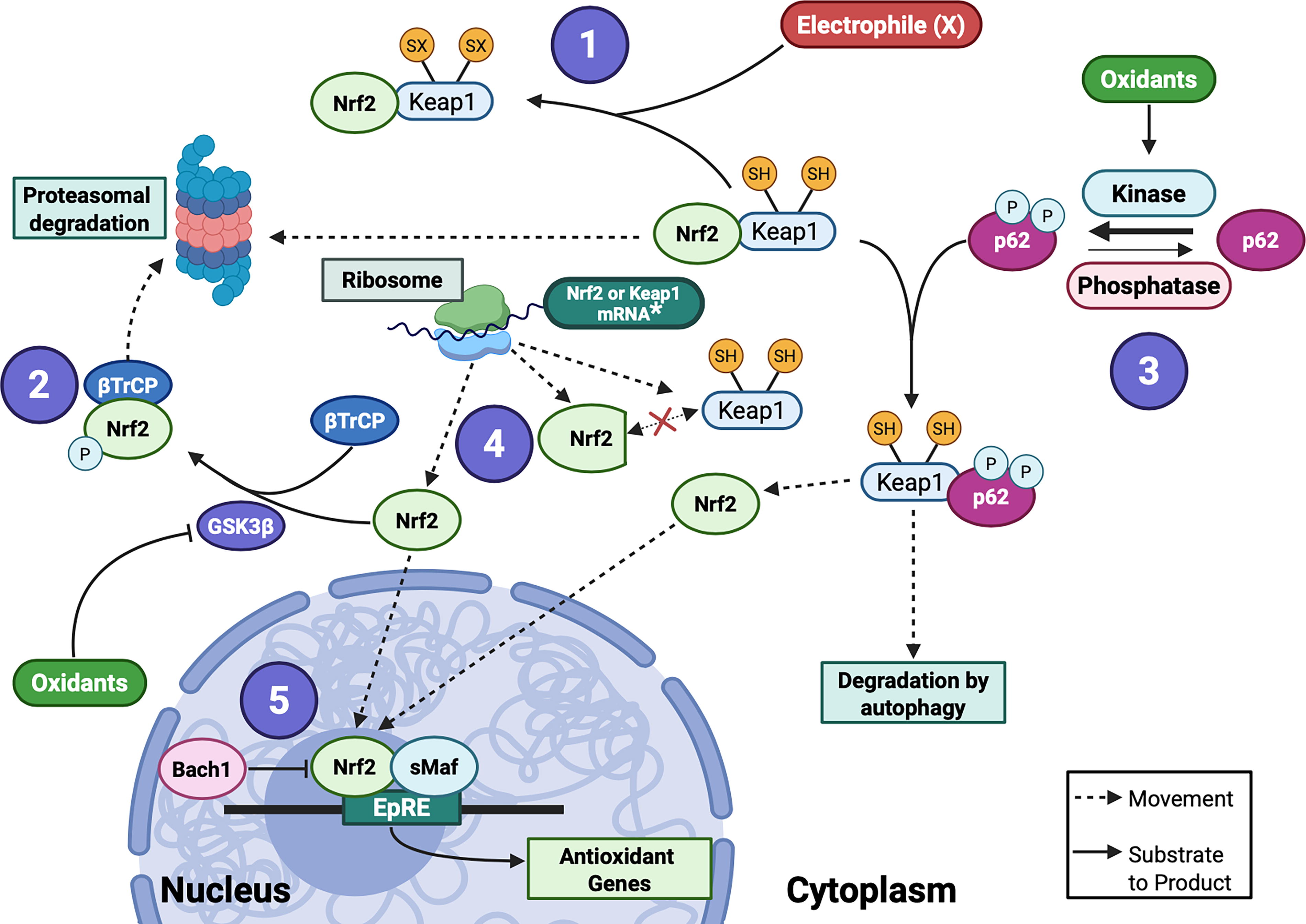
In brief, Nrf2 binds to Kelch-like ECH-associated protein (Keap1) under basal conditions, which facilitates its proteasomal degradation. In conditions of increased oxidative stress, Keap1’s cysteine residues react with electrophiles or undergo oxidation to form disulfides, leading to the release of Nrf2 (Fig. 16). Nrf2 then translocates to the nucleus, forming heterodimers with transcription factors such as c-Jun and Maf. The heterodimers bind to antioxidant response elements (AREs) of DNA to mediate the transcription of antioxidant and anti-inflammatory genes (Hayes and Dinkova-Kostova, 2014; Reddy, 2008). GSK3β then phosphorylates Nrf2, allowing its interaction with β-transducin repeat-containing protein (βTrCP). βTrCP binds and ubiquitylates Nrf2, sending it to degradation (Fig. 16). Alternatively, phosphorylation of the autophagy adaptor protein p62 sends Keap1 to degradation by autophagy, which also leads to the release of Nrf2 (Fig. 16). Finally, the transcriptional repressor BTB and CNC homology 1 (Bach1) also binds to AREs and suppresses Nrf2 function. Hence, the strategies to increase Nrf2 levels and downstream signaling can be focused on five categories: (1) modification of Keap1 using electrophiles; (2) disruption of the interaction between Nrf2 and βTrCP by inhibiting GSK3β activity; (3) controlling the phosphorylation status of p62; (4) de novo synthesis of Nrf2 that escapes degradation by Keap1; and (5) Bach1 inhibitors (Fig. 16).
Compounds derived from numerous dietary fruits and vegetables such as broccoli sprouts, grape seeds, tea, cocoa, curcumin, and turmeric have been found to stimulate Nrf2 signaling and promote antioxidant enzyme expression (Fig. 15) (Zhao et al., 2021). The general antioxidant mechanism relies on the oxidation of coumarins and polyphenols to form electrophilic quinones. These quinones subsequently bind and form adducts with the regulatory cysteines on Keap1, leading to Nrf2 activation (Fig. 16) (Reinisalo et al., 2015). Numerous dietary Nrf2 activators, such as curcumin, sulforaphane, and resveratrol, have been formulated as daily supplements. Some are currently undergoing clinical trials for the treatment of various diseases (Table 4).
Despite the promise of these strategies, one must proceed with caution. For example, Bardoxolone Methyl (BarM), a potent Nrf2 activator, has unexpected cardiovascular effects, including cardiac arrest in patients with type 2 diabetes and CKD (Chin et al., 2014; Van Laecke et al., 2015). The proposed mechanism of BarM-induced heart failure involves endothelin-1 pathway modulation, leading to increased endothelial permeability in human microvascular endothelium (Chin et al., 2014; Szczesny-Malysiak et al., 2020). There are still ongoing trials assessing BarM safety and benefits (Table 4).
Glucagon-like peptide-1
Glucagon-like peptide-1 (GLP-1) is an incretin hormone secreted by enteroendocrine L cells in response to nutrient ingestion. Upon binding to its receptor, GLP1-R, GLP-1 regulates plasma glucose by increasing insulin synthesis and secretion and inhibiting glucagon secretion. Additionally, GLP-1 possesses an anti-inflammatory and antioxidant function, exhibiting protective effects in various cells and tissues. GLP-1 can control ROS levels by activating Nrf2 through cAMP, PI3K, and PKCδ (reviewed in Ahmed et al., 2017 and Oh and Jun, 2017). Indeed, stimulation of pancreatic cells with exendin-4, a GLP1-R agonist, activates PKCδ-Nrf2 (Kim et al., 2017). Moreover, rats treated with exendin-4 and omeprazole showed decreased oxidative stress and increased Nrf2 expression (Patel et al., 2013).
GLP-1 analogs, including Albiglutide (Tanzeum), Dulaglutide (Trulicity), Exenatide (Bydureon, Byetta), Liraglutide (Saxenda, Victoza), Lixisenatide (Adlyxin), Semaglutide (Ozempic, Wegovy), and Tirzepatide (Mounjaro, Zepbound), have undergone extensive investigation across various pathologies. As of the latest update, 1146 trials involving these analogs can be found on the clinicaltrials.gov website, with many focusing on microvascular aspects (see Table 4). However, most of these studies are either still recruiting patients or have yet to publish updated results.
Adverse effects of GLP-1 analogs that have been reported include gastrointestinal disturbances, hypoglycemia, increased heart rate, and allergic reactions. Less common but more serious effects include pancreatitis, gallbladder disease, and gastroparesis (Filippatos et al., 2014).
NADPH oxidase inhibitors
Exacerbation of NADPH oxidase activity has been linked to microvascular pathologies such as arteriolar remodeling, endothelial dysfunction, and integrity of the BBB (reviewed in Li and Pagano, 2017). NADPH-oxidase-derived ROS can scavenge NO•, leading to increased vasoconstriction, or in some arteries can activate H2O2-induced vasodilation.
Nox2ds-tat (gp91ds-tat) is a peptide that selectively inhibits Nox2 with minimal effects on Nox1, Nox4, or XO activity. Nox2ds-tat blocks the p47phox-Nox2 interaction, preventing the translocation of the cytosolic activating complex to the membrane. Nox2ds-tat inhibits Nox2-derived ROS and tissue damage in vitro and in vivo in numerous organ systems (Li and Pagano, 2017). It reduces vasodilation in human coronary arterioles, positing Nox2 as the H2O2 enzymatic source that mediates bradykinin-mediated vasorelaxation (Larsen et al., 2009). Similarly, Nox2ds-tat attenuates basal and pressure-induced ROS in resistance arteries from hamster skeletal muscle (Keller et al., 2006). In other preclinical assessments, Nox2ds-tat decreases myogenic tone in afferent arterioles from hypertensive rats (Ren et al., 2010). The potential benefit of Nox2ds-tat was also explored in the cerebral microcirculation (Kazama et al., 2004; Park et al., 2007; Park et al., 2005), neovascularization during ischemic retinopathy (Al-Shabrawey et al., 2005), and in mesenteric resistance arteries from type 2 diabetic mice (Kassan et al., 2015).
Another peptide, NoxA1ds, blocks Nox1 activation without effects on Nox2, Nox4, Nox5, or XO (Ranayhossaini et al., 2013). While there is no evidence of NoxA1ds therapeutic effects on the microvasculature, it is expected to attenuate microvascular dysfunction due to well-known roles of Nox1 in vascular function (Rodríguez et al., 2015; Wind et al., 2010a).
Of note, different isoforms of NADPH oxidases localize to distinct membranes depending on the cell type, including plasma membrane and other membranous organelles, such as the endoplasmic reticulum, vacuoles, nucleus, and mitochondria (Hilenski et al., 2004; Vermot et al., 2021). The differential localization of NADPH oxidases enables precise regulation of ROS signaling in both normal physiology and pathophysiology, and targeting specific enzymes in a particular location represents a strategy to selectively modulate ROS therapeutically.
DPI and apocynin (4-hydroxy-3-methoxyacetophenone) are considered broad-spectrum Nox inhibitors with multiple nonspecific effects. DPI is also a nonselective inhibitor of flavin-dependent enzymes such as NOS, XO, and mitochondrial complexes (Li and Pagano, 2017). Apocynin can inhibit NADPH oxidases such as Nox1 and Nox2 by preventing p47phox translocation to the membrane (Stolk et al., 1994) and decreasing the expression of p47phox, p67phox, and gp91phox (Hur et al., 2010; Li et al., 2013). However, apocynin can also scavenge non-radical oxidant species such as HOCl− and H2O2 at higher concentrations (Cifuentes-Pagano et al., 2014). Despite the off-target effects, DPI and apocynin have been widely studied in various microvascular-related conditions. For instance, both drugs decrease ROS in cerebral arterioles from aged rats (Mayhan et al., 2008), and apocynin reverses neurovascular dysfunction in aged mice (Toth et al., 2014). In clinical studies, apocynin decreases ROS and reverses local microvascular endothelial dysfunction in the skeletal muscle of patients with obesity (La Favor et al., 2016). Although DPI and apocynin both have positive effects in reducing ROS and microvascular dysfunction, further investigation is necessary to elucidate if these responses are entirely dependent on NADPH oxidase signaling.
VAS2870 and VAS3947 are triazolopyrimidine derivatives lacking antioxidant properties that inhibit Nox-derived ROS without inhibiting XO or eNOS activity (Altenhöfer et al., 2012; Wind et al., 2010b). VAS2870 is considered a pan-Nox inhibitor (Nox1, Nox2, Nox4, and Nox5) (Wingler et al., 2012). VAS compounds can inhibit platelet activation and thrombosis in mouse mesenteric microvessels (Lu et al., 2019).
GKT137831 and GKT136901, pyrazolopyridine compounds, are considered dual Nox1/Nox4 inhibitors without inhibitory effects on other ROS-generating enzymes (Laleu et al., 2010). GKT137831 has been broadly studied in diabetic nephropathy and retinopathy, showing positive effects (Gorin et al., 2015; Lee et al., 2022). Most recently, it has undergone a phase 2 clinical trial that enrolled 200 patients with T2D with nephropathy, although results are not yet available (NCT02010242). GKT136901 has been shown to improve endothelial-dependent vasodilation in mesenteric arterioles expressing elevated Nox1 from obese (db/db) mice (Qiu et al., 2014), indicating the potential therapeutic utility of these inhibitors in treating microvascular dysfunction. However, recent work has shown that the GKT compounds are actually inactive as Nox inhibitors, instead interfering with peroxidase-dependent assays (Augsburger et al., 2019). In light of this, previous results using these compounds might not derive from Nox inhibition and should be reconsidered.
ML171 (2-acetylphenothiazine) is a small molecule that inhibits Nox1 at nanomolar concentrations. Higher concentrations of ML171 can also inhibit Nox2, Nox3, Nox4, and XO but with 20-fold lower potency than that for Nox1. Preclinical in vitro experiments have shown that ML171 inhibits ischemia-induced hyperpermeability in human brain microvascular endothelial cells (Dao et al., 2020). Although beneficial effects of ML171 have been reported on the macrovasculature in rodents (Côco et al., 2017; Wong et al., 2015), there are no reports of its effectiveness in microvascular dysfunction in vivo.
Although cytotoxicity remains low in in vitro assays, most drugs or peptides targeted to NADPH oxidases have not yet been tested for safety in human trials, with the exception of the GKT compounds. Because NADPH oxidases have been widely implicated in many disease processes as discussed above, truly specific inhibitors are theoretically excellent therapeutic strategies.
Challenges and limitations in targeting oxidative stress to treat disease
The clinical efficacy of antioxidant therapeutics has been underwhelming at best. Many limitations explain these results, including differences in the physiology between human and murine animal models. Erroneous assumptions about antioxidant function can also play a role. For instance, attempting to scavenge •OH is not practical due to the high reactivity of this radical. Instead, reducing the production of H2O2 to prevent the formation of •OH and consequent damage is more effective, but because H2O2 is critical to many physiological processes, preventing H2O2 formation is not desirable. Another prevalent misconception pertains to the relative effectiveness of small molecules in scavenging O2 •− or H2O2 within the cellular environment. Naturally occurring antioxidant enzymes interact with oxidants thousands to millions of times more quickly than small molecules, rendering them a much more efficient line of defense against oxidative stress than pharmacological interventions. Hence, the new generation of drugs like GLP-1 analogs, which enhance endogenous antioxidants, holds much more promise. Another limitation is the fact that antioxidants are often administered after the disease is established (e.g., atherosclerosis), but oxidant signaling contributes to the pathogenesis of the disease; thus, treatment is occurring too late. Moreover, early trials failed to measure the effectiveness of the antioxidant in reducing oxidative stress, potentially resulting in insufficient dosage or duration of therapy. Because oxidants tend to act locally, measurement of oxidants in blood may not truly reflect efficacy, making it challenging to testing efficacy. Harmful interactions with other medicines have also been challenges in translating these therapies to the clinic. An additional major disadvantage of targeting ROS is the low oral bioavailability of small molecules and peptides used as therapeutics. Many peptides are unstable and display a short half-life in the body.
Finally, as noted above, ROS also play a role in many physiological processes. An ideal drug should target the organ/tissue experiencing aberrant amounts of ROS without affecting healthy tissue, which is challenging for ROS-directed therapies. New emerging technologies for improving stability and delivery (i.e., nanoparticle formulations) are critical. Because of the multiple roles of oxidants, a thorough investigation into the disease etiology is crucial. In certain instances, oxidative stress may be a consequence rather than a cause of the pathology. Consequently, treatment with an antioxidant drug may mitigate symptoms without achieving a cure for the illness.
Conclusions and Future Directions
A tremendous body of basic science and preclinical work has established ROS and RNS as important players in microvascular function, both physiologically and pathophysiologically. ROS and RNS play diverse and intricate roles in microvascular physiology, functioning as signaling molecules in normal cellular processes such as proliferation, differentiation, and apoptosis, and thus regulating endothelial function, vascular tone, and angiogenesis. Conversely, excessive ROS/RNS leads to oxidative stress within the microvasculature, causing endothelial dysfunction characterized by increased vascular permeability, impaired vasodilatation, and exacerbated inflammation. These pathological changes induced by oxidative stress translate into microvascular remodeling, edema, rarefaction, microvascular spasm, microthrombi, and microvascular sludging (Fig. 13). Overall, these microvessel alterations are a hallmark of microvascular dysfunction observed in the clinic for the pathologies discussed in this review.
Scattered studies in patients suggest that ROS and RNS molecules, most notably NO• and H2O2, are also crucial in humans, especially for vasodilatation. However, the complexity and rapidity of the chemical interactions and the fundamental role of ROS and RNS in physiology have impeded progress on therapeutically targeting these molecules. Newer methods, such as CRISPR/Cas9 gene editing and RNA-based therapies that target specific enzymes, or strategies to boost tissue-specific endogenous antioxidant enzymes, hold more promise. Similarly, the development of more specific small molecule inhibitors of ROS-producing enzymes is imperative. The distinct subcellular distribution of ROS-producing enzymes, such as NADPH oxidases, underscores the importance of developing drugs that can be delivered in an organelle-specific manner. Likewise, developing nanotechnology-based approaches to deliver potential drugs to a tissue of interest may also improve our ability to prevent the harmful effects of ROS while preserving physiological functions. Repurposing therapeutic agents offers another promising approach to addressing microvascular diseases by utilizing well-characterized drugs to mitigate oxidative stress. For instance, therapeutic humanized antibodies currently used to treat inflammatory autoimmune disorders and cancer can indirectly decrease the oxidative stress induced by inflammation. Lastly, adopting a healthy lifestyle (healthy diet and regular exercise), managing risk factors, and seeking appropriate medical care can significantly reduce the risk of developing microvascular diseases and promote overall health.
An emerging area of interest involves the role of ROS in the modulation of microvasculature during cancer progression and cancer therapy. ROS are known to promote tumor formation and angiogenesis, and they also contribute to the cytotoxic effects of cancer treatments (Cheung and Vousden, 2022; Perillo et al., 2020). ROS produced by anticancer therapy have been linked to inflammation and vascular dysfunction, leading to both acute and long-term consequences in cancer survivors. Therefore, the new trend in cancer treatment is to integrate ROS modulation into cancer therapeutics to preserve cardiovascular health and improve the quality of life for cancer survivors (Shah and Rogoff, 2021; Terwoord et al., 2022).
While this review has focused on the role of ROS in the microvasculature, much of the conventional wisdom in this area comes from older literature based on extrapolation from studies in large arteries or from in vitro work on cells cultured from large arteries and veins. It is possible that systemic oxidant stress acts similarly on the microvasculature and the microvasculature, but given the local nature of redox signaling, that assumption may not hold true. Relatively little is known about the exact expression profile of oxidant and antioxidant enzymes in the microvasculature. Furthermore, there has been a notable lack of data regarding the phenotypic differences between microvessels and macrovessels, and there has been little consideration of how ROS and RNS may contribute to them (Table 5). The limited number of studies that exist do not compare these vessels in parallel, which means they vary in experimental parameters and cannot be definitively compared (Table 5).
A recently published substantial body of research has focused on single-cell RNA sequencing (scRNA-seq) and transcriptomics analysis comparing gene expression across different vascular beds, providing insight into the heterogeneity of endothelial cells across tissues (Barnett et al., 2024). Moreover, scRNA-seq combined with endothelial cell lineage-tracing methods has identified functionally distinct capillary populations in the brain, lung, liver, and kidney, and has outlined their transcriptional responses in various disease models (Cheng et al., 2024; Garcia et al., 2022; Groten et al., 2024; Rojas et al., 2024; Wälchli et al., 2024). These studies have revealed substantial differences in gene expression between micro- and macrovessels, as well as between venous and arterial beds, bringing previous inferences of microvascular function drawn from experimental models using macrovascular cells such as HUVECs into question. Conversely, these same techniques have added validity to the use of murine models of microvascular disease. Comparing scRNA-seq atlases from humans and mice reveals that endothelial cell populations are largely conserved across multiple organs in both species. Although there are some species-specific gene expression patterns in endothelial subtypes, the high level of similarity between humans and mice underscores the utility of mice as a model organism for vascular biology research (Phansalkar et al., 2021).
Another limitation in the field is the lack of rigorous assessment of the role of ROS during disease. Often, the association between ROS and a disease is based on indirect measurements, such as the oxidation of lipids, proteins, and DNA. These studies do not pinpoint the specific source or types of ROS involved, complicating the development of targeted therapeutic interventions.
It is imperative that we undertake a careful investigation of ROS signaling in specific microvascular beds. Newer methods of ROS detection, more powerful microscopic techniques, single-cell technologies, and bioinformatics analysis of large datasets hold promise for advancing the field. Understanding the redox-related mechanisms driving microvascular function and dysfunction, as well as the development of new specific therapeutics that can be targeted to specific tissues, is essential to ensure vascular health.
Footnotes
Acknowledgment
The authors used Biorender.com for creating the figures.
Authors’ Contributions
D.A.B.: Conceptualization, writing—original draft, review, and editing. Z.Z.: Writing—original draft. R.H.: Writing—original draft. E.K.D.: Visualization. P.K.M.: Conceptualization, writing—original draft, review, and editing. K.K.G.: Conceptualization, writing—original draft, review, and editing. A.V.: Conceptualization, writing—original draft, review, and editing.
Author Disclosure Statement
The authors declare that the research was conducted in the absence of any commercial or financial relationships that could be construed as a potential conflict of interest.
Funding Information
This publication was supported by HL169373, HL157311, and 5T32HL7745 awards from the National Institutes of Health.