



Abstract
Significance:
Melanin is no longer best understood as a passive UV filter. It is emerging as a programmable, redox-active pigment system that converts environmental energy and oxidative stress into lasting genomic and immunological consequences, placing pigment metabolism at the center of melanoma biology.
Recent Advances:
Oxidative melanogenesis activates nitric-oxide synthase (NOS), generating reactive nitrogen species that oxidize melanin fragments into electronically excited triplet carbonyls. These intermediates drive delayed cyclobutane pyrimidine dimers (dCPDs) hours after ultraviolet exposure, establishing a pigment-powered window of “dark mutagenesis.” In melanoma, this pigment-NOS-chemiexcitation axis forms a lineage-specific redox circuit linking oxidative metabolism to mutation density, nitrosylation signaling, and immune evasion. Genome-wide mapping further indicates that chemiexcitation concentrates damage within open, transcriptionally active chromatin.
Critical Issues:
Key gaps remain in defining how pigment oxidation state, NOS flux, and subcellular organization coordinate chemiexcitation and nitrosylation in vivo, and how these processes reshape DNA repair and interferon programs across melanoma states. Parallels in neuromelanin, the retinal pigment epithelium, and mucosal pigment systems are provocative but require direct mechanistic validation.
Future Directions:
Targeting pigment chemistry itself, through NOS inhibition, denitrosylation, and triplet-state quenching, offers a compelling translational strategy to suppress dCPDs, rebalance redox signaling, and restore immune function. Coupling these interventions to quantitative biomarkers of pigment oxidation, nitrosylation networks, and chemiexcitation activity may enable patient stratification and rational therapeutic sequencing, transforming melanin from a risk modifier into an actionable redox interface. Antioxid. Redox Signal. 44, 618–635.
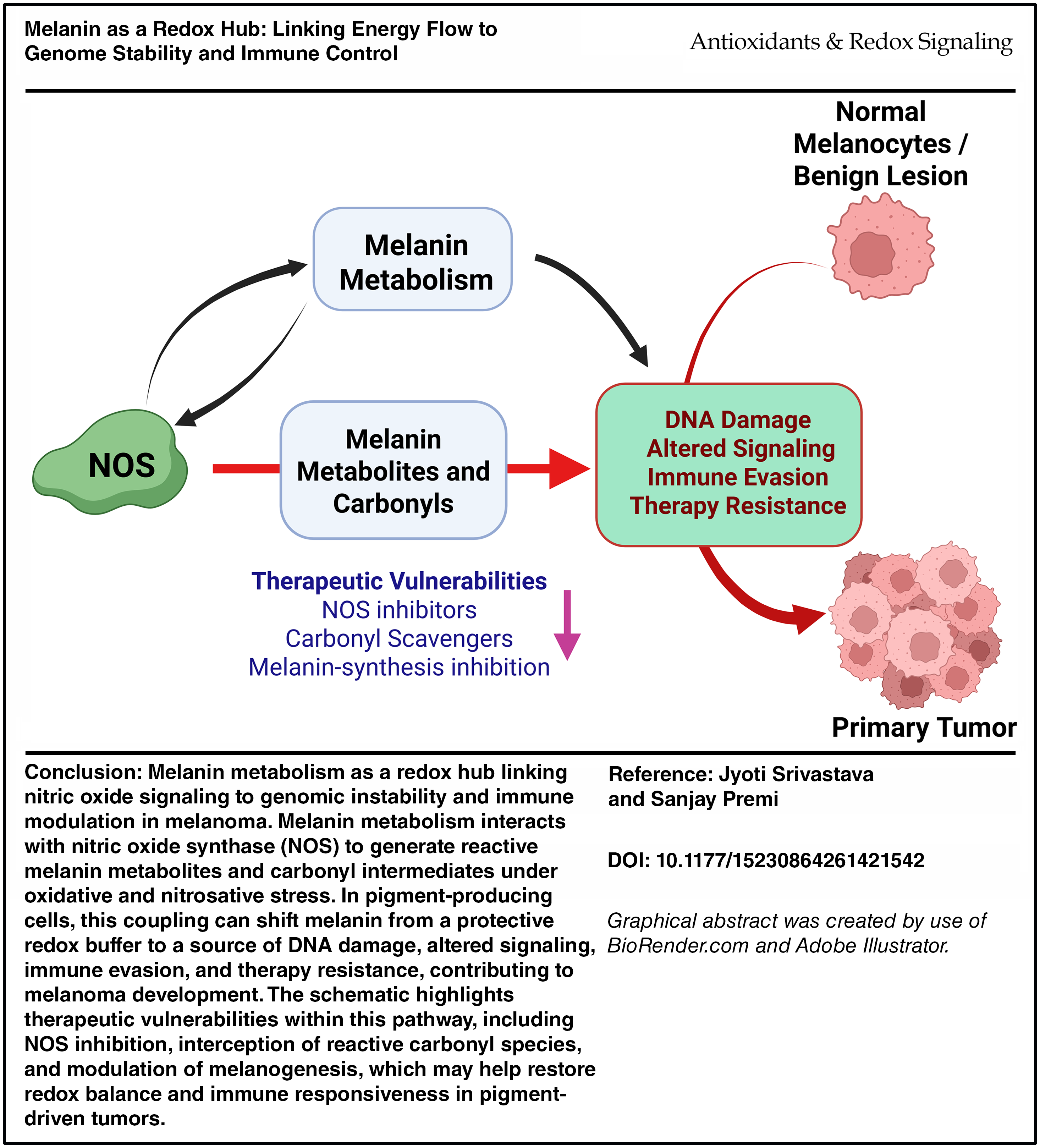
Graphical Abstract
Keywords
Introduction
Melanin as a redox-active system
Melanin is often viewed as nature’s sunscreen, protecting genomic integrity through broadband photon absorption, radical quenching, and ultraviolet (UV) attenuation in the epidermis (Brenner and Hearing, 2008). Yet, this optical model does not fully capture melanin’s intrinsic chemical reactivity. Rather than a static pigment, melanin is a redox-active polymer synthesized and packaged within melanosomes (membrane-bound organelles that store and traffic pigment), capable of storing, transforming, and redistributing redox potential in ways that influence genome stability, metabolic signaling, and immune tone.
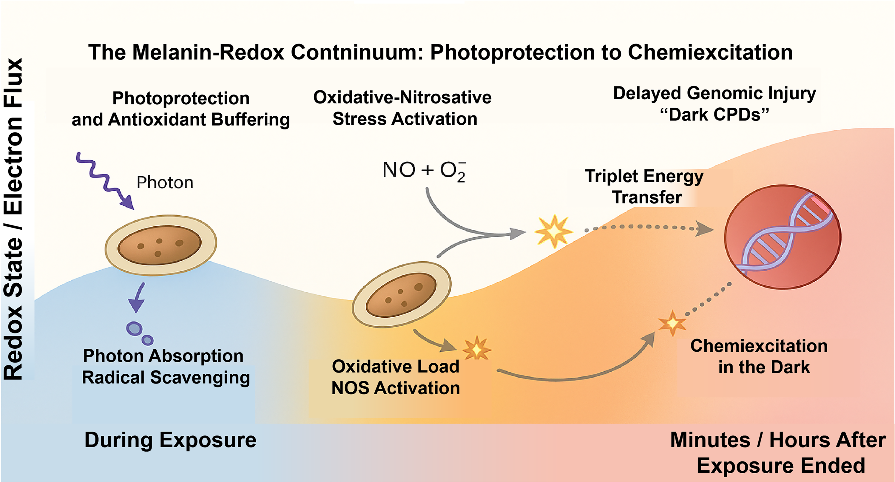
Melanoma, a malignancy of pigment-producing melanocytes, exposes the limits of a purely protective paradigm. After ultraviolet A radiation (UVA) exposure, cyclobutane pyrimidine dimers (CPDs) continue to accumulate for hours even in the absence of light, indicating that melanin’s oxidative state (Mouret et al., 2006) - not photon flux alone - can govern post-exposure DNA lesion formation (Premi et al., 2015). This post-irradiation phase reflects chemiexcitation, in which reactive nitrogen species (RNS) oxidize melanin fragments to triplet-state carbonyls that transfer electronic energy to DNA, producing “dark” CPDs (Fig. 1). The result is a temporally extended mutagenic window that converts photoprotection into a latent genotoxic pathway [Figures 1 - 4 in Premi et al., (2015)].
In this review, we propose that these principles may extend beyond cutaneous melanoma and exemplify a generalizable redox paradigm: melanin acts as a dynamic “redox hub” that conditions how pigment-bearing cells translate environmental energy into genomic and immunological outcomes. Pigment-rich organs such as the skin, retina, inner ear, and brain use melanin to buffer oxidative stress, while also risking redox imbalance that can contribute to disease (Kaufmann and Han, 2024; Maurya et al., 2023; Moreno-Garcia et al., 2021; Wang et al., 2006). Accordingly, melanin metabolism offers a framework connecting photobiology, cancer biology, immunology, and neuroscience.
Mechanistically, this shift is orchestrated through nitric oxide synthase (NOS) activation. Pigment synthesis and oxidative tone converge to induce NOS activity in melanocytes and stromal cells, generating nitric oxide (NO) and peroxynitrite that modify melanin, proteins, and nucleic acids (Hayden and Ghosh, 2011; Monteiro et al., 2015; Rizza and Filomeni, 2018; Tsatmali et al., 2000; Xu et al., 2018; Yarlagadda et al., 2017). Clinically, inducible NOS (iNOS) expression and nitrosative stress markers associate with poor prognosis, immune exclusion, and therapeutic resistance in melanoma (Pawelek, 2007; Srivastava et al., 2025; Tsatmali et al., 2000; Yadav et al., 2025; Yarlagadda et al., 2017). Together, these findings define a pigment–NOS–chemiexcitation axis that couples melanogenesis to genome instability and redox-driven immune modulation.
Beyond photochemistry, this circuitry intersects with master redox regulators. Nuclear factor erythroid 2-related factor 2 (NRF2), a central node in oxidative stress defense, integrates with melanocytic transcriptional programs and can modulate pigment output via the phosphoinositide 3-kinase-protein kinase B-microphthalmia-associated transcription factor (MITF) pathway (Ma, 2013; Ogawa and Ishitsuka, 2022; Shin et al., 2014). This intersection places melanin metabolism at the nexus of stress response and lineage identity. Even “amelanotic” metastases preserve expression of melanogenic enzymes (tyrosinase [TYR], TYR-related protein 1 [TYRP1], and DCT), supporting that melanocytic lineage programs maintain a persistent redox phenotype independent of visible pigment.
Because this framework draws on concepts that may be unfamiliar outside redox chemistry, we briefly define two terms. “Chemiexcitation” refers to the generation of electronically excited molecules through chemical reactions rather than direct photon absorption - a form of “chemical light” in which high-energy intermediates, such as triplet-state carbonyls, can donate energy to DNA and create CPDs long after UV exposure has ended (Brash, 2016; Goncalves et al., 2023; Premi et al., 2015; Premi and Brash, 2016). “Nitrosylation” (typically S-nitrosylation) describes the reversible addition of NO-derived groups to cysteine residues on proteins, altering activity, localization, or interactions, and functioning as a redox-regulated post-translational modification analogous to phosphorylation (Fernando et al., 2019; Hernansanz-Agustin et al., 2013; Rizza and Filomeni, 2018). Together, chemiexcitation and nitrosylation provide a chemical vocabulary linking melanin chemistry to nuclear and immune pathways in melanoma and related pigment systems.
In the substantia nigra, neuromelanin accumulates within dopaminergic neurons and can function both as an antioxidant sink and as a source of oxidative and inflammatory stress, contributing to selective neuronal vulnerability in Parkinson’s disease (Moreno-Garcia et al., 2021; Teleanu et al., 2022). In the retinal pigment epithelium (RPE), melanin buffers high photon flux and iron-dependent oxidation, yet photobleaching and age-related changes can convert this pigment from an antioxidant reservoir into a pro-oxidant driver of retinal degeneration (Kaufmann and Han, 2024; Maurya et al., 2023; Wang et al., 2006). These examples suggest that melanin-dependent redox balance - and its breakdown - may represent a recurring theme in neurodegeneration, ocular disease, and barrier-tissue immunity, not only in cutaneous oncology.
This redox-centric view reframes pigmentation as a metabolic program rather than a colorimetric trait. The evolutionary logic is conserved: melanin originated as a universal redox buffer in microbial photoprotection and persists in vertebrate tissues such as skin, retina, and brain, where oxidative energy must be absorbed and redistributed (Li et al., 2021; Ma et al., 2019; Yang et al., 2025). Neuromelanin and RPE melanin share this circuitry, emphasizing a cross-tissue pigment logic that couples environmental stress to redox adaptation. Recent work further suggests that chemiexcitation of melanin and melanin-like chromophores may operate in the retina and in neurotransmitter systems, raising the possibility that “dark” excited states also shape neurodegenerative and retinal pathologies (Goncalves et al., 2023).
Modern imaging advances now make this chemistry quantifiable. Melanin-specific positron emission tomography (PET) tracers ([18F]P3BZA, [18F]DMPY2) and melanocortin 1 receptor (MC1R)-targeted radiotherapies allow in vivo assessment of pigment oxidation and therapeutic response (Li et al., 2021; Ma et al., 2019; Yang et al., 2025). These tools transform pigment biology into a measurable pharmacologic landscape, converting melanin from a descriptive biomarker to an actionable variable. In parallel, redox and nitrosylation profiling, as well as emerging neuromelanin- and RPE-sensitive imaging modalities, provide opportunities to follow melanin-coupled redox signaling in the brain and eye, bringing pigment biology into mainstream discussions of neurodegeneration and ocular disease (Kaufmann and Han, 2024; Maurya et al., 2023; Moreno-Garcia et al., 2021; Teleanu et al., 2022; Wang et al., 2006).
To support interdisciplinary readers, we envision a brief “Concept Box 1” accompanying Figure 1 that summarizes key terms (chemiexcitation, triplet carbonyls, peroxynitrite, and S-nitrosylation) with simple schematics.
Concept Box 1. Essential Terms in Pigment-Linked Redox Biology
Enzymes producing nitric oxide (nNOS, eNOS, iNOS). Activated by oxidative stress, melanogenesis, NF-κB, STAT1/3, and NRF2. Provide the NO and peroxynitrite that fuel chemiexcitation and nitrosylation.
Strong oxidant formed by NO+ superoxide. Oxidizes melanin into chemiexcitation-competent fragments. Drives nitrosative stress and pigment fragmentation in melanoma and RPE.
Chemical, not photonic, generation of electronically excited molecules (triplet carbonyls). Responsible for Occurs in melanin, catecholamines, lipids, and other oxidized biomolecules.
High-energy reaction intermediates created when oxidized melanin fragments enter an excited triplet state. Directly transfer energy to DNA, generating CPDs without light. Central to melanin chemiexcitation across skin, retina, and brain.
CPDs formed post-irradiation through triplet-carbonyl energy transfer. Create a Enriched at open chromatin and enhancer elements.
Reversible addition of NO-derived groups to protein cysteines. Alters enzyme activity, signaling, and DNA repair. Hyper-nitrosylation in melanoma suppresses nucleotide-excision repair and interferon signaling.
The oxidation spectrum of melanin (reduced → semiquinone → quinone). Determines whether melanin is Predicts delayed CPD yield, NOS activation, and pigment-linked immune tone.
Radiotracers such as [18F]P3BZA and [18F]DMPY2. Bind melanin polymer and quantify its oxidation state in vivo. Allow non-invasive imaging of melanoma burden, pigment chemistry, and therapy response.
Molecules (AZ, MBPD, DBAS) that intercept excited melanin intermediates. Block chemiexcitation and prevent delayed CPDs. Foundation for
By integrating photobiology, redox chemistry, and tumor immunology, we propose a unified framework in which pigment metabolism shapes genomic stability and therapeutic outcome, with broader implications for cancer therapy, neurodegeneration, ocular disease, and systemic immunity.
Throughout this review, melanoma is treated as the experimentally resolved anchor for pigment-linked redox biology, whereas extensions to other pigment-bearing tissues are presented as biologically plausible but incompletely validated hypotheses.
For clarity of scope, this review follows a tiered structure: Sections—“Key Breakthroughs (2015–2025) Redefining Pigment-Linked Redox Biology” to “Melanin’s Protective Role and the Emergence of Paradox” summarize established cutaneous mechanisms and in vivo evidence; Sections—“Genomic Hotspots, Chromatin Landscapes, and the Emerging Footprint of Chemiexcitation” to “Melanin, Immunobiology, and the Tumor Microenvironment” examine NOS signaling, nitrosylation, and immune modulation; and Sections “Translational Outlook: Therapeutic Modulation of the Melanin-Redox Axis” to—“Outstanding Questions” address translational strategies and cross-tissue extensions presented as emerging concepts requiring further validation.
Key Breakthroughs (2015–2025) Redefining Pigment-Linked Redox Biology
Table 1 provides a chronological map of the key discoveries (2015–2025) discussed in this review. Sections “Chemiexcitation and delayed DNA damage” to “Pigment imaging and redox-theranostic advances” then organize these advances into three linked domains: chemiexcitation/delayed CPDs, NOS-nitrosylation with immune consequences, and melanin imaging/theranostics.
A Decade of Key Discoveries Linking Pigment Metabolism to Redox Biology (2015–2025)

CPD, cyclobutane pyrimidine dimer; NOS, nitric oxide synthase; PET, positron emission tomography; UV, ultraviolet.
Chemiexcitation and delayed DNA damage
Foundational studies established that melanin fragments oxidized by peroxynitrite generate electronically excited triplet carbonyls capable of transferring energy to DNA long after UV exposure (Premi et al., 2015). Recent work now validates this mechanism across cellular, ex vivo, and in vivo systems, confirming chemiexcitation as a physiological process. These findings are strongly supported in melanocytes, melanoma, and porcine skin. Potential parallels in the retina and neuromelanin systems remain hypothetical and require direct in vivo confirmation. In 2025, Yadav et al. (2025) demonstrated that melanin oxidation state dictates the magnitude of delayed CPD formation in both murine and human melanocytes, refining the link between presence of melanin, melanin oxidation, and DNA damage.
The most definitive in vivo confirmation of chemiexcitation was reported by Pospíšil et al. (2025). Using porcine skin, they showed that UVA exposure triggers formation of melanin-derived triplet carbonyls that produce CPDs for hours after irradiation. They validated this using time-resolved luminescence, photolyase rescue, and triplet-quenching pharmacology, providing the strongest biological confirmation that melanin acts as a post-photonic energy donor in intact tissue.
An independent line of evidence emerged from ACS Chemical Biology 2023 (Goncalves et al., 2023), showing that dopamine, serotonin, and melatonin analogs undergo oxidative chemiexcitation capable of generating CPDs even in the absence of light. This broadens chemiexcitation beyond melanin and suggests relevance to oxidative neurochemistry, neurodegeneration, and inflammatory redox cycling, but it does not yet establish in vivo CPD formation in the brain.
High-resolution CPD-seq and assay for transposase-accessible chromatin using sequencing (ATAC-seq) analyses revealed that delayed CPDs accumulate at open chromatin, enhancers, and transcription-factor binding sites, indicating that chemiexcitation follows chromatin accessibility, consistent with but more spatially precise than earlier CPD-seq work (Elliott et al., 2023; Figs. 1–2 in (Bohm et al., 2025; Hu et al., 2015; 2017; Jiang et al., 2021).
While lipid peroxidation-derived dioxetanes and reactive oxygen species (ROS)-driven lesions can also generate oxidative DNA damage, the chemiexcitation model currently provides the most coherent explanation for delayed, photolyase-rescuable CPDs occurring post-irradiation in pigmented systems. Thus, we conclude that UV mutagenesis is no longer limited to irradiation. Melanin continues to generate CPDs in darkness through chemically excited intermediates. Chemiexcitation is well supported in melanocytes and pigmented skin models; analogous excited-state chemistry has been demonstrated in neurotransmitter systems in vitro, raising testable hypotheses for other tissues.
NOS-driven nitrosylation and immune modulation
A second major advance centers on NOS as an upstream metabolic regulator linking pigmentation, DNA repair suppression, immune tone, and therapy resistance in melanoma. This section summarizes pathways with direct evidence in melanocytes and melanoma, while noting that parallels proposed in the retina, neuromelanin-rich regions, and cochlear pigment systems remain preliminary and require further validation.
Cancer Research 2025 showed that neuroblastoma RAS viral oncogene (NRAS)-mutant melanoma exhibits a NOS-driven nitrosylome, including S-nitrosylation of MAPK regulators and immunomodulators, driving MEK-inhibitor resistance and immune evasion (graphical abstract in Srivastava et al., 2025). Pharmacologic or genetic denitrosylation restored repair fidelity and sensitized tumors to targeted therapy, providing in vivo causal support for the role of nitrosylation in therapeutic outcome.
Complementary findings from Int J Mol Sci demonstrated that NOS/NO-mediated S-nitrosylation suppresses signal transducer and activator of transcription 1 (STAT1)-dependent interferon signaling and dampens immune activation (Chen et al., 2022; Garg et al., 2023). These results integrate with longstanding observations linking high iNOS expression to immune-excluded melanomas and poor prognosis (Ding et al., 2021). In melanoma, NOS thus emerges as a central metabolic–immune regulator: through nitrosylation, it links melanogenesis to DNA repair suppression, interferon silencing, metabolic rewiring, and therapy resistance. The evidence summarized in this subsection reflects mechanisms directly demonstrated in cutaneous models.
Pigment imaging and redox-theranostic advances
A third set of breakthroughs has transformed melanin from a descriptive histological feature into a quantifiable redox biomarker and theranostic target, enabling real-time measurement of pigment oxidation and lineage identity.
Melanin-targeted PET imaging has progressed rapidly, driven by second-generation dibenzamide tracers. A landmark report in Theranostics 2025 showed the first-in-human use of [18F]DMPY2, demonstrating high tumor-to-background ratios and sensitive detection of melanoma metastases (Yang et al., 2025). These agents enable in vivo quantification of pigment oxidation and distribution, marking a shift from static pigment histology toward dynamic, redox-responsive imaging. Parallel tracers such as [18F]P3BZA and related benzamide derivatives continue to refine melanin PET for both diagnosis and treatment monitoring.
MC1R-directed radionuclide therapy represents another major translational development. Preclinical studies show that MC1R-targeting peptides can deliver α-particle-emitting radionuclides to melanoma with high specificity. When combined with immune checkpoint inhibitors, this produces synergistic suppression of tumor growth (Li et al., 2021). These findings support the concept of pigment-lineage theranostics, where melanocytic differentiation markers - not only melanin polymer itself - serve as gateways for targeted intervention.
Finally, outside the skin, neuromelanin-sensitive MRI has matured into a robust biomarker of dopaminergic neuronal health. Recent studies show that loss or altered oxidation of neuromelanin in the substantia nigra correlates with Parkinson’s disease progression, redox imbalance, and neuronal vulnerability (He et al., 2023). This establishes melanin imaging as a cross-disciplinary tool that links pigment chemistry to neurodegeneration, emphasizing the systemic relevance of pigment-redox biology.
Based upon these findings, we suggest that melanin is now a measurable, imageable, and targetable redox organelle. Across PET, radiotheranostics, and MRI, pigment redox state is quantifiable in vivo, enabling translational strategies spanning oncology, ophthalmology, and neuroscience.
To delineate the evidentiary basis underlying the mechanistic framework presented in this review, Table 2 summarizes the principal pigment-linked redox processes discussed and stratifies them according to the level of experimental support, including in vivo validation, cell-based and biochemical demonstration, and cross-system hypothesis-generating analogies. This framework provides a structured reference for interpreting subsequent sections that extend the melanin-redox paradigm beyond cutaneous systems while maintaining clear boundaries between established mechanisms and emerging concepts.
Evidence Levels Supporting Melanin-Linked Redox Mechanisms Discussed in this Review

CPD, cyclobutane pyrimidine dimer; IFN, interferon; NOS, nitric oxide synthase; PET, positron emission tomography; RPE, retinal pigment epithelium; UVA, ultraviolet A radiation.
Melanin’s Protective Role and the Emergence of Paradox
While melanin evolved to dissipate photon energy and buffer oxidative stress, recent work reveals that this protection is conditional rather than absolute. This section examines the biochemical and kinetic basis of this transition and defines the conditions under which photoprotection gives way to chemiexcitation-driven genome injury. In melanocytes, this dark phase of DNA damage originates from melanin redox chemistry rather than photons: RNS oxidizes melanin fragments into electronically excited triplet carbonyls that transfer energy to DNA, forming delayed CPDs (dCPDs; Premi et al., 2015; Premi and Brash, 2016; Yadav et al., 2025). Quantitatively, delayed lesions constitute nearly half of total CPDs in pigment-competent cells, establishing melanin as a conditional genome-injury catalyst, protective when balanced but genotoxic when over-oxidized (Goncalves et al., 2023; Premi et al., 2015; 2019; Premi and Brash, 2016; Yadav et al., 2025).
Melanin’s extended π-electron network confers broad redox plasticity. Under physiological conditions, eumelanin buffers oxidative stress through radical scavenging and sequestration of redox-active metal ions, whereas pheomelanin is associated with pro-oxidant behavior, lipid peroxidation, and increased melanoma susceptibility (Napolitano et al., 2014; Nasti and Timares, 2015). This contrast underscores that the melanin paradox is not uniform across pigment subtypes. Yet, the same conjugated structure that dissipates excitation energy can, when over-oxidized, spawn reactive quinones, superoxide, and singlet oxygen (d’Ischia et al., 2015; Ito et al., 2000; Mo et al., 2022). During active melanogenesis, TYR-driven oxidation of DOPA and 5,6-dihydroxyindole intermediates generate semiquinones and hydrogen peroxide; under chronic UV or inflammatory stress, these intermediates propagate oxidative cascades rather than quench them.
This redox inversion defines the melanin paradox: a pigment evolved for defense that becomes a catalyst of injury. Peroxynitrite and related oxidants promote melanin over-oxidation and fragmentation, consistent with a dioxetane→triplet-carbonyl mechanism that rationalizes CPD formation in the absence of photons (Brash, 2016; Goncalves et al., 2023; Premi et al., 2015; Premi and Brash, 2016; Yadav et al., 2025). While alternative pathways such as lipid peroxidation-derived dioxetanes and ROS-driven lesions can also generate oxidative DNA damage, chemiexcitation currently provides a leading explanation for photolyase-rescuable, post-irradiation CPDs in pigmented systems. By converting stored oxidative potential into chemical excitation, melanin effectively transforms from a protective polymer into a photochemical energy donor. UVA1 remains the most efficient initiator of melanin-mediated delayed CPDs, highlighting the interplay between dose, oxidative state, and post-exposure chemistry (Premi et al., 2015).
Within cutaneous models, pigmented melanocytes display elevated basal NOS activity and peroxynitrite flux that modify melanin’s redox potential. NO and peroxynitrite participate in dopaquinone cycling and melanosome maturation, closing a feed-forward loop in which melanogenesis primes NOS, and NOS oxidants in turn regenerate reactive pigment fragments (Reszka et al., 1998; Romero-Graillet et al., 1997; Sasaki et al., 2000). At equilibrium, melanin acts as a photon sink; beyond that threshold, it becomes a self-reactive redox capacitor that channels stored energy back into DNA (Fig. 1).
A brief note on scope is that this chemistry extends beyond pigment. Neurotransmitters such as serotonin, dopamine, and melatonin can undergo peroxynitrite-driven chemiexcitation, forming triplet carbonyls that induce CPDs even in darkness (Goncalves et al., 2023). Lipids, sugars, and α-aminoketones can likewise form dioxetanes during peroxidation or Maillard-type reactions (Bechara et al., 2007; Timmins et al., 1997; Wondrak et al., 1995), revealing a broader “hidden photochemistry” of oxidative stress of which melanin is the most visible exemplar. However, evidence for CPD formation outside cutaneous or pigment-competent systems remains preliminary and should be regarded as hypothesis-level rather than established biology.
Mapping studies show that delayed CPDs accumulate preferentially within open, transcriptionally active chromatin, implying that metabolic activity and repair engagement heighten local vulnerability (Bohm et al., 2025; Elliott et al., 2023; Jiang et al., 2021). Thus, carcinogenic potential is determined not only by UV dose but also by the pigment’s redox state long after exposure. NO-driven oxidation converts melanin from an energy sink to an energy donor, producing a second wave of genome injury long after sunlight is gone. Figure 1 illustrates this transition from photoprotection to chemiexcitation, establishing the central theme of this review: pigmentation as a dynamic redox continuum that integrates environmental stress, metabolism, and genomic stability. Understanding where this delayed mutagenesis occurs in the genome reveals how pigment chemistry imprints mutation patterns characteristic of melanoma.
Genomic Hotspots, Chromatin Landscapes, and the Emerging Footprint of Chemiexcitation
UV damage does not occur uniformly across the genome. Instead, DNA lesions are sculpted by chromatin structure, transcriptional activity, and nucleosome dynamics that define regions of selective vulnerability (Frigola et al., 2021; Hu et al., 2015; 2017; 2019; Sabarinathan et al., 2016). Genome-wide CPD-seq and excision repair sequencing analyses have revealed that both lesion formation and repair preferentially occur within accessible chromatin marked by active transcription, DNase hypersensitivity, and enhancer signatures. This spatial bias provides a mechanistic framework for understanding why melanoma mutational spectra are concentrated at regulatory elements and transcription-factor footprints (Bohm et al., 2025; Elliott et al., 2023; Poetsch et al., 2018).
Delayed, melanin-dependent CPDs (dCPDs) follow this same architectural logic. CPD-seq comparisons demonstrate that post-UV chemiexcitation lesions accumulate within open, transcriptionally engaged regions, whereas immediate, photon-induced CPDs distribute more broadly (Frigola et al., 2021; Hu et al., 2015; 2017; 2019; Poetsch et al., 2018; Premi et al., 2019). The correlation between delayed CPDs and ATAC-seq peaks suggests that chemiexcitation preferentially directs stored redox energy toward DNA already poised for transcription and repair (Fig. 2). Rather than random mutagenesis, pigment chemistry appears to reinforce pre-existing genomic hierarchies, focusing damage at loci critical for lineage identity and signal regulation. These data indicate that pigment-driven lesion formation is functionally coupled to transcriptional activity rather than random exposure, suggesting functional coupling between energy metabolism and chromatin state.
This selectivity implies that melanin-mediated chemiexcitation and nucleotide-excision repair (NER) compete for the same chromatin territories. In repair-proficient cells, transcriptionally active regions experience recurrent cycles of injury and restoration, a process predicted to increase local mutational burden at promoters and enhancers. Consequently, the genome’s most active domains become its most fragile, linking metabolic energy flow to mutational evolution.
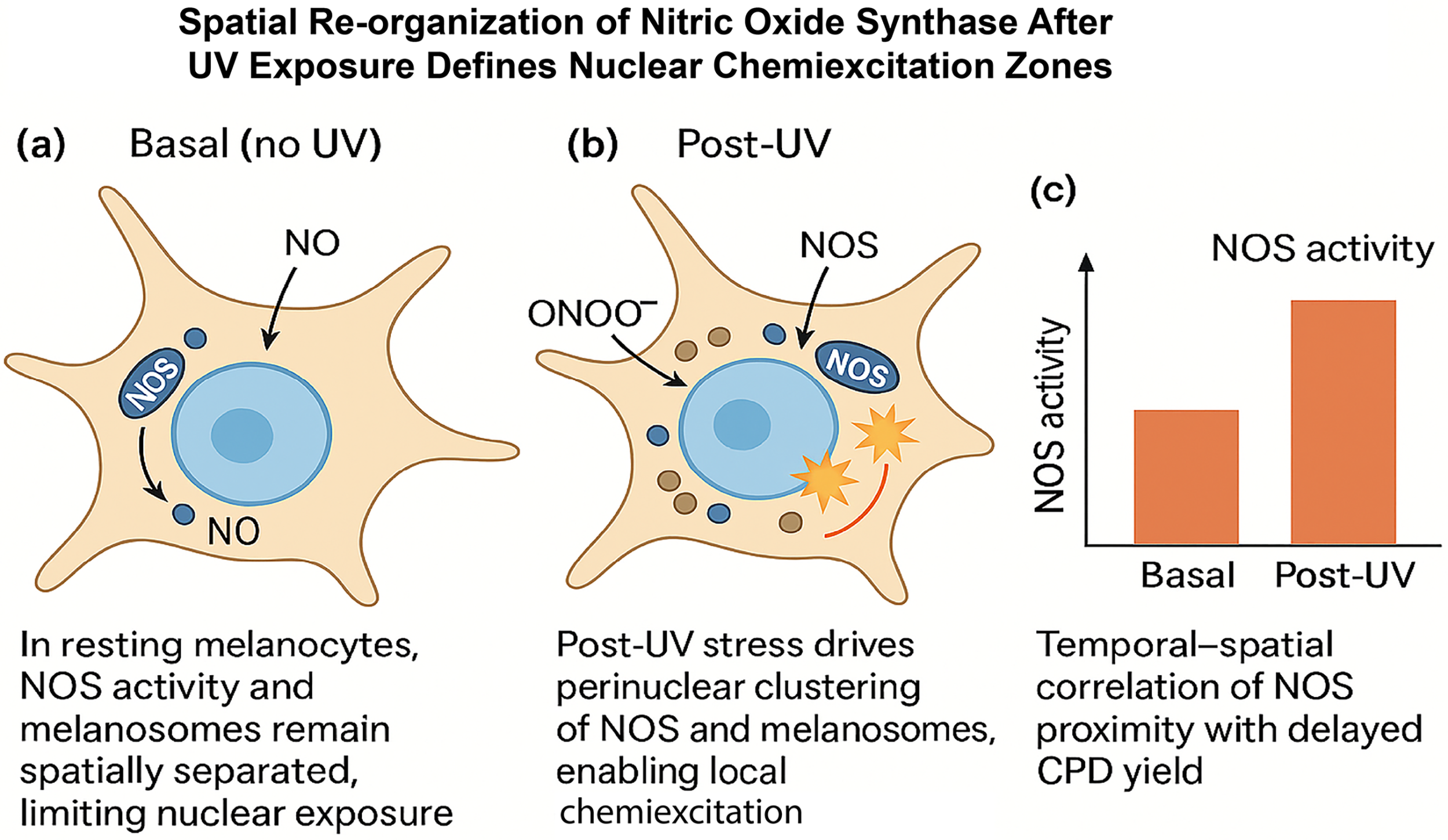
Mechanistic insights support this spatial coupling. In resting melanocytes, NOS resides near the endoplasmic reticulum and plasma membrane, spatially segregated from melanosomes. Following oxidative or UV stress, studies in melanocytes indicate that both NOS and melanosomes migrate toward the nucleus, forming perinuclear zones enriched in peroxynitrite and triplet-carbonyl formation. Within these microdomains, energy transfer from electronically excited melanin fragments to DNA yields dCPDs concentrated near euchromatin borders (Fig. 3). This model provides a spatial framework that explains the overlap between chemiexcitation signatures and active chromatin maps and reinforces the concept that the subcellular geography of redox reactions dictates genomic fate.

Together, these findings redefine pigment-driven DNA damage as an organized, not stochastic, process. The intersection of melanin localization, NOS activation, and chromatin architecture converts metabolic flux into patterned genomic injury. Chemiexcitation thus bridges photobiology and epigenetics, an energetic conduit through which pigmentation imprints mutation landscapes and fuels melanoma evolution.
Melanin as a Redox Engine: NOS, Nitrosylation, and Immune Crosstalk
The redox activity of melanin is inseparable from NO signaling. In pigment-producing cells, oxidative melanogenesis and inflammatory cues converge on NOS, establishing a self-reinforcing circuit that couples pigment turnover to nitrosative stress, protein modification, and immune modulation. This section focuses on how NOS activation and downstream S-nitrosylation translate pigment chemistry into durable changes in DNA repair, signaling networks, and tumor–immune interactions.
Oxidative intermediates and inflammatory cytokines activate NOSs through nuclear factor kappa-light-chain-enhancer of activated B cells (NF-κB), STAT1/3, and NRF2 signaling (Fecker et al., 2002; Hayden and Ghosh, 2011; Ma, 2013; Ogawa and Ishitsuka, 2022; Romero-Graillet et al., 1997; Sasaki et al., 2000; Shin et al., 2014). As a result, pigment turnover and NOS activity form a self-reinforcing circuit: melanogenesis primes NOS, and NOS-derived oxidants remodel pigment chemistry. In melanocytes, basal NOS activity exceeds that of neighboring keratinocytes and fibroblasts, maintaining a persistent nitrosative tone that influences melanosome maturation, mitochondrial respiration, and calcium signaling (Weller, 1999). Analogous pigment–NO interactions have been described in neuromelanin and the RPE; however, their mechanistic roles in immune regulation remain less well defined than in melanoma (Moreno-Garcia et al., 2021; Zucca et al., 2023).
This coupling establishes the biochemical foundation for chemiexcitation. NO reacts with superoxide to form peroxynitrite, which oxidizes melanin fragments into electronically excited triplet carbonyls capable of energy transfer to DNA (Premi et al., 2015; Premi and Brash, 2016; Yadav et al., 2025). The resulting delayed CPDs integrate redox metabolism with genomic injury, extending mutagenic potential long after UV exposure. Persistent nitrotyrosine accumulation in pigment-competent cells indicates ongoing NOS activity even in the dark (Denat et al., 2014; Moreno-Garcia et al., 2021). This relationship between NOS activity, pigment oxidation, and delayed DNA damage is directly supported in melanocytes and melanoma models.
In melanoma, iNOS expression scales with melanin density and oxidative load (Ding et al., 2021; Massi et al., 2009; Pawelek, 2007; Yadav et al., 2025; Yarlagadda et al., 2017). Even amelanotic metastases retain expression of TYR, TYRP1, and DCT (Journe et al., 2011; Sturm et al., 2024), demonstrating that melanocytic lineage programs preserve this redox circuitry independent of visible pigment. Through continuous NO flux and melanin oxidation, the lineage maintains an endogenous source of genome injury and signal rewiring, a hallmark of metabolic persistence rather than pigment abundance, reflecting lineage-specific redox programming rather than pigment abundance alone.
The downstream consequence is S-nitrosylation, a reversible modification of cysteine residues that alters protein function (Foster et al., 2009; Li et al., 2025; Srivastava et al., 2025; Yarlagadda et al., 2017). In normal melanocytes, transient S-nitrosylation fine-tunes TYR activity and melanosome maturation. In melanoma, excessive NOS flux drives widespread hyper-nitrosylation of DNA repair enzymes (poly(ADP-ribose) polymerase 1, XRCC1, and OGG1), chromatin remodelers, and immune regulators (Ding et al., 2021; Massi et al., 2009; Sharma et al., 2021; Srivastava et al., 2025). This suppresses NER, dampens interferon responses, and impairs immunogenic cell death (ICD), reinforcing survival and therapy resistance. Importantly, pharmacologic or genetic denitrosylation restores repair capacity and resensitizes NRAS-mutant melanomas to MEK inhibition (Liang et al., 2024; Sharma et al., 2021; Srivastava et al., 2025).
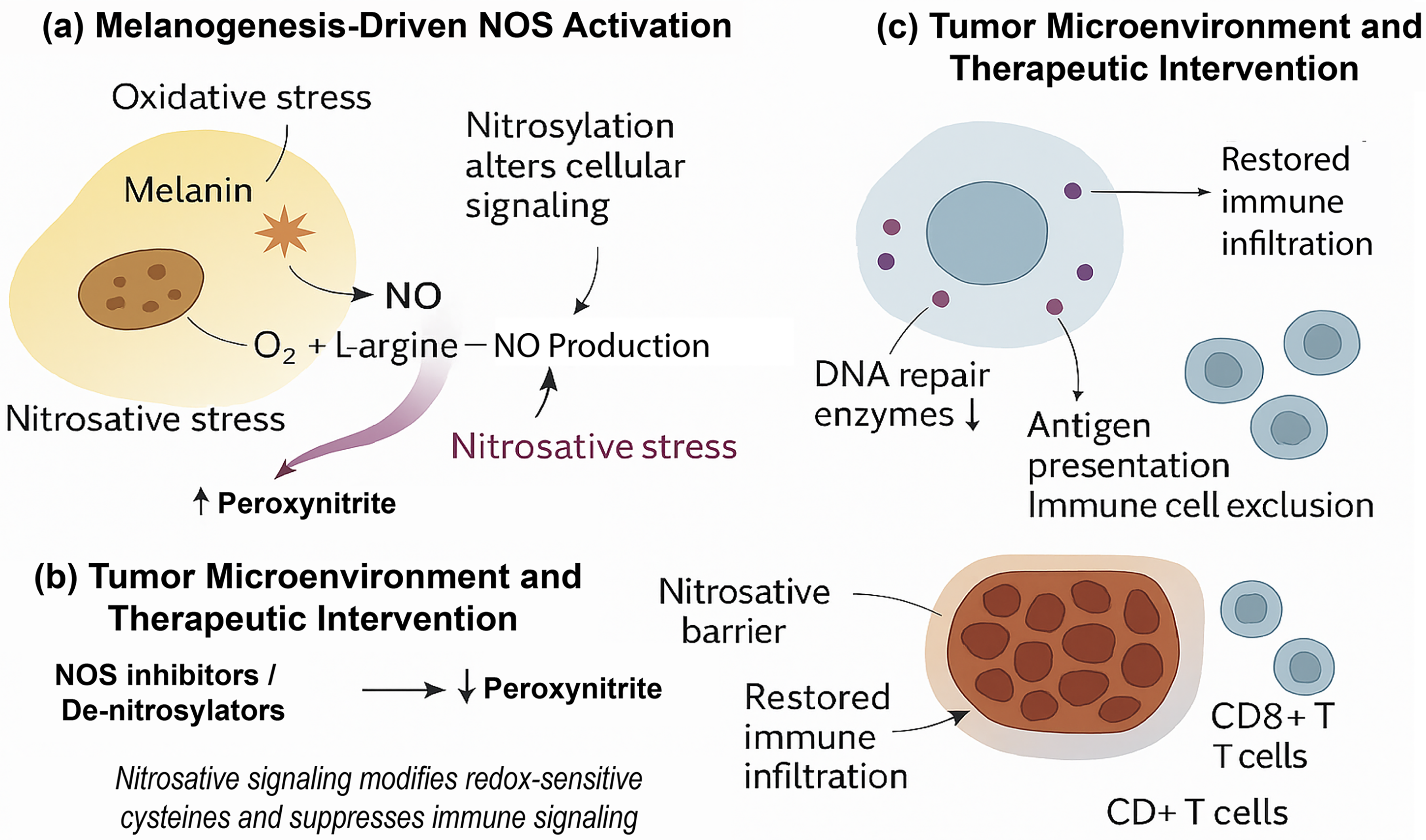
Functionally, pigment-proximal nitrosative stress shapes tumor–immune interactions. High iNOS and peroxynitrite concentrations are localized to pigment-dense tumor zones, producing nitrated chemokines and modified T-cell receptors that hinder effector infiltration (Capietto and Delamarre, 2022; Molon et al., 2011; Tanese et al., 2012; Tcyganov et al., 2022). These reactive-nitrogen barriers correspond spatially to oxidative hotspots identified by imaging and biochemical mapping (Fig. 4). NOS inhibition or peroxynitrite scavenging restores chemokine gradients and synergizes with checkpoint blockade in preclinical melanoma and colon models (Ekmekcioglu et al., 2017; Greiner et al., 2025; Grimm et al., 2013; Jimenez et al., 2024).
Viewed as a system, the melanin–NOS–nitrosylation axis links three scales of biology:
Metabolic: oxidative pigment synthesis continuously generates reactive oxygen and nitrogen species. Chemical: peroxynitrite oxidation of melanin fragments drives chemiexcitation and delayed DNA damage. Immunological: S-nitrosylation and nitration remodel repair enzymes and cytokine networks to promote immune exclusion.
When balanced, this circuitry maintains photoprotection and redox homeostasis; when over-driven, it connects energy metabolism to mutation and immune escape. In melanoma, this axis is supported by direct genetic, biochemical, and in vivo evidence. The architecture of this self-sustaining redox circuit (Fig. 4) underpins the dual identity of melanin as both antioxidant reservoir and generator of stress, transforming a photoprotective polymer into a lineage-specific engine of genomic and immunological adaptation.
Melanin, Immunobiology, and the Tumor Microenvironment
Beyond intracellular signaling, pigment metabolism shapes the tumor microenvironment by regulating antigen presentation, cytokine gradients, and immune cell access. In melanoma, these effects are directly linked to melanin-dependent redox and nitrosative pathways; in other pigment-bearing tissues, related principles are emerging but remain less mechanistically defined. This section examines how pigment state, vesicular trafficking, and NO signaling collectively influence immune visibility and immune privilege across tissue contexts.
Pigment state as an immunophenotypic axis
In melanoma, pigmentation defines an axis between immune recognition and immune evasion. MITF-high, melanin-rich cells present lineage antigens and attract cytotoxic lymphocytes (de Vries et al., 1997; Mandelcorn-Monson et al., 2003; Rosenberg, 1999), whereas AXL-high, depigmented or dedifferentiated states suppress antigen presentation and resist interferon signaling (Tsoi et al., 2018; Willemsen et al., 2024). This inverse relationship between pigment synthesis and immune invisibility situates redox tone as a determinant of tumor immunophenotype. Oxidative and nitrosative stress, through NOS, NRF2, or NF-κB activation, push tumors along this axis (Haq et al., 2013; Jessen et al., 2020). Related but less direct observations have been reported in other pigment-containing systems: RPE cells downregulate MHC under oxidative stress (Taylor et al., 2021), and neuromelanin-containing neurons reduce antigen display as a neuroprotective adaptation (Zucca et al., 2014; 2023). These findings are largely correlative or context-specific and are presented here as conceptual parallels rather than mechanistically equivalent pathways.
Together, these findings reveal pigmentation as a rheostat of immune visibility: when the oxidative burden of melanogenesis rises, cells favor a low-antigen state; when redox tone subsides, differentiation and antigenicity return. Clinically, interventions that reduce oxidative pressure or NOS activity can restore pigment, antigen expression, and T-cell engagement (Capietto and Delamarre, 2022; Greiner et al., 2025; Molon et al., 2011; Tanese et al., 2012; Tcyganov et al., 2022).
Melanosomes and extracellular pigment communication
Pigment metabolism influences immunity not only through intracellular signaling but also via extracellular communication. Melanocytes and melanoma cells release melanosomes and pigment-laden vesicles containing lipids, cytokines, and redox mediators (Parikh et al., 2024; Pawelek, 2007; Prosperi et al., 2024). These vesicles are internalized by keratinocytes, fibroblasts, and macrophages, where they reshape antioxidant defenses and cytokine networks (Parikh et al., 2024). In the eye, RPE-derived vesicles modulate complement activation and macrophage recruitment (Manai et al., 2024), while, in the brain, neuromelanin released from dying neurons activates microglia through TLR and NF-κB pathways (Zhang et al., 2011). While the molecular composition of these vesicles differs by tissue, their shared feature is the transmission of pigment-associated redox information to immune cells.
This traffic of pigment-laden vesicles constitutes a biochemical quorum-sensing system, in which reduced melanin fragments promote repair and regeneration, whereas oxidized or nitrosylated species dampen inflammation and enforce tissue quiescence by redistributing redox information across the microenvironment. Although this model is best supported in skin, similar signaling logic has been proposed - though not yet fully validated - in ocular and neural contexts.
Nitrosative editing and immune privilege across organs
NO signaling is a unifying thread linking pigment to immune privilege. In the RPE, iNOS and eNOS regulate leukocyte adhesion, barrier permeability, and photoreceptor survival (Sripathi et al., 2012; Taylor et al., 2021). In the inner ear, melanin within the stria vascularis buffers free radicals and suppresses immune infiltration, contributing to auditory protection (Ito et al., 2022). In the meninges and substantia nigra, neuromelanin interacts with microglial NOS to control neuroinflammatory thresholds (Wilms et al., 2003; Zucca et al., 2014; 2023). Across these settings, pigment oxidation has been proposed to function as a rheostat defining when immune surveillance is permitted and when tolerance must prevail. However, the mechanistic depth and experimental validation of this process vary substantially between tissues. Pathology emerges when this equilibrium collapses. In the eye, chronic oxidative stress converts protective NO signaling into inflammatory uveitis; in the brain, persistent neuromelanin oxidation is associated with microglial hyperactivation; in the skin, direct experimental evidence supports the same chemistry underlying immune exclusion in melanoma. These represent variations on a shared theme - melanin-associated nitrosative editing - with melanoma providing the most mechanistically resolved example to date.
Melanin as a cross-tissue immune-modulatory interface
Viewed across biology, melanin functions as a multiscale regulator of immunity. Its biosynthesis couples oxygen consumption to antioxidant release, influencing tissue redox balance; its polymeric matrix sequesters metals and xenobiotics, shaping local inflammatory tone; its vesicle export distributes redox information among neighboring cells; and its NOS-coupled nitrosylation communicates directly with nuclear and mitochondrial pathways governing DNA repair and apoptosis.
In health, this circuitry preserves tissue integrity - absorbing radiation, neutralizing radicals, and restraining inflammation. In disease, it sustains chronic stress or immune escape. Across cancers, infections, and neurodegeneration, melanin marks the anatomical crossroads where metabolism meets immunity. Recognizing this continuity does not imply mechanistic equivalence across tissues but rather highlights a shared redox logic that is most fully defined in melanoma and remains to be experimentally resolved elsewhere. Therapeutically, manipulating this interface - by modulating NOS activity, melanin oxidation state, or vesicular signaling - offers a means to toggle tissues between immune silence and activation, extending beyond melanoma to ocular, neural, and mucosal immunity.
Translational Outlook: Therapeutic Modulation of the Melanin-Redox Axis
Melanin’s chemistry has moved beyond photobiology toward therapeutic relevance. The pigment that once defined photoprotection now delineates a network of redox vulnerabilities that can be pharmacologically tuned. Because melanogenesis couples oxidative flux, NO signaling, and immune tone, targeting this circuitry offers a unified strategy to modulate DNA damage, tumor metabolism, and immunogenicity in melanoma and other pigment-rich contexts. The translational challenge, therefore, is not to erase pigmentation but to redirect its chemistry - transforming melanin from a passive chromophore into a controllable redox interface.
Redox modulation as precision therapy
The pigment–NOS–nitrosylation axis provides multiple druggable nodes. NOS inhibitors (e.g., NG-Nitroarginine methyl ester, N-Nitro-
Chemiexcitation inhibitors and post-exposure photoprotection
Beyond NOS targeting, inhibitors of chemiexcitation represent a new class of “molecularly active, post-exposure photoprotectants.” Compounds such as acetyl zingerone (AZ), 3-(4-Methoxy-benzyl)-Pentane-2,4-dione, and related triplet-state quenchers block the transfer of excited energy from melanin intermediates to DNA (Brash, 2016; Premi and Brash, 2016). These agents operate in the post-exposure window, extinguishing delayed CPDs and oxidative afterglow - offering protection that begins after sunlight ends. Their mechanism differs fundamentally from UV filters: they intercept endogenous energy, converting chemiexcitation from a mutagenic process into a dissipative one. Ongoing trials and preclinical studies with AZ analogs indicate that triplet-quenching pharmacology could complement both sunscreens and DNA repair-enhancing agents to reduce cumulative genomic injury (Brash, 2016; Cabello et al., 2023; Premi and Brash, 2016; Vico et al., 2007).
In parallel, melanin metabolism can be visualized and quantified in vivo using PET tracers such as [18F]P3BZA and [18F]DMPY2, enabling real-time assessment of pigment oxidation and therapy response (Li et al., 2021; Ma et al., 2019; Yang et al., 2025). These tools render redox modulation measurable, providing pharmacodynamic readouts rather than surrogate biomarkers.
Reversing immune exclusion by redox reprogramming
Tumor immune evasion often reflects redox imbalance rather than antigenic loss. NOS-driven peroxynitrite production nitrates chemokines and T-cell receptors, physically excluding immune effectors from the pigment-dense core (Capietto and Delamarre, 2022; Molon et al., 2011; Tanese et al., 2012; Tcyganov et al., 2022). Correcting this imbalance, via NOS blockade, peroxynitrite neutralization, or metabolic rewiring, restores chemokine gradients and re-opens tumors to immune attack, complementing checkpoint blockade. Early combination strategies pairing NOS inhibition with anti-PD-1 therapy demonstrate additive effects in preclinical melanoma and colon models (Ekmekcioglu et al., 2017; Greiner et al., 2025; Grimm et al., 2013; Jimenez et al., 2024).
More broadly, therapies that modulate pigmentation state may alter immune tone. Pharmacologic induction of differentiation (via MITF upregulation) restores antigenicity, while suppression of NRF2 or iNOS signaling re-establishes interferon responsiveness. Thus, pigment reprogramming and immune activation are mechanistically linked in melanoma.
Integrating redox and therapy design
Integrating pigmentation biology into oncology reframes therapy design. Redox modulation intersects with every major treatment modality, phototherapy, radiotherapy, targeted inhibitors, and immunotherapy. Melanin’s redox network is not an epiphenomenon of lineage but a governing variable that dictates therapeutic sensitivity and resistance.
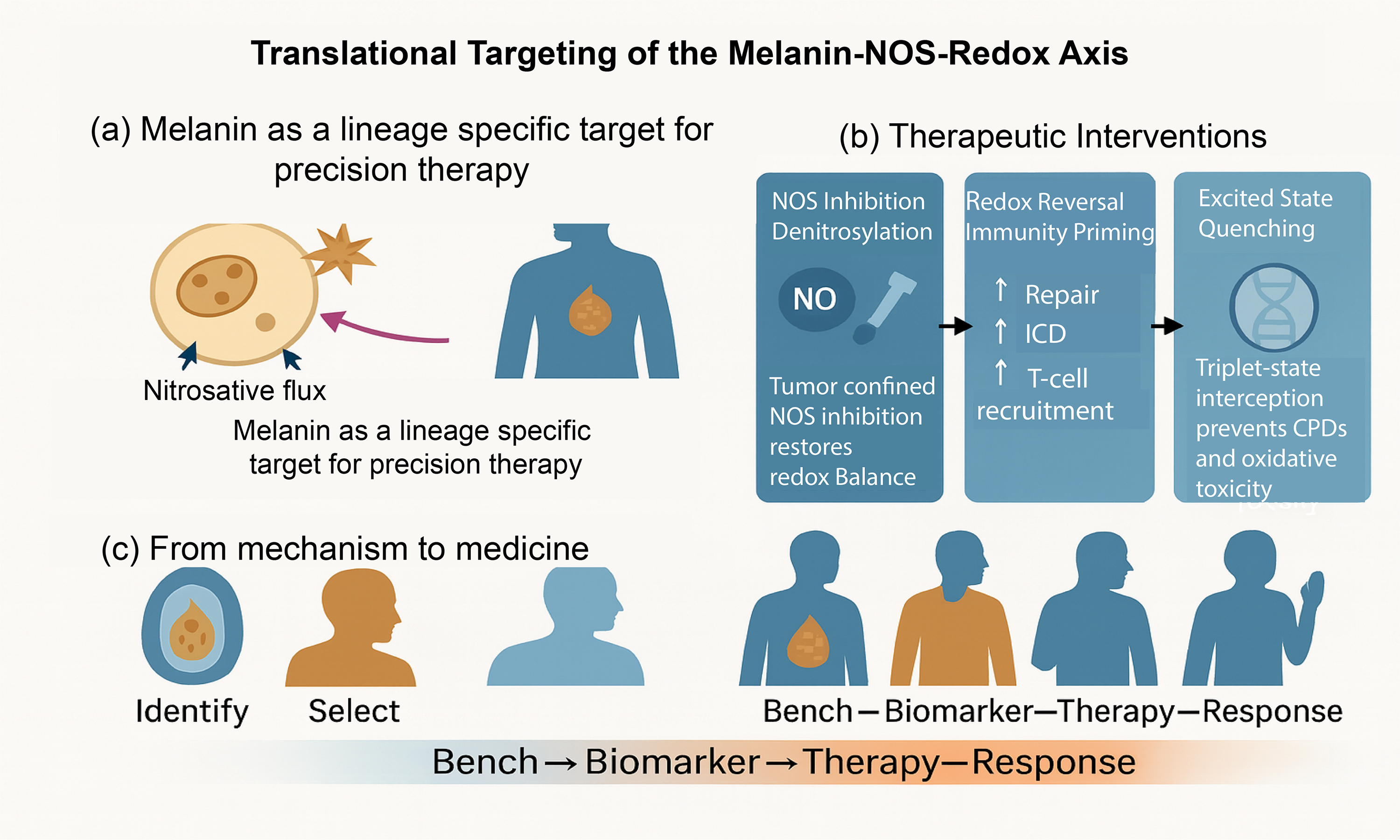
Conceptually, melanoma therapy may evolve from cytotoxicity toward redox choreography, fine-tuning the interplay between oxidative flux, NOS activity, and immune activation. Redox-responsive nanoparticles, controlled ROS generators, and denitrosylating agents could serve as adaptive levers, shifting the tumor microenvironment from reductive stagnation to oxidative awakening. By targeting melanin’s chemistry rather than its abundance, therapeutic intervention aligns with lineage-specific biology rather than against it.
In summary, melanin’s redefinition from photoprotector to redox modulator expands therapeutic imagination. Manipulating its oxidative, nitrosative, and chemiexcitative states could reconcile protection with precision, creating interventions that operate after light, across redox scales, and through immune pathways. This convergence of pigment biology and redox pharmacology marks a translational horizon - one where energy flow itself becomes the therapeutic target.
Practical directions and clinical sequencing
Therapeutically, the lessons emerging from pigment biology are pragmatic as much as conceptual. The first is one of specificity: NO is indispensable for vascular and neuronal function, so the success of this approach will depend on melanin- or MC1R-guided delivery that confines NOS modulation to the tumor. The second lesson is measurement. Melanin imaging and nitrosylation profiling now provide the means to quantify engagement of this axis in vivo, marking a shift from descriptive redox biology to trackable pharmacodynamics. A third principle concerns sequencing. Short-term NOS or denitrosylation priming may condition tumors for subsequent targeted or immune therapy, although optimal order remains to be defined. In practice, a stepped design - NOS or denitrosylation priming, followed by kinase inhibition and immune engagement - could transform transient responses into durable control. Finally, the field must recognize that manipulating pigmentation is not cosmetic: it is metabolic and immunological surgery at the level of redox tone. Recasting melanin as a therapeutic scaffold, rather than a risk factor, may redefine how we treat melanoma and, ultimately, how we understand pigment biology itself. Translating the chemistry of pigment metabolism into therapeutic opportunity requires viewing melanin not only as a biochemical end product but also as a dynamic redox interface. This section synthesizes mechanistic and translational insights by outlining how pigment-linked NO signaling, oxidative stress, and excited-state intermediates can be therapeutically modulated to restore immune and redox balance in melanoma. Figure 5 integrates this framework - from molecular targets to clinical implementation - highlighting pigment metabolism as both a disease mechanism and a lineage-specific therapeutic entry point.
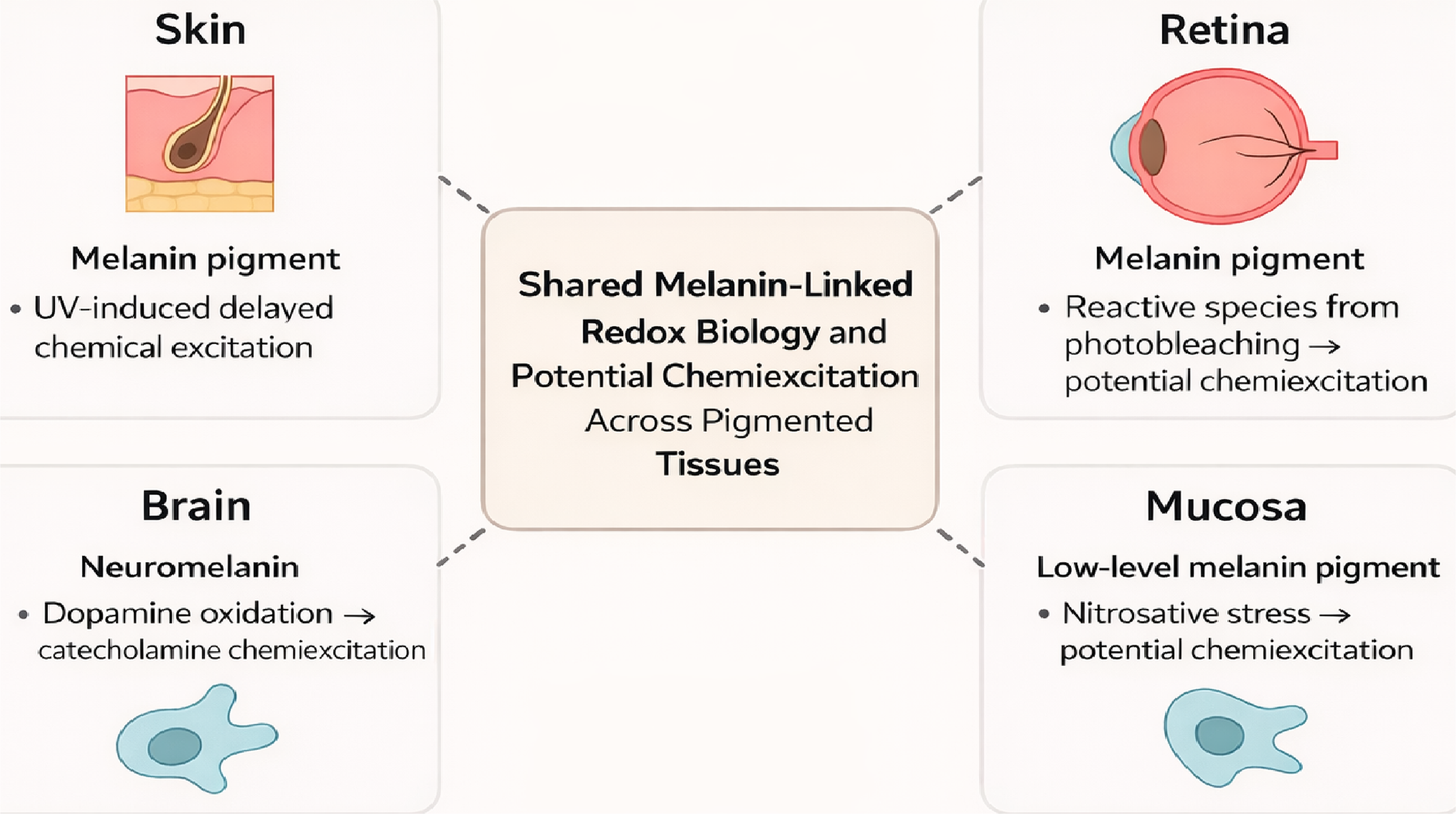
Cross-tissue redox biology of melanin
Melanin is often treated as a skin-specific pigment, but its chemistry runs much deeper. Pigmented cells in the retina, brain, inner ear, and mucosal surfaces all use melanin as a way to manage redox pressure and environmental stress. Across them, melanin helps absorb energy, buffer radicals, and engage NO-driven redox signaling. This shared logic is why pigment biology can connect conditions as distant as melanoma, retinal aging, and neurodegeneration.
A central theme emerging from recent work is that melanin engages related oxidative and nitrosative chemistries across tissues, even when downstream biology diverges. When pressure builds, melanin can interact with NO and superoxide to form peroxynitrite, oxidizing the pigment and generating reactive intermediates. In the skin, this process is strong enough to produce electronically excited carbonyls that damage DNA in the dark - a phenomenon now known as chemiexcitation. In other tissues, the same upstream chemistry is present, even if the downstream outcomes differ. The retina shows signs of oxidized melanin with altered energy handling. Neuromelanin-rich neurons generate excited intermediates through dopamine oxidation. Mucosal melanocytes, though lightly pigmented, sit at the interface of redox stress and immune signaling and may be capable of similar chemistry under inflammatory conditions.
These parallels remain hypothesis-generating rather than experimentally established. Nevertheless, seeing these tissues through a shared chemical lens is powerful. It suggests that pigment is not simply a color-producing molecule but a built-in redox interface, helping cells sense and respond to stress. It also raises new questions: Could excited-state reactions or nitrosative modifications contribute to retinal degeneration or neuroinflammation? Do oxidized pigment fragments act as signals across tissues? And can the therapeutic strategies being developed for melanoma - NOS inhibition, denitrosylation, and triplet-state quenching - be adapted for diseases where pigmentation has never been considered relevant?
Figure 6 summarizes a cross-tissue model in which melanin couples oxidative and nitrosative stress to downstream functional consequences, with melanoma providing the strongest mechanistic support. Recognizing melanin as a shared redox system does not imply uniform outcomes, but it opens new opportunities for cross-disciplinary research and reframes pigment biology as a unifying biochemical principle rather than a tissue-restricted phenomenon.
Perspectives
The melanin continuum - spanning pigment synthesis, redox flux, NO signaling, chemiexcitation, and immune modulation - defines an integrated biochemical system that shapes both tissue resilience and malignant adaptation. Viewed holistically, this circuitry unites photobiology, metabolism, redox chemistry, and immunology into a shared energetic framework through which cells sense and respond to stress. Its mechanistic foundation is most clearly established in cutaneous melanocytes and melanoma, with emerging evidence suggesting that related redox logic may operate in the retina and brain, where pigment chemistry contributes to oxidative balance, signaling, and repair.
In melanoma, this continuum links lineage identity to therapeutic vulnerability. The same pigment machinery that protects the genome from light can, under sustained oxidative or nitrosative pressure, support tumor persistence through redox buffering, nitrosylation, and immune exclusion. This duality - protection versus persistence - represents not a contradiction but a tunable biological principle. Modulating melanin’s redox state may therefore influence not only UV-associated mutagenesis but also therapy resistance and immune responsiveness in pigment-rich tumors.
Recent technological advances now make this vision experimentally tractable and increasingly accessible for translational investigation. Single-cell redox profiling, live-cell photonic imaging, and proteomic mapping of S-nitrosylated networks now enable spatial and temporal resolution of pigment-linked redox signaling. In parallel, melanin-targeted PET tracers ([18F]P3BZA, [18F]DMPY2) and paramagnetic MRI approaches allow noninvasive assessment of pigment state, oxidative load, and therapeutic response. Together, these tools permit direct testing - rather than inference - of whether redox correction, NOS inhibition, or chemiexcitation quenching can reprogram the tumor microenvironment.
Beyond melanoma, the concept of melanin-driven chemiexcitation raises broader, largely unanswered questions. Whether analogous excited-state reactions contribute to other oxidative pathologies, including neurodegeneration, vascular disease, or aging, remains speculative. However, the growing ability to detect and modulate electronically excited intermediates suggests that an additional layer of oxidative signaling may link diverse disease states through shared chemical principles.
Future therapeutic strategies are therefore likely to focus on controlling melanin’s electronic and redox state rather than its abundance. By tuning redox balance, quenching triplet intermediates, or regulating NOS coupling, it may be possible to reconcile photoprotection with anti-tumor immunity. In this view, melanin emerges not as a static pigment but as a programmable redox-active polymer operating at the interface of chemistry, metabolism, and immunity. Understanding and harnessing this system may enable new redox-based diagnostics and therapies, transforming a symbol of protection into a framework for precision intervention in cancer and beyond.
Outstanding questions
Melanin is increasingly recognized as a conserved redox-active system that operates across diverse tissues - including skin, retina, cochlea, and the central nervous system - where it shapes genomic stability, immune tone, and oxidative resilience. While mechanistic insight is most advanced in cutaneous melanocytes and melanoma, many aspects of pigment-driven redox biology remain unresolved, particularly how chemiexcitation, nitrosylation, and pigment oxidation may contribute to disease beyond the skin. Emerging evidence suggests that these processes could play roles in neurodegeneration, retinal aging, mucosal immunity, and chronic inflammatory states, but the molecular logic underlying these connections remains incompletely defined. Addressing the questions below will help unify pigment biology across photobiology, cancer research, immunology, ophthalmology, and neuroscience.
Key Open Questions
How does the balance between eumelanin and pheomelanin influence NOS activation, peroxynitrite chemistry, and nitrosative stress in melanoma, and to what extent are comparable redox relationships present in the RPE, cochlea, and neuromelanin-containing brain regions? What molecular sensors, transcriptional regulators, and metabolic checkpoints integrate pigment synthesis with immune modulation in melanoma and are analogous regulatory circuits operative in other pigment-bearing tissues - such as interferon signaling in melanoma, complement regulation in the RPE, or microglial activation in the brain? Can triplet-state quenchers, NOS inhibitors, or denitrosylation agents prevent delayed (“dark”) DNA damage and nitrosative injury in vivo, without disrupting the essential physiological roles of pigment in photoprotection, neuromodulation, visual function, and barrier immunity? To what extent do chemiexcitation, triplet-carbonyl formation, and nitrosylation occur in non-cutaneous melanized tissues such as the retina, substantia nigra, and inner ear - and do these reactions generate DNA damage, inflammatory signaling, or other functional consequences relevant to neurodegeneration, retinal aging, or cochlear inflammation? Can integrated pigment-redox signatures - combining melanin oxidation state, NOS flux, nitrosylation patterns, and chemiexcitation markers - serve as context-dependent biomarkers of oxidative stress, tissue vulnerability, disease progression, or therapeutic responsiveness across melanoma, ocular disease, neurodegeneration, and aging? Do pigment-containing extracellular vesicles and melanin-derived fragments act as redox or immunological messengers across tissues, and how might these circulating pigment-associated signals influence inflammatory thresholds in tumors, immune-privileged sites, and mucosal barriers? What other biologically relevant chromophores (e.g., catecholamines, lipofuscin, and lipid peroxides) undergo similar excited-state or nitrosative chemistry during aging or chronic inflammation - and how do these pathways converge with, or diverge from, melanin-driven redox processes?
Together, these questions underscore that melanin is not merely a pigment but a cross-tissue redox interface whose chemistry intersects with genome maintenance, immunity, sensory function, and neural health. Resolving these gaps will help establish pigment metabolism as a unifying framework for understanding oxidative stress across biological systems and will define where mechanistic continuity exists versus where tissue-specific adaptations dominate. Such insight may reveal new therapeutic targets for cancer, neurodegeneration, retinal disease, and inflammatory disorders.
Authors’ Contributions
S.P. conceived the overall concept and scope of the review, developed the central framework, secured funding support, and wrote the article. J.S. contributed substantially to the intellectual development of the review through figure design, drafting and editing multiple sections of the text, article organization, and critical revision for clarity, balance, and accuracy. Both authors discussed the content throughout the writing process, reviewed and approved the final version, and agreed to its submission.
Footnotes
Acknowledgments

Author Disclosure Statement
The authors declare no competing interests.
Funding Information
This work was supported by the National Cancer Institute (NCI) of the National Institutes of Health (NIH) under award number R21ES035196-01 and by the Department of Defense (DoD) Melanoma Research Program, Team Science Award under award numbers HT94252410751 and HT94252410752 (Log #ME230101). Additional institutional support was provided by the Support Account (02-25999-19-13), Miles for Moffitt Pilot Funds (09-33661-22-02), the Molecular Medicine Program (#30-20458-04-59), and the Melanoma Center of Excellence (#09-33835-23-02), all at the H. Lee Moffitt Cancer Center and Research Institute.