
Abstract
This study investigated the safety and effect of oxymatrine (OMT) and/or diammonium glycyrrhizinate (DG) on allergic contact dermatitis (ACD) induced by 1-fluoro-2,4-dinitrofluorobenzene (DNFB) in ICR mice. Mice were topically smeared with vehicle (control) or DNFB on their ear and skin to induce ACD. The mice were randomized and injected with saline as the model, treated intraperitoneally with dexamethasone (DEX), 45 or 90 mg·kg−1 OMT and/or DG daily beginning one day post the first smearing for two weeks. The body weights, the severity of ear and skin inflammation, the levels of serum IgE, IL-4, and IFNγ, creatinine and urea as well as plasma sodium and potassium in individual mice were measured. In comparison with the control group, the model group did not change the body weights, but developed severe skin and ear inflammation with increased ear thickness, accompanied by many inflammatory infiltrates in the lesions and high levels of serum IgE, IL-4, and IFNγ. Combination of OMT and DG prevented the OMT- or DG-altered body weights in mice. While treatment with either OMT or DG moderately reduced the skin and ear inflammation, their thickness and inflammatory infiltrates, combination of OMT and DG further significantly increased their anti-inflammatory effects in mice. A similar pattern of inhibitory effect on the levels of serum IgE, IL-4, and IFNγ was observed in the different groups of mice. Combination of OMT and DG also prevented the OMT-, DG-, or DEX-altered plasma sodium or potassium levels in mice. Therefore, combination of OMT and DG significantly increased anti-inflammatory effects on ACD induced by DNFB in mice and attenuated DG- or OMT-related adverse effects.
Impact statement
Diammonium glycyrrhizinate (DG) and oxymatrine (OMT) have similar anti-inflammatory, anti-allergic, anti-tumor, immunomodulatory, and other pharmacological properties. Our previous study has shown that when DG and OMT are combined, DG can attenuate both high-dose (347.44 mg·kg−1) and regular-dose (90 mg·kg−1) OMT-induced mortality and adverse effects (such as body weight loss and hyponatremia). Furthermore, OMT can similarly attenuate the adverse effects (such as body weight gain, hypernatremia, and hypokalemia) induced by regular dose (90 mg·kg−1) of DG. Accordingly, we tested whether combination of OMT and DG would increase anti-inflammatory activities and reduce their adverse effect in a mouse model of allergic contact dermatitis (ACD) induced by 1-fluoro-2,4-dinitrofluorobenzene (DNFB). Our findings indicated that combination of OMT and DG significantly increased anti-inflammatory effects on ACD induced by DNFB in ICR mice and attenuated adverse effects of DG or OMT alone.
Keywords
Introduction
Allergic contact dermatitis (ACD), also referred to contact hypersensitivity (CHS), is a chronic inflammatory skin disease characterized by skin hyper-responsiveness and progressive deterioration. 1 The pathogenesis of ACD remains unclear. Theoretically, ACD is mediated by allergen-specific T cell-mediated delayed type hypersensitivity triggered by multiple allergens in the environment, accompanied by the production of immunoglobulin E (IgE) and the dysregulation of T-helper (Th) 1 and 2 responses.2–4 During the pathogenic process of hypersensitivity, many inflammatory cells, such as lymphocytes, macrophages, and eosinophils, infiltrate into the skin lesions and secrete cytokines and chemokines, creating an inflammatory cascade to mediate the skin damage. 5 The incidence of ACD is increasing recently due to environmental pollution. Although the available therapeutic strategies, such as steroid therapy and immunosuppressive agents, can temporarily improve ACD-related symptoms in patients with ACD, long-term applications of these drugs can cause severe adverse effects. 6 Hence, development of new therapeutic strategies is of significance in management of patient with ACD.
Diammonium glycyrrhizinate (DG), a salt of glycyrrhetinic acid (GA), has been widely used for the treatment of chronic hepatitis and human immunodeficiency virus infection.7,8 DG has anti-inflammatory effect in animal models of ulcerative colitis and cerebral ischemic-reperfusion (IR) injury.9,10 DG has been used for the treatment of dermatitis, eczema, and other diseases in the clinic.11,12 However, GA has been reported to induce a severe pseudoaldosteronism complicating with hypertension, hypokalemia, and potential suppression of renin and aldosterone after high doses or long-term administration.13–15
Oxymatrine (OMT) is a quinolizidine alkaloid extracted from Chinese herb Sophora flavescens Ait. OMT has anti-inflammatory, anti-allergic, antioxidant, antiviral, and anti-cancer activities16–22 and has been extensively used for the treatment of viral hepatitis, cancer, cardiac diseases, and skin diseases (such as atopic dermatitis and eczema) in China.16,23–26 In addition, OMT also has anti-hypertension 27 and immunomodulatory effects.28,29 However, some studies have shown that OMT can have adverse effects, such as diuresis and weight loss.30,31
Our previous study has shown that when DG and OMT are combined, DG can attenuate both high-dose (347.44 mg/kg) and regular-dose (90 mg/kg) OMT-induced mortality and adverse effects (such as body weight loss and hyponatremia). 32 Furthermore, OMT can similarly attenuate the adverse effects (such as body weight gain, hypernatremia, and hypokalemia) induced by regular dose (90 mg/kg) of DG. 32 Accordingly, we tested whether combination of OMT and DG would increase anti-inflammatory activities and reduce their adverse effect in a mouse model of ACD induced by 1-fluoro-2,4-dinitrofluorobenzene (DNFB). Our findings indicated that combination of OMT and DG significantly increased anti-inflammatory effects on ACD induced by DNFB in ICR mice and attenuated adverse effects of DG or OMT alone.
Materials and methods
Chemicals and drugs
Oxymatrine (molecular formula, C15H24N2O2; molecular weight, 264.36; CAS, 16837–52-8; high performance liquid chromatography (HPLC) purity >98%) and diammonium glycyrrhizinate (molecular formula, C42H62O16.2H3N; molecular weight, 856.997; CAS, 79165–06-3; HPLC purity >98%) were purchased from Nanjing Zelang Biological Technology (Nanjing, China) and their structures are shown in Figure 1(a). DNFB (molecular formula, C6H3FN2O4; molecular weight, 186.10; CAS, 70–34-8; purity, 99%) was purchased from Shandong Xiya Chemical Industry (Jinan, China). Acetone (molecular formula, CH3COCH3; molecular weight, 58.08; CAS, 67–64-1; purity, 99%) and olive oil were purchased from Nanjing Zelang Biological Technology. Dexamethasone sodium phosphate injection was purchased from Xi’an Lijun Pharmaceutical (Xi’an, China). The IgE, IFN-γ, and IL-4 enzyme-linked immunosorbent assay (ELISA) kits were purchased from Shenzhen Xinbosheng Biological Technology (Shenzhen, China).
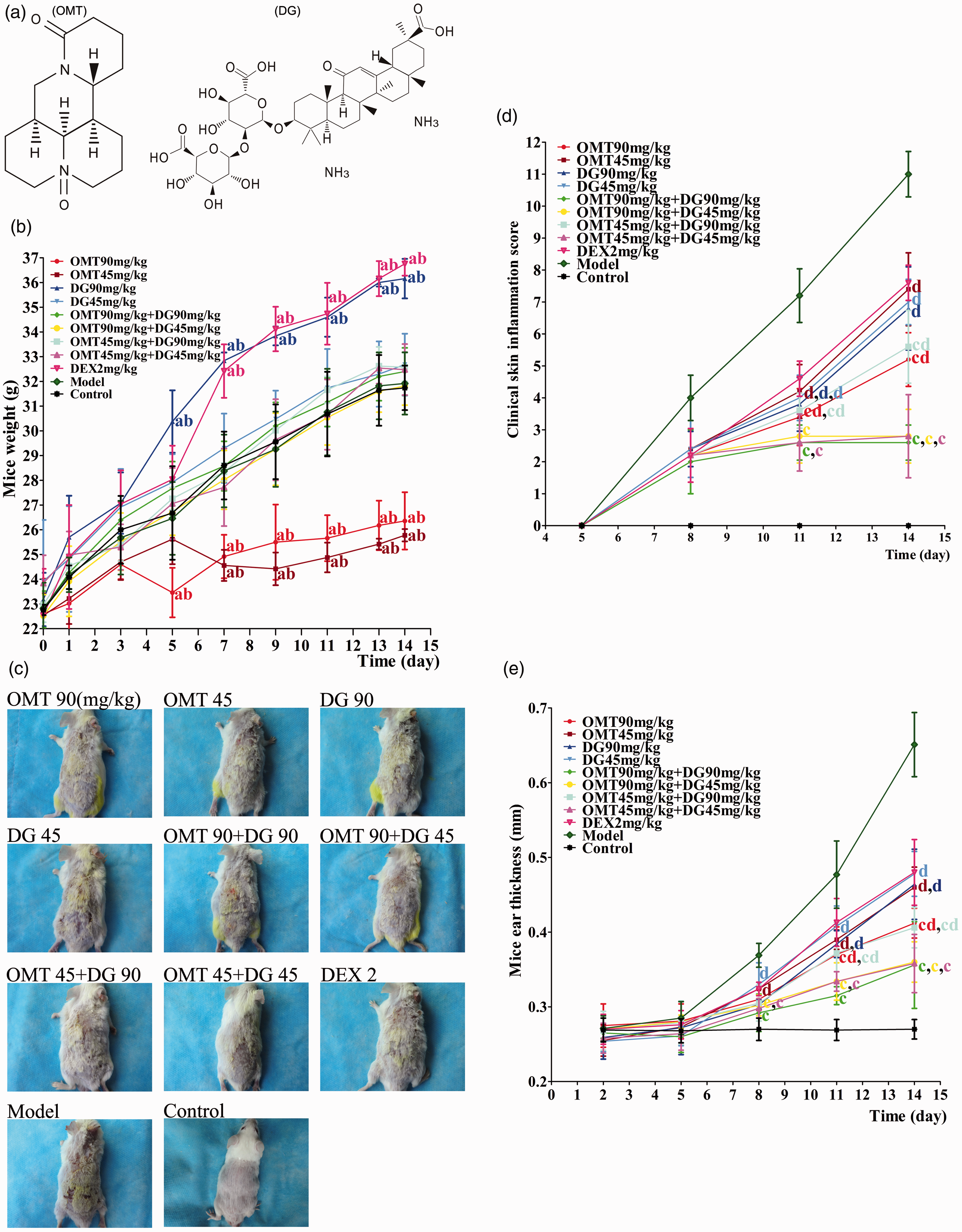
Combination of OMT and DG mitigates the DNFB-induced skin inflammation in mice. ICR mice were smeared with DNFB for five times and after first smearing, the mice were randomized and treated intraperitoneally with saline (model), or the indicated doses of OMT and/or DG daily for 14 days. Their body weights, skin inflammation and ear thicknesses were measured longitudinally. (a) The structures of OMT or DG. (b) The body weights of the different groups of mice. (c) Representative images of skin inflammation in different groups of mice (on day 14). (d) The inflammation scores of individual groups of mice. (e) The ear thicknesses. Data are representative images or expressed as the mean values of individual groups (n = 5 per group). The skin inflammatory scores and ear thicknesses of all ACD mice were significantly greater than the control while the skin inflammatory scores and ear thicknesses in all drug-treated groups were significantly less than that in the model group (statistical markers not shown in Figure 1(d) and (e)). Letters on the graphs indicate the following statistical significant group comparisons: aP< 0.05 vs. the control group; bP< 0.05 vs. the model group; cP< 0.05 vs. the DEX 2 mg·kg−1 group; dP< 0.05 vs. the combination (OMT+DG, 45 + 45 mg·kg−1). OMT: oxymatrine; DG: diammonium glycyrrhizinate; DEX: dexamethasone. (A color version of this figure is available in the online journal.)
Animals
Male ICR mice at six weeks of age and weighing 18–22 g were obtained from the Laboratory Animal Center of Ningxia Medical University (Yinchuan, China). The mice were housed in a specific pathogen-free facility at 23 ± 2°C, a relative humidity of 60% with a light–dark cycle of 12 h and free access to food and water. The mice were food-fasted overnight before experiment. The experimental protocol was approved by the Medical Ethics Committee of the Ningxia Medical University General Hospital (issue date: 6 April 2016; file number: 2016–177).
Methods
DNFB-induced ACD and treatment
Male mice (n = 55) were randomly divided into 11 groups (five mice each group). The mice were induced for ACD, as described previously.33,34 Briefly, individual mice were topically applied with 25 µL of 0.15% DNFB in acetone/olive oil (3:1) onto the outer and inner surfaces of mouse right ear and 100 µL onto shaved back skin on days 1 and 4. On days 7, 10, and 13, the sensitized mice were challenged with 0.2% DNFB onto their skin surface. The mice were randomized one day after the first smearing and injected intraperitoneally with saline (Model), 45 or 90 mg·kg−1 OMT and/or DG or 2 mg·kg−1 dexamethasone (DEX, Positive control) daily for 14 consecutive days. The doses of OMT and DG were based on previous studies for their efficacy and toxicity.32,35–38 A group of control healthy mice received vehicle smearing and injected with saline (n = 5). Their body weights were measured daily beginning one day before induction throughout the experimental period using an electronic balance (YP601N electronic balance, Shanghai Precision Science Instrument, Shanghai, China). The ear thicknesses of individual mice were measured 39 using a gauge digital micrometer (Hengxing Measuring tools, Shanghai, China) on the day post each DNFB application. The severities of inflammation, such as erythema (hemorrhage), edema, excoriation (erosion), and dryness (scaling) in individual mice were scored as 0 (absent), 1 (mild), 2 (moderate), and 3 (severe) in a blinded manner.33,40 A total score (minimum 0 and maximum 12) of each mouse was recorded daily. At the end of the experiment (on day 14), the mice were subjected to light anesthetization with diethylether, and their blood samples were withdrawn retro-orbitally for preparing plasma and serum samples, which were stored at −80°C until further processing. The levels of plasma sodium, potassium, creatinine, and urea were measured using a Siemens ADVIA 2400 Autoanalyzer (Siemens AG, Munich, Germany).
Enzyme-linked immunosorbent assay
The levels of serum IgE, IFN-γ, and IL-4 in individual mice were measured by ELISA using specific kits, according to the manufacturer’s instructions.
Histologic analysis
The ear and back skin samples were taken in the same manner at the end of the experiment, and fixed in 10% neutral buffered formalin, followed by paraffin-embedded. The tissue sections (3–4 μm) were stained with hematoxylin and eosin (H&E). The numbers of inflammatory infiltrates in individual samples were measured in a blinded manner. The number of inflammatory cells in five fields (magnification × 200) with the most infiltrates of each mouse was calculated in a blinded manner.
Statistical analysis
All data are expressed as the mean ± SD. The difference among groups was analyzed by repeated one-way ANOVA and least significant difference (LSD) test using the SPSS version 17.0 statistical software package (IBM, Armonk, NY). A P-value of <0.05 was considered statistically significant.
Results
Combination of OMT and DG significantly mitigates the severity of inflammation in ACD mice
OMT and DG can benefit patients with dermatitis.23,32 To investigate the impact of OMT and DG combination on the severity of inflammation, ICR mice were induced for ACD, randomized and treated with OMT and/or DG for 14 days. In comparison with the healthy control, treatment with vehicle in the model group did not significantly change the body weights (P > 0.05) while treatment with DG (90 mg·kg−1) alone or DEX significantly increased the body weights beginning at five or seven days post induction, respectively (P < 0.05 for all, Figure 1(b)). In contrast, treatment with OMT (90 mg·kg−1) alone or OMT (45 mg·kg−1) significantly reduced the body weights in mice beginning at five or seven days post induction, respectively (P < 0.05 for all). However, treatment with both OMT and DG, regardless their doses, did not change the body weights in mice (P > 0.05), relative to that of either group of mice receiving monotherapy.
During the experimental period, we observed that some mice developed the skin inflammation, characterized by skin erythema, edema, dryness, and excoriation on day 8 post induction and some mice developed severe skin lesions at the end of the observation period (Figure 1(c)). Quantitative analysis indicated that all experimental groups of mice, but not the healthy control, developed varying degrees of skin inflammation beginning on day 8 post induction. The inflammation scores in the model group gradually increased throughout the observation period. In comparison with that in the model group, treatment with OMT or/and DG or DEX (2 mg·kg−1) significantly decreased the skin inflammation scores beginning on day 8 post induction and later time points (P < 0.05 for all, Figure 1(d)). More interestingly, treatment with a high dose of OMT (90 mg·kg−1) or any combination of both drugs significantly mitigated the skin inflammation scores, relative to the DEX group of mice beginning on day 11 and later time points (P < 0.05 for all). In addition, treatment with both OMT and DG significantly reduced the skin inflammation scores, compared with the mice receiving either OMT or DG monotherapy (P < 0.05 for all). A similar pattern of ear thicknesses was observed in the different groups of mice at varying time points (Figure 1(e)). These data clearly demonstrated that combination of OMT and DG additively enhanced their therapeutic effects on skin and ear inflammation in ACD mice.
Combination of OMT and DG decreases the DNFB-induced ear and skin inflammation in ACD mice
To understand the therapeutic action of OMT and DG, we measured inflammation in the skin and ear lesions of ACD mice by histology. As shown in Figure 2(a), the ear tissues in the control group displayed a clear structure of the epidermis without obvious inflammation in the superficial dermis. In contrast, the ear tissues in the model group exhibited hypertrophy, hyperkeratosis, partial crusts and ulceration, epidermal hyperplasia, obvious edema sponge, acanthosis, superficial dermal vessels with many inflammatory infiltrates, possibly by neutrophils and lymphocytes. Quantitative analysis indicated that the numbers of inflammatory infiltrates in the ear lesions of ACD mice were significantly greater than in the control mice (P < 0.05 for all) and the numbers of inflammatory infiltrates in the ear lesions of the model group of mice were also significantly higher than that of any of the drug-treated mice (P < 0.05 for all, Figure 2(b)). In comparison with that of the DEX group, treatment with a high dose of OMT or DG or with any of combination of OMT and DG, but not with a lower dose of OMT or DG, significantly reduced the numbers of inflammatory infiltrates in the ear lesions of mice (P < 0.05 for all). Interestingly, the inhibitory effect of treatment with both lower dose of OMT and DG on the numbers of inflammatory infiltrates was significantly stronger than that of treatment with a lower dose of OMT or DG as well as with a lower dose of OMT and higher dose of DG in mice (P < 0.05 for all). Similar patterns of histological changes and inflammatory cell infiltration were observed in the skin tissues of the different groups of mice, except that the anti-inflammatory effect of treatment with a high dose of DG was similar to that of DEX in mice (Figure 3). Collectively, such data demonstrated that combination of OMT and DG significantly mitigated the DNFB-induced ear and skin inflammation in ACD mice.
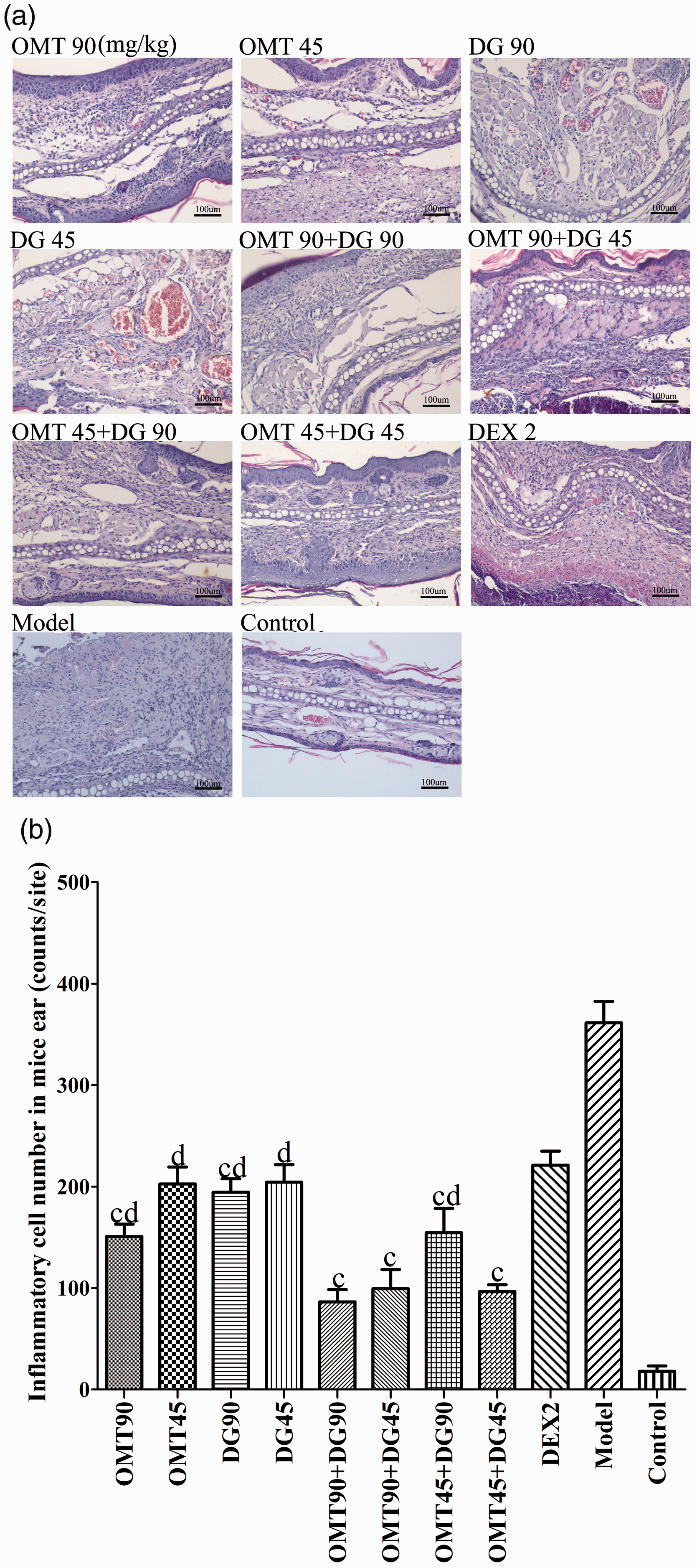
Histologic analysis of inflammatory infiltrates in the ear lesions of mice. At the end of the experiment (on day 14), the different groups of mice were sacrificed and their ear tissues were collected. The ear tissue sections were stained by H&E. The numbers of inflammatory infiltrates in individual sections were counted in a blinded manner. Data are representative images or expressed as the means ± SD of each group (n = 5 per group) from three separate experiments. (a) The representative images (magnification × 200). (b) Quantitative analysis of the inflammatory infiltrates. The numbers of inflammatory infiltrates in the ear tissues of all ACD mice were significantly greater than the control while the numbers of inflammatory infiltrates in all drug-treated groups were significantly less than that in the model group (statistical markers not shown). cP< 0.05 vs. the DEX 2 mg·kg−1 group; dP< 0.05 vs. the combination (OMT+DG, 45 + 45 mg·kg−1). (A color version of this figure is available in the online journal.)
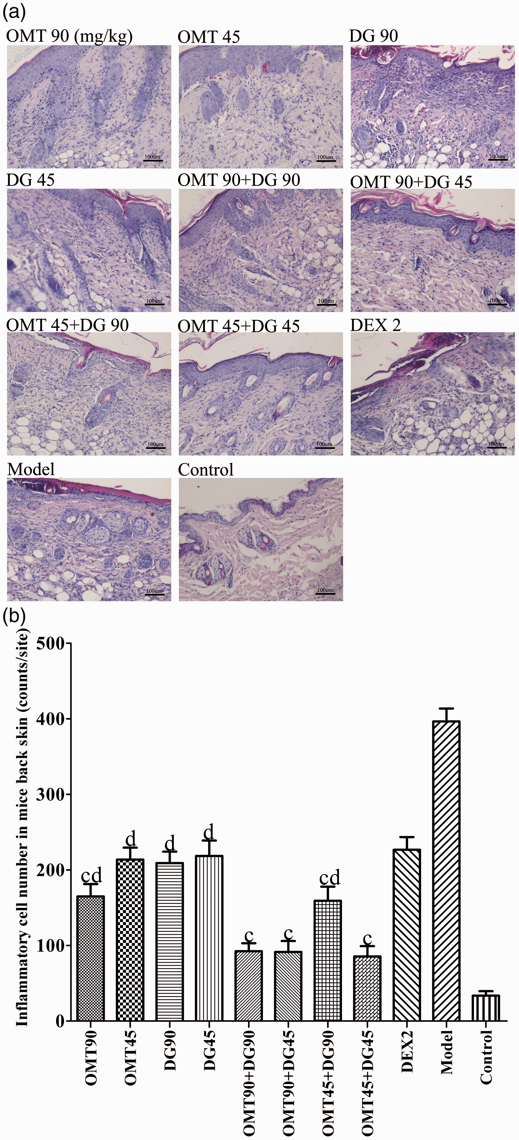
Histologic analysis of inflammatory infiltrates in the skin lesions of mice. At the end of the experiment (on day 14), the different groups of mice were sacrificed and their skin tissues were collected. The skin tissue sections were stained by H&E. The numbers of inflammatory infiltrates in individual sections were counted in a blinded manner. Data are representative images or expressed as the means ± SD of each group (n = 5 per group) from three separate experiments. (a) The representative images (magnification × 200). (b) Quantitative analysis of the inflammatory infiltrates. The numbers of inflammatory infiltrates in the skin tissues of all ACD mice were significantly greater than the control while the numbers of inflammatory infiltrates in all drug-treated groups were significantly less than that in the model group (statistical markers not shown). cP< 0.05 vs. the DEX 2 mg·kg−1 group; dP< 0.05 vs. the combination (OMT+DG, 45 + 45 mg·kg−1). (A color version of this figure is available in the online journal.)
Combination of OMT and DG significantly decreases the levels of serum IgE, IFNγ, and IL-4 induced by DNFB in mice
High levels of IgE, Th1, and Th2 responses are associated with the development and progression of ACD.2–4 To further understand the role of combined OMT and DG in regulating the DNFB-induced inflammation in mice, the levels of serum IgE, IFNγ, and IL-4 in individual mice were measured by ELISA. First, the levels of serum IgE in all ACD mice were significantly higher than that in the healthy control (P < 0.05 for all, Figure 4). Second, the levels of serum IgE in all drug-treated mice were significantly lower than that in the model group of mice (P < 0.05 for all), indicating that treatment mitigated the DNFB-induced IgE responses in mice. In comparison with the DEX-treated mice, treatment with a high dose of OMT or any combination of OMT and DG, but not other monotherapies significantly reduced the levels of serum IgE in ACD mice (P < 0.05 for all). More importantly, the inhibitory effect of combined lower dose of OMT and DG (45 mg·kg−1 of each) was similar to that of combined OMT and DG (90 mg·kg−1 of each, 90 mg·kg−1 of OMT, and 45 mg·kg−1 of DG), but significantly stronger than that of any of the monotherapies in ACD mice (P < 0.05 for all). Similar patterns of serum IFNγ and IL-4 were detected in the different groups of mice, except that treatment with a lower dose of OMT or each dose of DG had stronger inhibitory activity against the DNFB-induced IL-4 responses than DEX treatment (P < 0.05 for all). Thus, treatment with both OMT and DG significantly mitigated the DNFB-induced ACD-related IgE, IFNγ, and IL-4 responses in mice.
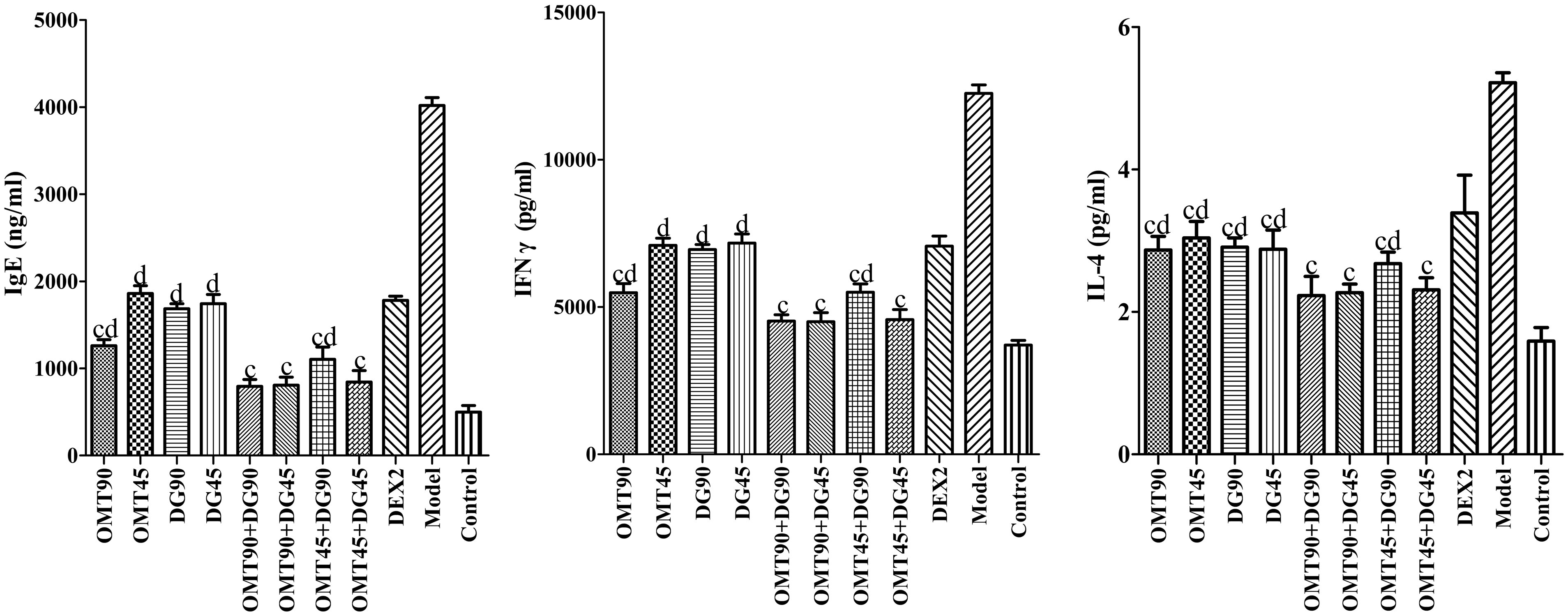
Measurement of serum IgE, IFN-γ, and IL-4 levels in individual mice. At the end of the experiment (on day 14), peripheral blood samples were collected from individual mice and the levels of serum IgE, IFN-γ, and IL-4 in individual mice were measured by ELISA using specific kits. Data are expressed as the means ± SD of each group (n=5 per group) from three separate experiments. The levels of serum IgE, IFNγ and IL-4 in all ACD mice were significantly higher than the control while the levels of serum IgE, IFNγ, and IL-4 in all drug-treated groups were significantly lower than that in the model group (statistical markers not shown). cP< 0.05 vs. the DEX 2 mg·kg−1 group; dP< 0.05 vs. the combination (OMT+DG, 45 + 45 mg·kg−1).
Effects of OMT, DG, and combination of OMT and DG on plasma levels of sodium, potassium, creatinine, and urea in mice
High doses of OMT or DG as well as DEX may change the balance of plasma electrolytes, such as sodium and potassium. Finally, we determined the levels of plasma sodium and potassium in individual mice. As shown in Figure 5, there was no significant difference in the levels of plasma sodium and potassium between the model and healthy control groups of mice. Treatment with a high dose of OMT significantly reduced the levels of plasma sodium while treatment with high doses of DG increased the levels of plasma sodium in mice, relative to that in the model or healthy control group (P < 0.05 for both). Furthermore, treatment with either DEX or a high dose of DG significantly decreased the levels of plasma potassium in mice, relative to that in the model or healthy control group (P < 0.05 for both). However, treatment with both OMT and DG did not change the levels of plasma sodium and potassium in mice (P > 0.05 for both). In addition, there was no significant difference in the levels of plasma creatinine and urea among the different groups of mice (P > 0.05 for all, Figure 6). Therefore, combination of OMT and DG mitigated the high dose of OMT- or DG-altered plasma sodium and reduced potassium levels in mice treated with high-dose DG.
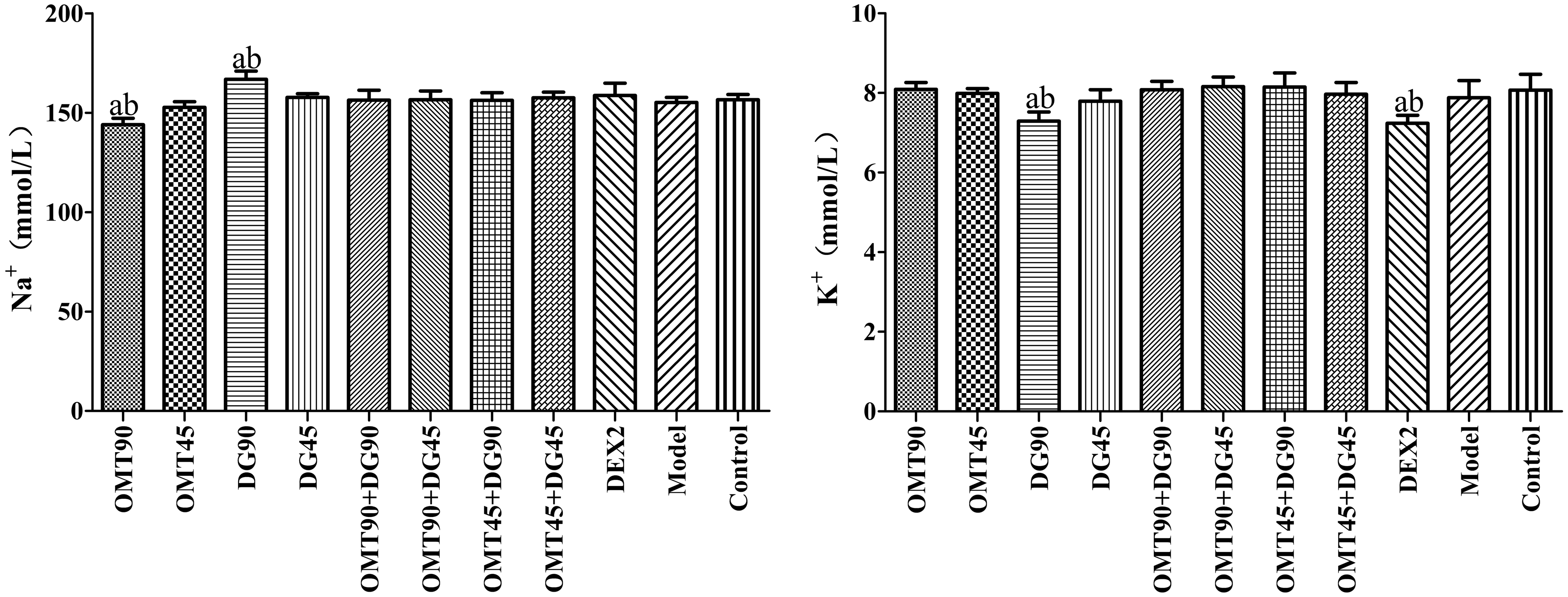
Measurement of plasma sodium and potassium in individual mice. At the end of the experiment (on day 14), peripheral blood samples were collected from individual mice and the levels of plasma sodium and potassium in individual mice were measured using a Siemens ADVIA 2400 autoanalyzer. Data are expressed as the means ± SD of each group (n = 5 per group) from three separate experiments. There was no significant difference in the levels of plasma sodium and potassium between the model and control groups. aP< 0.05 vs. the control group; bP< 0.05 vs. the model group.
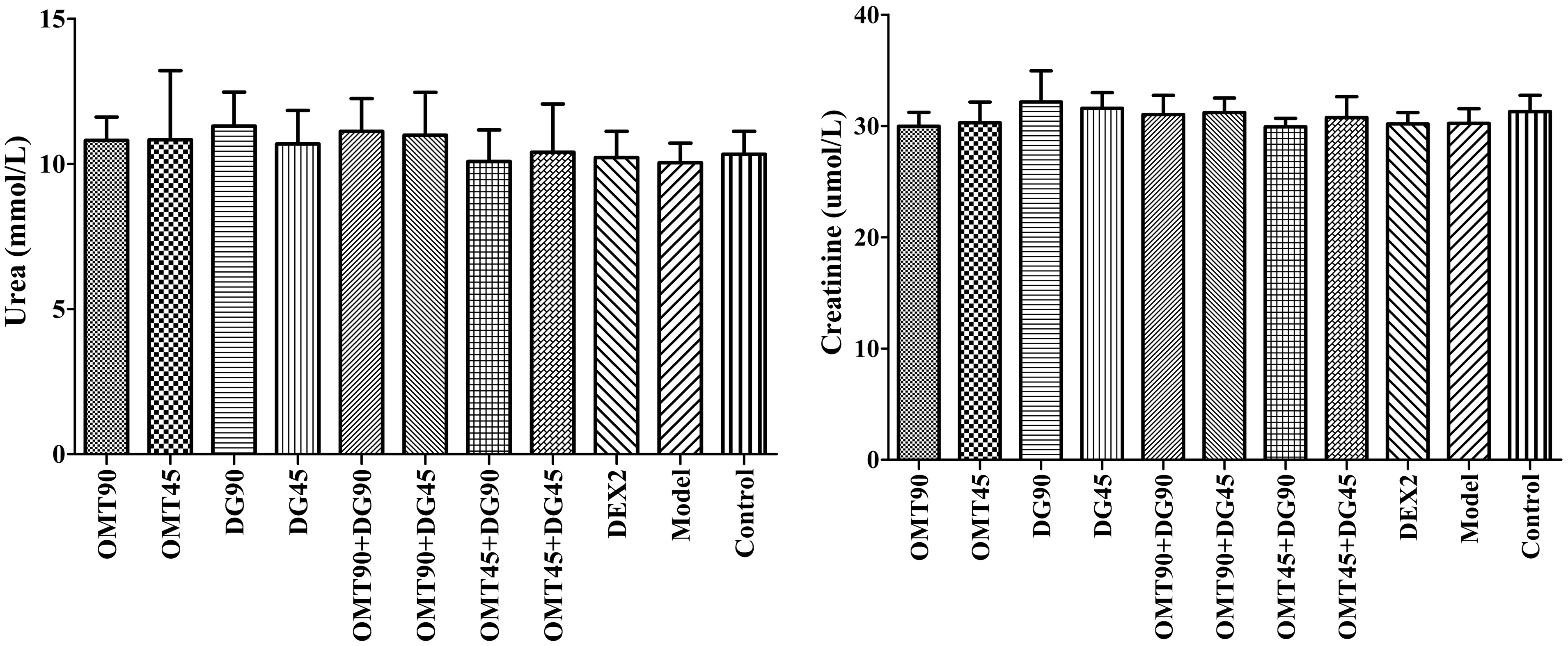
Measurement of serum creatinine and urea in individual mice. At the end of the experiment (on day 14), peripheral blood samples were collected from individual mice and the levels of serum creatinine and urea in individual mice were measured using a Siemens ADVIA 2400 autoanalyzer. Data are expressed as the means ± SD of each group (n = 5 per group) from three separate experiments. There was no significant difference among the different groups of mice.
Discussion
Given that OMT and DG have anti-inflammatory, anti-allergic and immunomodulatory activities, this study investigated whether combination of OMT and DG could increase their anti-inflammatory activities and reduce their adverse effects in a mouse model of ACD induced by DNFB. We found that combination (OMT+DG, 90 + 90, 90 + 45, 45 + 45 mg·kg−1) significantly inhibited the DNFB-induced inflammation and their therapeutic effects were superior to OMT, DG alone or DEX (2 mg·kg−1) in ACD mice. Our data extended previous observations that combination of Kushen and Gancao or Glycyrrhizin and Matrine inhibits inflammatory chronic liver diseases and fibrosis. 41 To the best of our knowledge, this was the first study to demonstrate that combination of OMT and DG effectively inhibited inflammation and improved clinical symptoms of ACD. These suggest that combination of OMT and DG may be valuable for the treatment of ACD and other chronic inflammatory diseases in the clinic.
In this study, we found that the model group of mice developed severe inflammation and increased the ear thickness, accompanied by many inflammatory infiltrates in the lesions and high levels of serum IgE, IL-4, and IFNγ. The increased skin and ear thicknesses should reflect inflammatory hyperplasia and edema of the skin. Our data support the notion that allergen-specific T cells and other inflammatory cells, such as macrophages and neutrophils infiltrate into the skin lesions to secrete inflammatory cytokines and chemokines, creating a pro-inflammatory cascade to drive the pathogenic process of ACD. More importantly, we found that while treatment with either OMT or DG moderately mitigated inflammation in the skin and ear lesions of mice, combination of OMT and DG further significantly reduced the inflammation in ACD mice, accompanied by decreasing the levels of serum IgE, IL-4, and IFNγ. It is notable that the levels of serum IFNγ were many folds higher than that of IL-4 in mice with ACD. These data were consistent with previous observations42,43 and indicated that pro-inflammatory Th1 responses were crucial for the development of ACD in the mouse DNFB model of ACD. The significantly reduced levels of IFNγ by combination of OMT and DG suggest that both OMT and DG may inhibit Th1 responses, leading to reduced inflammation in this model.
Previous studies have shown that long-term or high dose of OMT or DG can cause severe adverse effects, such as pseudoaldosteronism, diuretic, weight loss, sodium and water retention and potassium excretion, respectively.13–15,30,31 Actually, we did observe that treatment with a high dose of OMT reduced the body weights and plasma sodium levels in mice while a high dose of DG, or DEX, increased the body weights and reduced the plasma potassium levels in ACD mice. However, combination of OMT and DG prevented the OMT- or DG-altered body weights and plasma sodium and potassium levels in ACD mice. These data were consistent with previous observations that treatment with both Glycyrrhizin and Matrine reduces sodium and water retention in animals.41,44 We are interested in further investigating how OMT or DG monotherapy alters body weights and plasma sodium and potassium levels in future studies. Nevertheless, these data indicated that combination of OMT and DG antagonized each compound-related adverse effects. These, together with better anti-inflammatory effects, suggest that combination of OMT and DG may be safe for the treatment of inflammatory diseases, such as ACD.
In summary, our data indicated that combination of OMT and DG significantly inhibited inflammation, reduced the severity of ACD and prevented single compound-related adverse effect in mouse model of DNFB-related ACD. If it is validated in the clinic, our findings suggest that combination of OMT and DG may be a safe strategy for the treatment of inflammatory diseases, such as ACD.
Footnotes
Authors’ contributions
H-jS and H-bS contributed equally to this paper. All authors participated in the design, interpretation of the studies and analysis of the data and review of the manuscript. H-jS and H-bS participated in research design; H-jS, H-bS, and QG conducted experiments; QG and J-wS provided new reagents or analytic tools; H-jS, H-bS, J-wS, and QZ performed data analysis; H-jS, H-bS, and QZ wrote the manuscript.
DECLARATION OF CONFLICTING INTERESTS
The author(s) declared no potential conflicts of interest with respect to the research, authorship, and/or publication of this article.
FUNDING
This work was supported by the Ningxia Autonomous Region Key R&D Program (Special Project for Foreign Scientific and Technological Cooperation) (project number: 2019BFG02008).