



Abstract
This case report discusses the unusual presentation and ultrasound features of a solitary fibrous tumour of the face. Solitary fibrous tumour is an uncommon form of soft tissue tumour which, although seen predominantly within the lung pleura, can occur throughout the body in sites such as the peritoneum, mediastinum and head and neck. Ultrasound is an excellent imaging modality in the assessment of soft tissue masses in the head and neck. The ultrasound features demonstrated by this example of solitary fibrous tumour are reviewed. This report also highlights that ultrasound alone is ultimately limited in reaching a definitive diagnosis. The roles of other investigations such as ultrasound-guided biopsy and cross-sectional imaging are discussed.
Case presentation
A 24-year-old Bengali woman was referred by her general medical practitioner with a right cheek swelling that had been present for one month. The mass had recently increased in size and had become painful to touch. The patient gave no history of trauma, and a course of oral antibiotics prescribed by her general medical practitioner had failed to improve her symptoms. There was no previous history of cheek swelling. The patient’s medical history was unremarkable.
Clinical examination revealed a firm, tender swelling of the right cheek. The mass was non-pulsatile, did not transilluminate and there was no overlying skin change. No punctum was evident. There was no palpable cervical lymphadenopathy. Intraoral examination of the soft tissues and dentition was normal. The clinical diagnosis was that of a benign epidermoid cyst or lipoma.
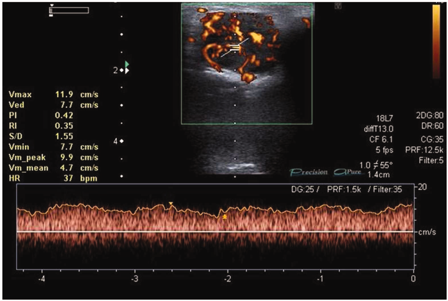
Ultrasound (US) of the right cheek demonstrated a well-defined hypoechoic 27 mm × 19 mm × 22 mm mass overlying the right maxillary buttress (Figure1). US power Doppler demonstrated marked internal vascularity, and spectral analysis confirmed that the mass was non-pulsatile (Figure 2). The US findings contradicted the clinical diagnosis, instead raising the possibility of a vascular anomaly. The remainder of the neck was unremarkable. Two US-guided fine-needle aspiration (FNA) biopsies were performed but were inconclusive, so US-guided core biopsy was undertaken using an 18-gauge Temno Evolution Adjustable Biopsy System (CareFusion, USA) under local anaesthesia.
Ultrasound power Doppler in transverse section demonstrating a hypoechoic mass with internal vascularity Spectral Doppler ultrasound confirming non-pulsatile nature of the hypoechoic mass
Histopathological analysis of the sample identified small vessels lined by plump endothelial cells mixed with larger structures. There was no evidence of mitoses, and although characterisation was difficult from the biopsy, the overall appearance was in keeping with a low-grade/benign vascular lesion.
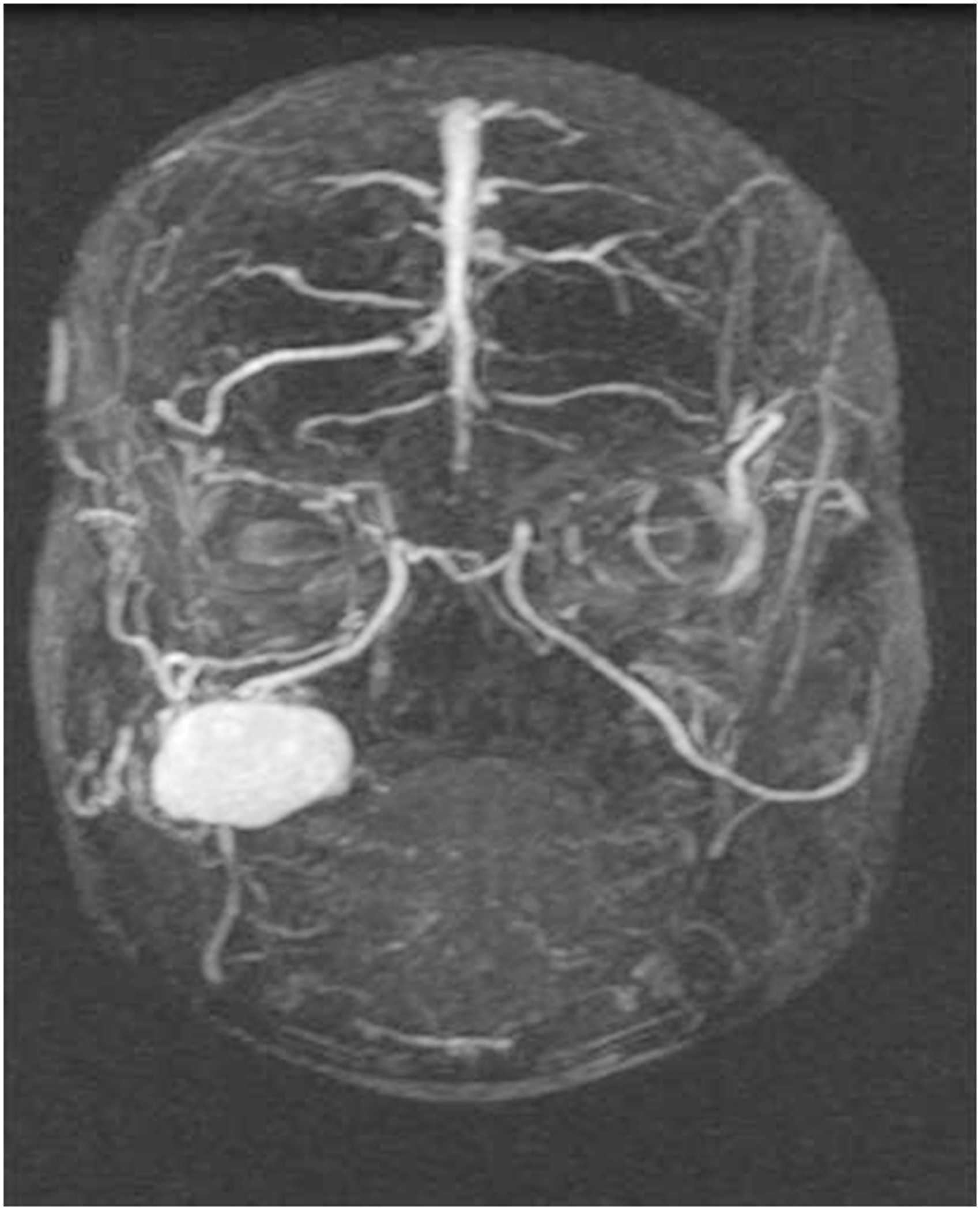
A contrast-enhanced magnetic resonance image (MRI) of the face demonstrated a well-defined, round, soft tissue mass in the right cheek abutting the maxilla. Post contrast, the mass demonstrated heterogeneous high signal due to flow voids within (Figure 3). Time-Resolved Imaging of Contrast KineticS (TRICKS) sequences (a vascular study) were performed and demonstrated a venous sac, drained by the angular and superficial temporal veins (Figure 4). The overall findings of the MRI were consistent with that of a venous vascular anomaly.
Axial T1 weighted post contrast MRI demonstrating an enhancing right cheek mass MRI TRICKS demonstrating a venous sac covering the right cheek, appearance suggestive of a vascular anomaly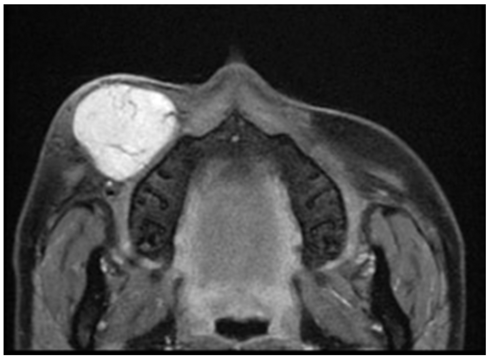
Given the progression in size of the lesion, surgical excision of the lesion was performed under general anaesthesia via an extra-oral, nasolabial fold approach. At the time of surgery, the lesion was noted to be well localised with no evidence of feeding vessels and was completely excised.
Macroscopic histological examination of the lesion demonstrated an encapsulated, tan-coloured mass, comprising mildly pleomorphic spindle cells that were arranged in a storiform pattern with numerous branching vessels with a staghorn form. Immunostaining revealed strong positivity for CD34, CD99 and bcl-2 with patchy expression of smooth muscle actin. There was no evidence of expression of S100, desmin or MUC4. Overall, the appearances were in keeping with solitary fibrous tumour (SFT) with complete excision and no histological features to suggest malignancy.
The patient made a good postoperative recovery and had no evidence of local recurrence 12 months later.
Discussion
SFT was first described by Lietaud and later reported by Klemperer and Rabin, 1 who described SFT as a lesion arising from lung pleura. They initially categorised these pleural tumours as either local, diffuse mesothelioma or SFT. 1 SFT are said to attribute their existence to mesenchymal cells. 2 Although seen predominantly within the lung pleura, SFT can occur throughout the body in extra pleural sites such as the peritoneum and mediastinum. Witkin and Rosai 3 first described the presence of SFT in the head and neck region. They account for <0.1% of all neoplasms of the upper respiratory tract and affect males and females equally. 4 Within the head and neck region, they typically occur in the oral and nasal cavity but can also occur in the orbit, tongue, larynx, parapharyngeal space, thyroid gland, salivary glands, paranasal cavities and skull base. 5 Although previously reported within the literature, the presence of SFT within the cheek is rare. 6 The origin of SFT tumours is not known, and although the majority are regarded as benign tumours, 7 they are known to have an uncertain malignant potential. Malignant SFT cases account for 10–15% of those reported and when present, these have shown to have recurrent and distant metastases.7,8 The current case showed no features to suggest a malignant process.
Presentation of SFT is variable, and the majority of reported symptoms are of a painless, smooth, non-tender, slow-growing mass. 7 This patient presented with a painful lump, presumably due to compression of the infraorbital nerve, but this was not possible to identify on the imaging investigations. In the head and neck, symptoms are commonly related to local compressive symptoms and can be diagnostically challenging to distinguish from other soft tissue pathology. 9 The overlying skin above SFT is usually normal in appearance. 7
SFT radiographic findings are variable and non-specific.9,10 The use of US in the initial assessment of these lesions has not been documented in the head and neck region. US is a quick, non-invasive imaging modality that is very useful in the initial assessment of superficial masses within the head and neck and can potentially differentiate between cystic and solid, benign and malignant, and allows assessment of vascularity, as well as guiding biopsy. In this case, US was able to exclude an epidermoid cyst or lipoma (contrary to the clinical findings), as the mass contained internal hyperechoic stria and marked internal vascularity. Doppler US and spectral US findings changed the differential diagnosis to a vascular anomaly. Within the thorax, SFT is hypoechoic and despite being a vascular soft tissue lesion, rarely demonstrates flow on Doppler imaging. 10 This case was unusual, as it demonstrated internal vascularity.
This case highlights how SFT, a soft tissue tumour, may mimic other benign disease such as vascular tumours. Although other differential diagnoses for lesions with similar US features include vascular metastases, sarcoma, liposarcoma and angiosarcoma, these would all be very unusual in a patient of this age and at this site. It is important to recognise that although US changed the initial clinical diagnosis, and potentially surgical management, it was ultimately limited in reaching a definitive diagnosis. This case demonstrated how US-guided biopsy may be useful in the head and neck region. Although US-guided FNA is commonly performed in the head and neck, the use of US-guided core biopsy tends to be reserved for cases where FNAs have been inconclusive (as in this case) or as a diagnostic tool in lymphoproliferative disorders. In this patient, core biopsy was able to exclude malignancy, thus narrowing down the list of differential diagnoses, but it did not identify SFT and demonstrates the difficulty of achieving accurate histological diagnosis on limited material.
The use of computed tomography is reported in the literature, 10 but in this case, MRI was performed and demonstrated heterogeneous enhancement on post contrast T1-weighted MRI. A heterogeneous, high signal pattern following intravenous contrast may be observed in a range of pathologies, but in this case, based on the clinical and radiological findings, it was reported as being due to the presence of multiple flow voids, consistent with a vascular anomaly. Similar findings have been identified in the literature.2,11 The use of dynamic contrast-enhanced MRI, in this case, TRICKS sequences, in the investigation of SFT, has been reported in the literature. 2 TRICKS MRI is a form of contrast-enhanced dynamic vascular imaging useful in the assessment of vascular anomalies. It may identify a mass as either an arterial or venous malformation. Yang et al. 2 suggest that SFT demonstrates a wash out pattern post contrast, which may be a useful diagnostic clue. This was not seen in this case. Two prominent draining veins were identified, the angular vein medially and a branch of the superficial temporal vein laterally. Conversely, the surgical findings revealed no evidence of an obvious feeder or draining vessel. Although a useful imaging modality, MRI is prone to artefacts, and the TRICKS sequences may be subject to interpretation errors. In this patient, there was no evidence of bone remodelling or destruction, features of SFT that have been previously reported. 10 Although MRI did not add any further information to that already supplied by the US, the use of cross-sectional imaging in the assessment of SFT is advocated,9,10 as it may add additional information regarding bone involvement, which may be subtle.
As in this case, SFT is often macroscopically tan or grey coloured, firm, well circumscribed and encapsulated. 12 Extrapleural lesions can reportedly range in size from 5 to 8 cm, 13 and although nasal lesions may attain this size, oral SFTs are usually smaller than this. Microscopically, SFT was previously diagnosed as ‘Haemangiopericytoma,’ but this term refers to the branching ‘staghorn-like’ vascular pattern and is not specific for SFTs and is seen in a variety of tumour types. This pattern of vascularity may explain the reasons why a vascular tumour was raised within the differential diagnosis.
The mainstay of treatment for SFT is surgical excision and regular close postoperative follow-up is recommended.7,9,14 In the few papers that document follow-up, recurrence of SFT after complete excision at the primary site is rare, 14 although recurrence six years following initial resection has been reported.14,15 Incomplete removal has been shown to increase the risk of recurrence and may necessitate further treatment. 14 No additional treatment for malignant SFT with complete excision margins is currently advocated. 14
Conclusion
This report presents a rare soft tissue tumour in an unusual site. US is a useful initial investigation in the assessment of swellings of the head and neck. Although SFT is rare, the authors suggest that it should be included in the list of differential diagnoses when presented with a well-defined mass that demonstrates internal vascularity, as its management may differ from other lesions with similar US features. US-guided biopsy can help the clinician in narrowing the list of differential diagnoses, in particular the exclusion of malignancy. US may demonstrate complete disease extent but is ultimately limited and further cross-sectional imaging may often be indicated.