



Abstract
Ultrasound assessment of the postpartum uterus has a significant role to play in the evaluation of a large proportion of symptomatic puerperal women. Often the imaging modality of choice for excluding retained placental tissue, correct application of postpartum ultrasound could enable more accurate identification of women requiring surgical intervention, with consequent reduction in patient morbidity and clinical workload. This article aims to review the current understanding and application of ultrasound in the puerperium and evaluate the current evidence investigating the physiological and pathological findings of the postpartum uterus and its contents.
Introduction
Portable and departmental ultrasound scanning has become an increasingly utilised investigative tool in the diagnosis of puerperal complications. It must be emphasised, however, that an understanding of the normal ultrasonographic appearance of the postpartum uterus is a prerequisite for the accurate diagnosis of puerperal pathology.
Common indications for postpartum ultrasound
The relative safety profile and patient acceptability rates of both transabdominal and transvaginal ultrasound scanning often renders it the first-line imaging modality of choice when post partum pathology is suspected. There are a number of possible indications for referral to ultrasound, such as suspected retained products of conception or pelvic sepsis, which often present with excessive or erratic bleeding.
Primary postpartum haemorrhage (PPH) is traditionally defined as the loss of at least 500 ml of blood from the lower genital tract within 24 hours of delivery or any blood loss less than 500 ml resulting in maternal haemodynamic compromise.1–4 Although the most common cause of primary PPH is uterine atony, care must be taken to exclude retained products of conception (placenta and membranes) or intrauterine blood clots as an additional or primary cause. 4 Bedside ultrasound scanning can therefore be a useful diagnostic adjunct to clinical examination; clearly, ‘off-ward’ ultrasound assessment is rarely indicated in this emergency setting.
The presence of increased or abnormal genital tract bleeding occurring between 24 hours and 12 weeks postpartum is classed as secondary PPH and is often a sign of underlying endometritis or retained products of conception.4,5 Abnormal bleeding up to 6 weeks is the more commonly used definition of secondary PPH in the UK and, in developed countries, 2% of postnatal women are admitted to hospital with such symptoms. 6 Up to 50% of these women will undergo surgical evacuation as per the guidance published by the Royal College of Obstetricians and Gynaecologists, who recommend the use of surgical measures when there is excessive or ongoing bleeding, irrespective of ultrasound findings.4,6 This is based on the understanding that although pelvic ultrasound can aid the exclusion of retained products of conception, the appearance of the immediate postpartum uterus can be unreliable.7,8 It is our belief that a better understanding of postpartum ultrasound findings could enable more accurate identification of women requiring surgical intervention, with consequent reduction in surgical complications, disruption to the woman and her family and cost to healthcare services.
The ‘normal’ postpartum uterus
Immediately following delivery, the uterus undergoes rapid involution, driven primarily by the action of endogenous or synthetic oxytocin. The average mass of the term uterus post-delivery is 1000 g, at which time, the uterine fundus may be palpable at the level of the umbilicus. The uterine mass decreases by approximately 50% by day 7 post-partum to 500 g. At 2 weeks postpartum, the uterine size has decreased further, returning to the true pelvis and is no longer palpable abdominally; after 6 weeks, the uterus has decreased to 50–100 g, a size consistent with a non-pregnant state. However, the final uterine size will remain larger than the original nulligravid state. The postpartum endometrium regenerates at a rapid pace. At day 7 postpartum, endometrial glands are already formed, and by day 16, the endometrial lining is almost completely restored throughout the uterine cavity, with the exception of the placental bed site. 9
Historically, the vast majority of this knowledge was obtained from histological analysis of post mortem specimens, when puerperal maternal death rates were higher; 10 fortunately, in more recent times, ultrasound has been the mainstay in furthering our knowledge of postpartum uterine physiology and appearances.
Ultrasound appearances of the postpartum uterus
The ultrasound appearances of the process of normal uterine involution are relatively well documented in the literature (Figure 1),
11
but a large proportion of these studies are now over 15 years old.8,11–13
Diagram depicting the uterus throughout an uncomplicated puerperium. (a) Days 1 to 3: The uterus is retroverted. The cavity appears as a white line in the upper segment, and the lower segment/cervical region is expanded with fluid and solid components. (b) Day 7: The uterus is axial. Copious fluid or mixed components are visible within the whole cavity. (c) Day 14: The uterus is anteverted and the cavity resembles the findings of those at day 7. (d) Days 28 to 56: The uterine size is much decreased. The cavity is empty and appears as a thin white line. Reprinted with permission from Dr Mulic-Lutvica (Ultrasonic evaluation of the uterus and uterine cavity after normal, vaginal delivery. Ultrasound Obstet Gynecol 2001; 18: 491–498).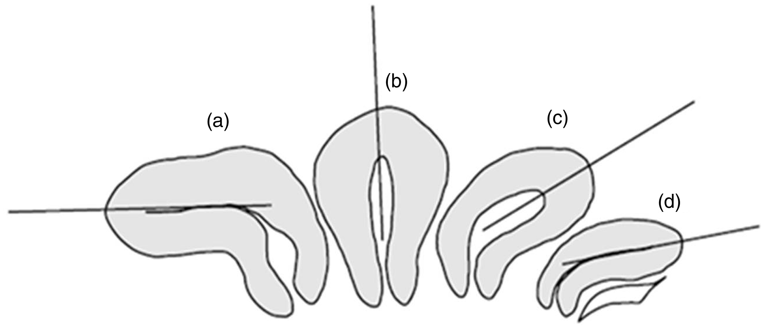
A more up to date assessment of the postpartum uterus, not only assessing the anteroposterior (AP) diameter of the uterus but also the endometrial thickness suggests that mode of delivery can affect the rate of uterine involution; for example, the decrease in endometrial thickness over the second to sixth week postpartum following a term vaginal delivery was found to be significantly greater than that following term caesarean section. 14 The findings were also similar when comparing gestational age, whereby the decrease in the AP diameter of the uterus was less after a preterm delivery versus a term delivery. Although based on a small number of cases, this study supports the theory that the process of uterine involution will vary with both mode and timing of delivery.
Despite numerous histological and ultrasonographic studies assessing uterine involution, there are conflicting data describing the typical ultrasound appearances of the uterine cavity and its contents postpartum, and also the relevance of such findings to clinical practice. To aid understanding of the physiological and pathological characteristics of the postpartum uterus, ultrasonographic assessment of the uterine cavity can be broadly divided into two categories: (a) the immediate post partum period, i.e. within the first 24 hours and (b) 24 hours post-delivery through to the end of the puerperium.
Ultrasound appearances immediately postpartum (within 24 hours)
In a prospective observational study of 94 women, Deans et al. sought to establish if there was any correlation between transabdominal ultrasound findings and patient morbidity. 15 In the first 24 hours after delivery, ultrasound revealed an unexpectedly large volume of echogenic material within the uterine cavity, in particular within the lower segment of the uterus, where mean volumes were as great as 54.8 cm3. However, when followed up for symptomatology, there was no correlation between the presence of this material and the development of postpartum morbidity, such as pyrexia, PPH or prolonged hospital stay. This suggests that the presence of large volumes of intrauterine echogenic material in the first day post-delivery can be accepted as normal.
Similarly, an American study undertook immediate ultrasonographic assessment following placental delivery but with concomitant manual exploration and sponge curettage of the uterine cavity, within 2 minutes of the scan. 16 Following histological assessment of the intrauterine material, the sensitivity, specificity, positive and negative predictive value of ultrasound in detecting retained products in their study was 44%, 92%, 58% and 87%, respectively. Of those patients with histologically confirmed retained products of conception, the majority in fact had a normal endometrial cavity on ultrasound scan (37.5%). An echogenic mass was seen in 25%, a heterogeneous mixed density mass in 21% and intrauterine fluid alone was visible in 16.6%. The vascularity of these intrauterine masses was, however, not assessed. Thus, it can be concluded that in the absence of colour Doppler assessment, the appearances of retained products immediately following delivery are highly variable and cannot be correlated with a need for intervention.
A more recent prospective observational study of 30 patients assessed the ultrasonographic appearances of the uterine cavity following lower segment caesarean section (LSCS) at 1 hour, 3 hours and again at 24 hours post delivery. 17 Unsurprisingly, as the operating surgeon undertook systematic manual examination of the uterine cavity following delivery of the placenta in each case, the incidence of intrauterine material visible on scan was low (3%, n = 1). This particular patient did not exhibit postpartum pathology, and once again questions the significance and clinical relevance of such findings.
Ultrasound appearances after the first 24 hours postpartum
As previously mentioned, the postpartum uterus is often found to contain an accumulation of clinically insignificant debris and fluid, mainly in the lower segment initially and then within the whole uterine cavity by the middle of the puerperium. 11
In the commonly cited study by Edwards and Ellwood, 40 women were systematically scanned at weekly intervals postpartum, starting from day 7 up until day 21. 8 They observed that in women with normal postpartum bleeding, there was an echogenic mass in 51% on day 7, in 21% on day 14 and in 6% on day 21, and found no difference in either the heaviness or bleeding duration between women with and without an echogenic mass at each of these three scans. Thus, they hypothesise that either ‘an echogenic mass does not always represent retained products of conception, or that products of conception are commonly retained and are therefore of little clinical significance in many cases’. However, there is no evidence that Doppler assessments of these products were undertaken, nor is there any clarification by the authors as to whether the description of an echogenic mass also includes mixed-echo patterns, a finding which other studies have suggested is an insignificant postpartum occurence. 18 Care must therefore be taken to interpret ultrasound scan findings in this clinical context; the specific finding of an echogenic mass in the setting of secondary PPH is likely to be associated with retained placental tissue and requires surgical intervention, whereas mixed-echo patterns are not, and can be managed expectantly, with early resolution of symptoms. 18
Van Den Bosch's group evaluated the application of colour Doppler in aiding identification of placental remnants.
19
Their cross-sectional study of 385 postnatal women revealed areas of enhanced vascularity in 32 women (8.3%), and 26 women (6.75%) had retained placental products on scan. Although no comment was made regarding patient morbidity, a high incidence of histological confirmation was obtained following surgical curettage (19 of 20 cases), suggesting that the use of colour Doppler may be of practical diagnostic value. Figures 2 to 6 demonstrate the use of colour Doppler in aiding the diagnosis of retained products of conception, later confirmed on histological analysis.
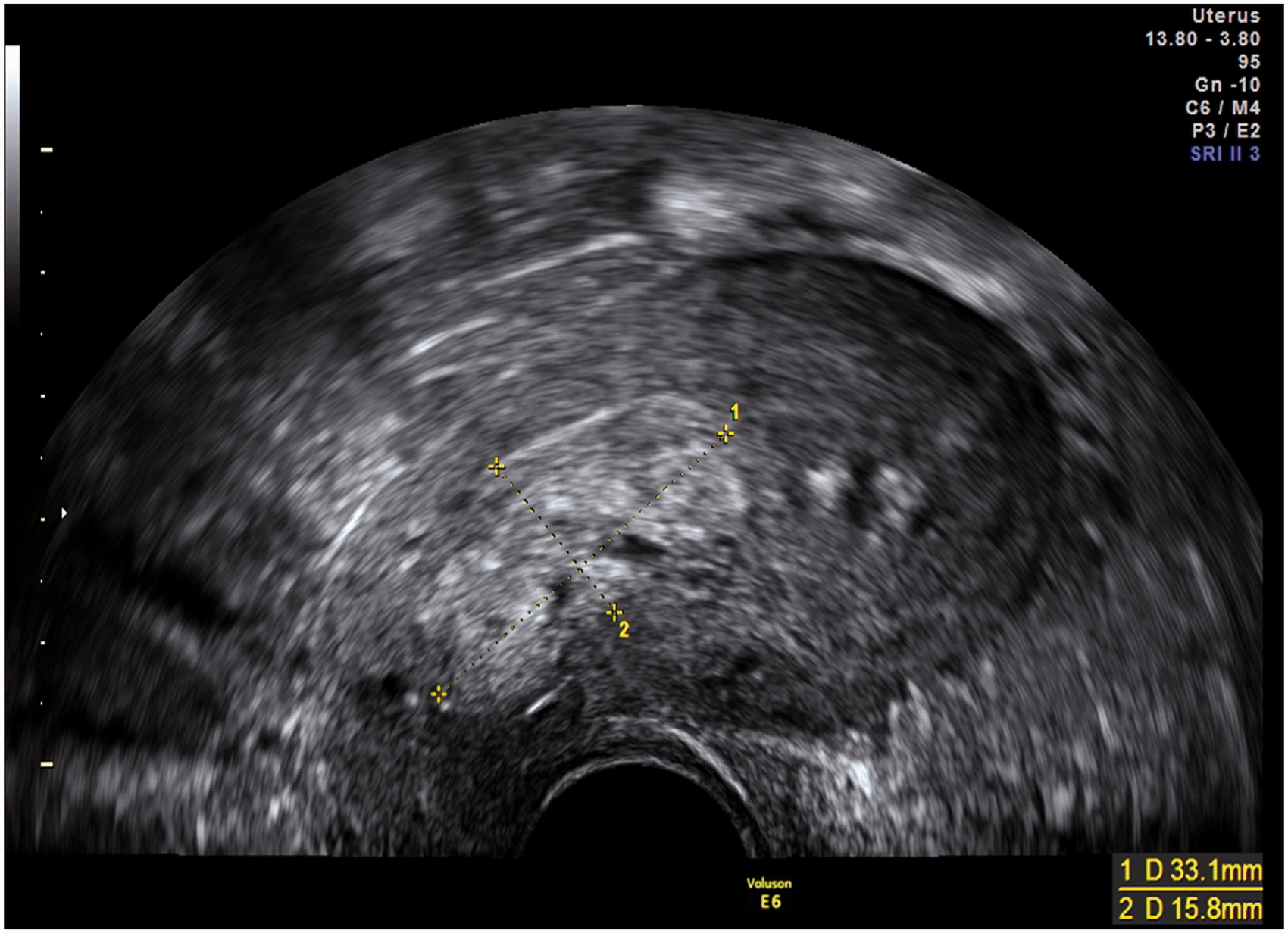
Echogenic mass within the endometrial cavity seen in the sagittal plane on transvaginal scan 6 weeks post vaginal delivery.
Rare postpartum ultrasound findings
Rarer postpartum ultrasound findings are summarised below.
Caesarean section associated findings
With rising rates of caesarean section delivery, our understanding of what are normal and abnormal post-caesarean ultrasound findings is improving. Following an uncomplicated procedure, the uterine incision can be seen as an iso- or hypoechoic region when compared with myometrium, and when imaged in the sagittal plane on transvaginal scan, is centrally located between the uterus and bladder.20–22 Depending on probe orientation, the uterine sutures can be identified as linear or point-like hyperechoic foci, and small haematomas (<15 mm) along the suture line can be considered as normal.20–22
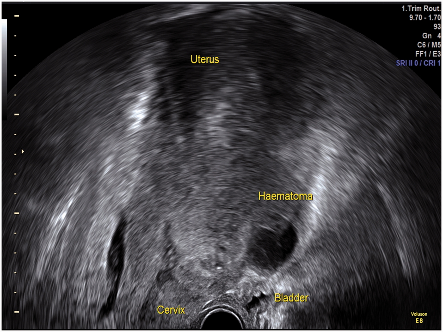
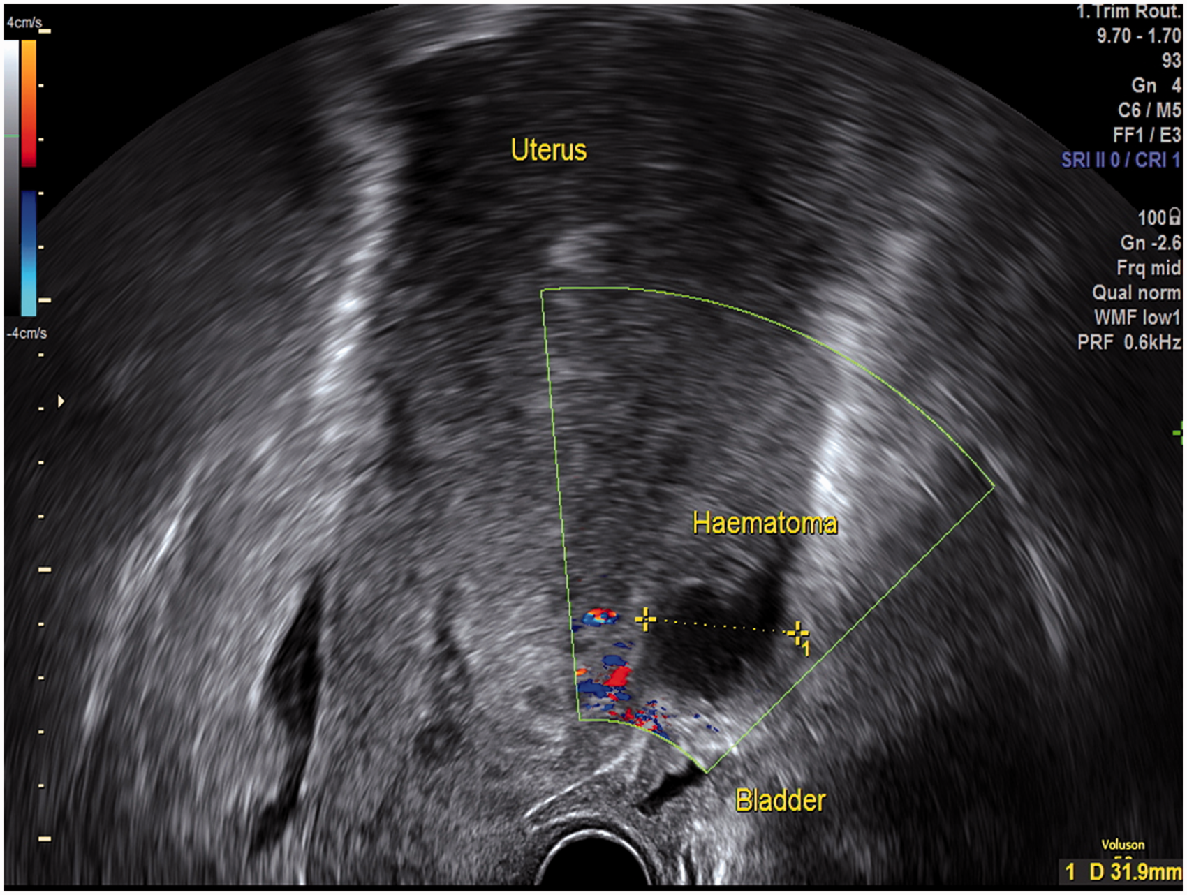
If adequate haemostasis has not been achieved intraoperatively, the immediate post-operative period may be complicated by the formation of a ‘bladder flap’ haematoma. During a LSCS, the visceral peritoneum is incised between the uterus and bladder, and reflected inferiorly. It is in this space that a haematoma may form, and will be seen as a non-vascular mass of mixed echogenicity, anterior to the uterus and posterior to the bladder, and has been described as a ‘bladder flap’ haematoma in the literature (Figures 7 and 8).
20
These may or may not be contained by the overlying peritoneum, and in the latter scenario will lead to the detection of haematoperitoneum on scan.
Application of colour Doppler demonstrates trophoblastic blood flow to the region of the echogenic mass, aiding diagnosis of retained products of conception. Echogenic mass seen within the lower uterine segment in the sagittal plane on transvaginal scan performed in a symptomatic patient 8 weeks post vaginal delivery. Echogenic mass within the lower uterine segment in the same patient (Figure 4) as seen in the transverse plane on transvaginal scan. Application of colour Doppler demonstrates trophoblastic blood flow to the region of the echogenic mass in the transverse plane on transvaginal scan (as seen in Figure 5) aiding diagnosis of retained products of conception. Post caesarean section haematoma originating from the anterior uterine wall, posterior to the urinary bladder, seen in the sagittal plane on transvaginal scan 7 days post procedure. Colour Doppler shows a non-vascular mixed echogenicity haematoma originating from the anterior uterine wall, posterior to the urinary bladder (sagittal plane on transvaginal scan).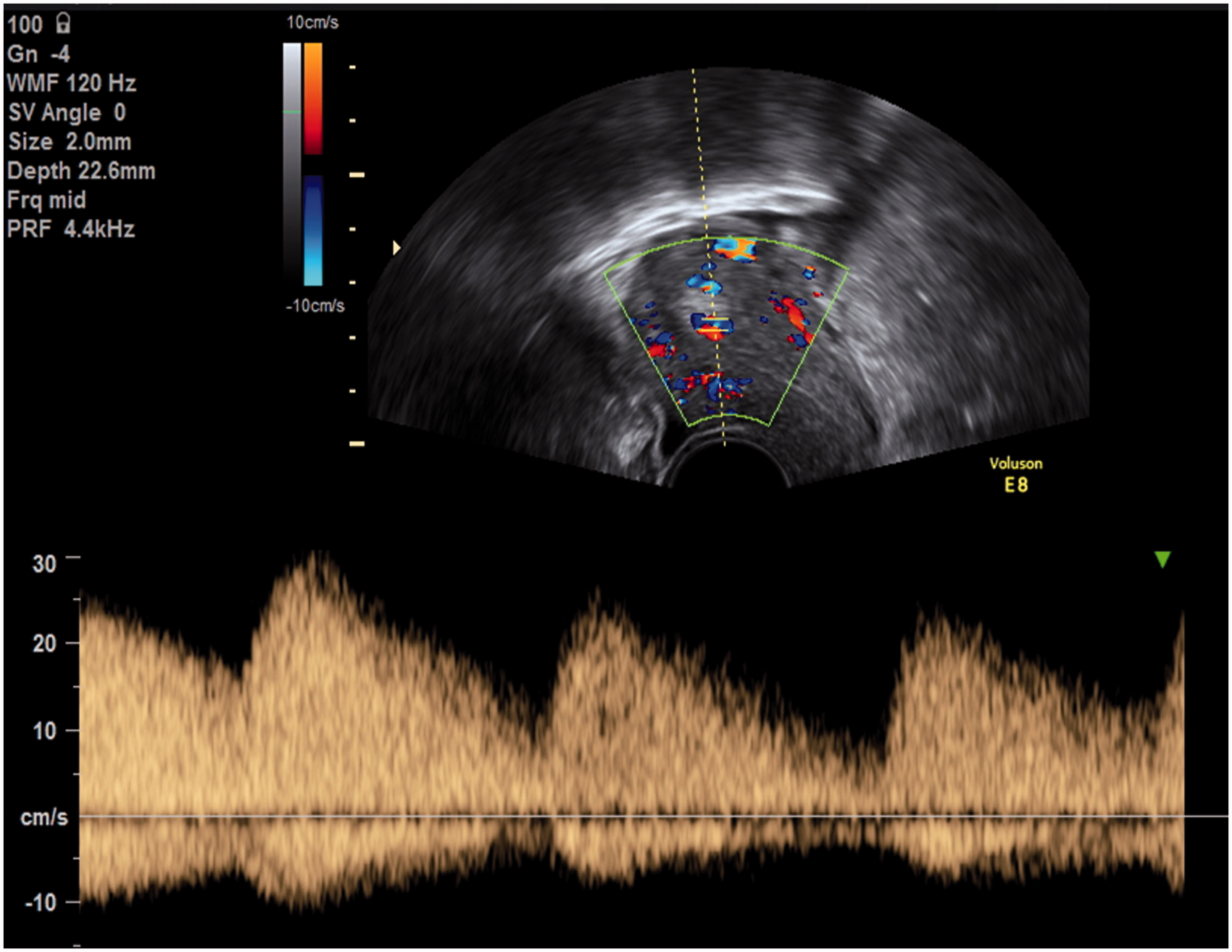

Pelvic sepsis
There are very few studies in the literature specifically reporting the ultrasound findings expected in postpartum endometritis. In contrast, cases of pelvic sepsis with underlying abscesses are more frequently described.20,23,24 In the event that superimposed infection of a pelvic or bladder flap haematoma occurs, ultrasound can be used to identify the formation of a pelvic abscess. The abscess will appear as a well-circumscribed fluid collection with or without internal septations, containing internal debris. The presence of gas, seen only when gas-producing organisms are present, will cause multiple highly echogenic foci, leading to ‘dirty’ posterior shadowing on scan. 20
Arteriovenous malformations (AVMs)
Since the first report of a uterine AVM almost 90 years ago, 25 these rare pelvic lesions are now increasingly reported in the literature, with over 200 cases.
Classically an acquired anomaly, uterine AVMs are most commonly associated with trophoblastic disease, pelvic surgery (e.g. myomectomy), endometrial curettage, uterine malignancy and caesarean scar pregnancy. A congenital aetiology has been postulated, particularly in cases where there is multi-organ involvement and the presence of multiple AVMs. They are most prevalent in women of reproductive age, rarely occurring in the nulligravid. Thus, it has been hypothesised that pregnancy contributes to the pathogenesis of uterine AVMs, 26 where necrosis of chorionic villi leads to the incorporation of venous sinuses into areas of myometrial scarring.
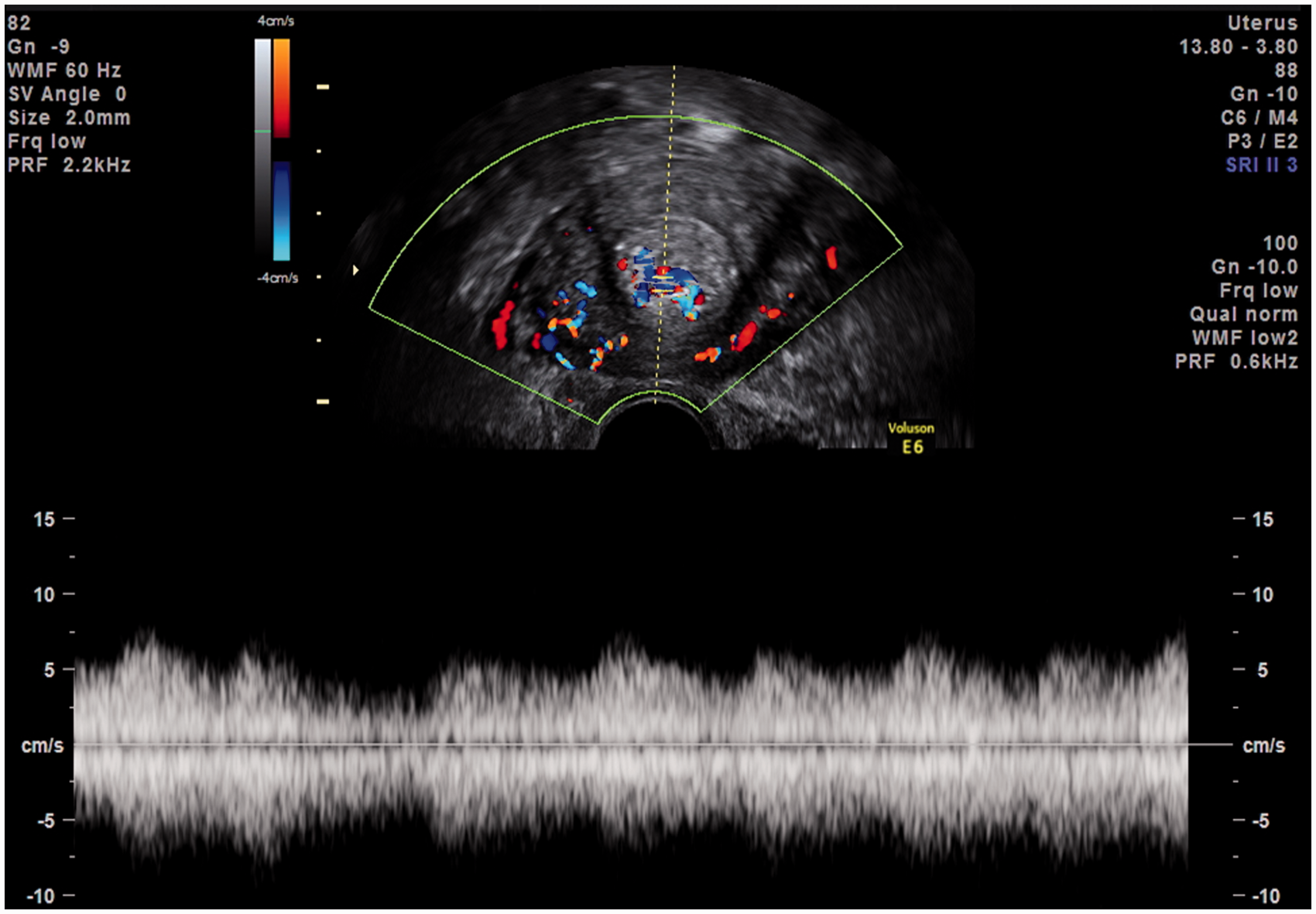
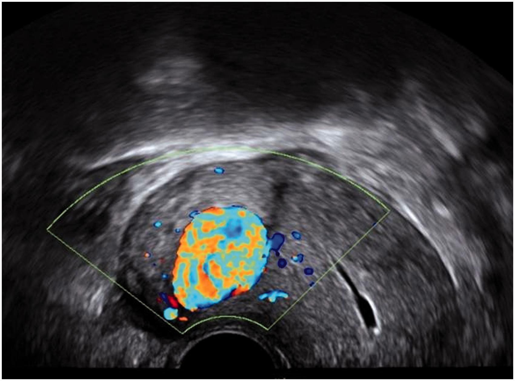
Uterine AVMs can present with either primary or secondary PPH, and rarely, a pulsatile pelvic mass. The volume of blood loss can be extensive and swift, leading to rapid haemodynamic compromise. Although the current gold standard diagnostic test is pelvic angiography, the use of Doppler ultrasound can successfully identify these vascular lesions. The typical appearance is of a highly vascular localised area within the myometrium (Figures 9 and 10). Pulsed Doppler evaluation will usually reveal a low-resistance blood flow with a broad waveform, high peak velocities and signs of turbulence.
26
Treatment is typically with selective embolisation of the feeding vessel, and less often surgical excision of the lesion.
Colour Doppler reveals localised, highly vascular AVMs within the myometrium identified on transvaginal scan (sagittal plane). Colour Doppler reveals localised, highly vascular AVMs within the myometrium identified on transvaginal scan (sagittal plane).
Conclusions
There are numerous studies in the literature describing postpartum appearances of the uterus and its contents. However, very few of these differentiate between the findings of an echogenic mass or mixed echo patterns, nor do they utilise colour Doppler to aid identification of retained placental tissue. Furthermore, there appears to be no convincing correlation between the presence of intrauterine material with patient symptomatology and morbidity. Only through accurate assessment and characterisation of such ultrasound findings can diagnostic confusion and ultimately, inappropriate and potentially dangerous surgical management be avoided.
Footnotes
Declaration of Conflicting Interests
The author(s) declared no potential conflicts of interest with respect to the research, authorship, and/or publication of this article.
Funding
The author(s) received no financial support for the research, authorship, and/or publication of this article.
Ethical approval
Not applicable.
Guarantor
Asli˙ Üçyiğit
Contributorship
JJ conceived the article topic and undertook the initial literature review. AÜ wrote the first draft of the manuscript and completed a further extensive literature review. Both authors reviewed and edited the manuscript and approved the final version of the manuscript.