




Abstract
The purpose of this study was to investigate the diagnostic performance of the head–perineum distance, angle of progression, and the head–symphysis distance as intrapartum ultrasound parameters in the determination of an engaged fetal head. Two hundred and one women in labour underwent both ultrasound and digital vaginal examination in the estimation of fetal head station. The transperineal ultrasound measured head–perineum distance, angle of progression, and head–symphysis distance for values correlating with digital vaginal examination head station. Using station 0 as the minimum level of head engagement, correlating cut-off values for head–perineum distance, angle of progression, and head–symphysis distance were obtained. Receiver operating characteristics were used in determining the diagnostic performance of these cut-off values for the detection of fetal head engagement. With head–perineum distance of 3.6 cm the sensitivity and specificity of sonographic determination of engaged fetal head were 78.7 and 72.3%, respectively. A head–symphysis distance of 2.8 cm also had sensitivity and specificity of 74.5 and 70.8%, respectively, in determining engagement, whilst an angle of progression of 101° was consistent with engagement by digital vaginal examination with 68.1% sensitivity and 68.2% specificity. Ultrasound shows high diagnostic performance in determining engaged fetal head at a head–perineum distance of ≤3.6 cm, head–symphysis distance of ≤2.8 cm, and angle of progression of ≥ 101°.
Introduction
Establishing engagement of the fetal head is important in active labour, as non-engagement at some point of cervical dilatation would indicate the need for caesarean section. 1 – 3 It is therefore useful to know if head station is within the expected range of a given cervical dilation in the first stage of active labour.4,5 Where instrumental vaginal delivery is being considered in the second stage of active labour, confirming the engagement of the fetal head is also a requirement.6,7
Conventionally, the determination of head station and engagement are done by digital vaginal examination (digital VE). However, apart from the reported cases of maternal discomfort8,9 and the increased risk of infection associated with this traditional method, 10 there are also reports of high levels of subjectivity and misdiagnosis. 11 A number of publications have reported sonographic parameters that could serve as an alternative non-invasive method for determining fetal head station and with the potential of diagnosing fetal head engagement. The most commonly reported sonographic parameters for head station are the head–perineum distance (HPD) and the angle of progression (AoP). 12 However, whilst the diagnostic value of HPD in determining a non-engaged fetal head had been reported, the HPD value for an engaged fetal head had not been reported. Again, in spite of the increasing number of publications reporting significant correlation between the sonographic AoP and the digital VE head station, we know from a recently published systematic review that the diagnostic value of the AoP for detecting an engaged fetal head was not yet reported. 12 In addition, other reported sonographic parameters for determining head station, such as the head–symphysis distance (HSD), had not been assessed for diagnostic performance in determining fetal head engagement.
The primary objective of this paper therefore was to investigate the diagnostic performance of the HPD, AoP, and the HSD in the determination of an engaged fetal head.
Methods
This is a prospective cross sectional study which was conducted at the labour and delivery ward of a Teaching Hospital in Ghana between April and September 2016. The study was approved by the institutional ethics review board, and informed consent was obtained from all study participants. It included 201 consenting pregnant women at a gestational age of 37–42 weeks. The inclusion criteria were singleton gestation, cephalic presentation, and spontaneous labour. Women in labour with the following conditions were excluded: induction of labour, breech presentation, multiple pregnancy, polyhydramnios, sonographically detected fetal abnormalities, and previous caesarean section.
Digital VE was performed by the managing clinician on duty to determine the fetal head station of consenting participants, along with other routinely measured parameters for assessing labour progress. These examiners were specialists with at least five years of experience in intrapartum care. Station was estimated by assessing the relationship between the level of the ischial spines and the leading edge of the fetal head. 13 Engagement referred to the leading edge of the fetal head being at the level of the ischial spines or further below (i.e. stations 0 to +5).11,13 Therefore, non-engagement implied that the leading edge of fetal head was above the ischial spines (i.e. stations −1 to −5). All digital VEs were performed in the absence of uterine contraction.
Immediately after the digital VE, transperineal ultrasound was performed by an independent ultrasound investigator with over 10 years of experience, who was blinded from the digital VE findings, to measure the HPD, AoP, and HSD. The ultrasounds were performed with a mobile ultrasound unit (P 300, Siemens-Acuson, Italy), using a 2–5 MHz curvilinear transducer.
The transperineal ultrasound examination was performed by placing the curvilinear transducer at the perineal space between the labia and the anus. With the transducer held in the sagittal plane over the perineal region, the fetal head was displayed on the monitor often with part of the symphysis pubis showing. The transducer was tilted slightly to direct the sound beam towards clear visualisation of the symphysis pubis in its longest axis (see Figure 1). In some cases slight rotational manoeuvres were necessary for obtaining the longest axis of the symphysis pubis.
The symphysis pubis in long axis.
The image was frozen upon visualisation of the symphysis pubis to obtain measurements for the AoP. This was measured by drawing a line through the long axis of the symphysis pubis, using the ‘distance’ calliper on the machine. By clicking on another distance calliper, a second line was then drawn from the inferior edge of the symphysis pubis to form a tangent with the leading edge of the fetal head as shown in Figure 2. Afterwards, a goniometer was then used in measuring the angle formed by the two drawn lines obtaining the AoP as described by Barbera et al.
14
The AoP measurement. AoP: angle of progression.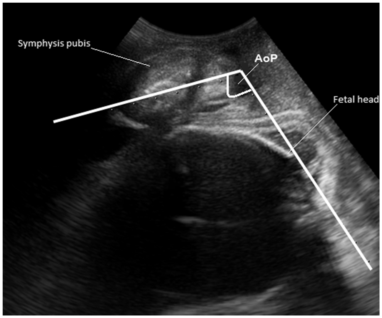
The HSD was also obtained from the same plane as the AoP by placing the distance calliper at the inferior edge of the symphysis pubis to obtain a perpendicular distance from the midline in the symphysis pubis to the fetal head in centimetres as described by Youssef et al.
15
(see Figure 3).
The HSD measurement. HSD: head-symphysis distance.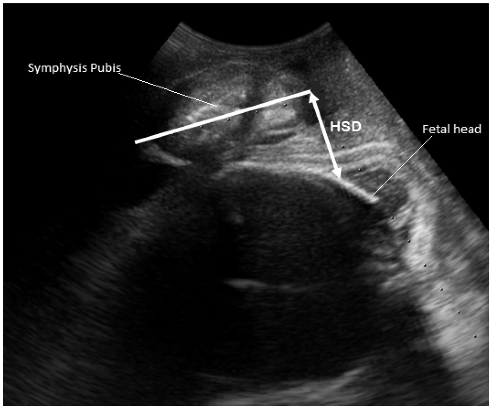
From the sagittal plane, the transducer was rotated 90° anticlockwise for a transverse plane image. The transducer was gripped firmly to prevent it from sliding or tilting. Gentle pressure was then applied until the hard pelvic bone could be felt. The image was then frozen to measure the HPD as the distance from the fetal head to the surface of the perineum as described by Eggebø et al.
16
(see Figure 4).
The HPD measurement. HPD: head–perineum distance.
Data were entered into an Excel Spread Sheet. Data were then expressed as mean ± SD and 95% confidence interval. P-value less than 0.05 was considered for statistical significant difference. Receiver operating characteristics (ROC) curve was performed to assess the diagnostic performance of the HPD, AoP, and HSD in comparison with digital VE findings on fetal head station for the determination of engagement. Data analysis was done with XLSTAT version 2015 for Windows.
Results
Out of the total 201 parturients who participated in the study, data analysis was possible in 196 participants, due to some missing information on five participants. Participants were in the age range of 20–39 years, which included 47% nulliparous women, 22% primiparous, and 31% multiparous. Their average gestational age before spontaneous onset of labour was about 39 weeks + four days. Also, their body mass index was in the range of 20–42 kg/m2 with the average being approximately 28 kg/m2.
One hundred and eighty-eight out of the 196 participants which represents 96% were in the digital VE head station range of −2 to +2. The highest percentage of participants were diagnosed by digital VE as being at station 0, followed by station −1. Generally, 46% were diagnosed by digital VE as non-engaged at various stations above the maternal ischial spines, whilst 54% were diagnosed as engaged at various stations below the maternal ischial spines.
Mean levels of HPD, HSD, and AoP in relation to fetal head station by digital VE

AoP: angle of progression; CI: confidence interval; digital VE: digital vaginal examination; HPD: head–perineum distance; HSD: head–symphysis distance; SD: standard deviation.
Also, a HSD of 3 cm was the upper limit of station 0, whilst an HSD of 2.6 cm was the lower limit of station 0. Lastly, an AoP of 98° was the lower limit of station 0, whilst an AoP of greater than 105° was the upper limit of station 0.
Figures 5 to 7 and Table 2 show the diagnostic performance of HPD, HSD, and AoP as sonographic methods of assessing fetal head engagement. Using the ROC curve, the threshold for HPD below which head engagement would be diagnosed was 3.6 cm, as it was the average HPD value corresponding to station 0. On the basis of this threshold, area under ROC curve (AUC), the sensitivity, specificity, positive predictive value, and negative predictive value of ultrasound of HPD for diagnosing engaged fetal head were 0.7946, 78.7%, 72.3%, 49.0%, and 92.0%, respectively.
ROC curve showing the diagnostic performance of HPD on engagement. AUC = 0.7946. AUC: area under ROC curve; HPD: head–perineum distance; ROC: receiver operating characteristic. ROC curve showing the diagnostic performance of HSD on engagement. AUC = 0.8265. AUC: area under ROC curve; HSD: head–symphysis distance; ROC: receiver operating characteristic. ROC Curve showing the diagnostic performance of AoP on engagement. AUC = 0.7729. AoP: angle of progression; AUC: area under ROC curve; ROC: receiver operating characteristic. Diagnostic performance of HPD, HSD, and AoP on fetal head engagement AoP: angle of progression; FN: false negative; FP: false positive; HPD: head–perineum distance; HSD: head–symphysis distance; NPV: negative predictive value; PPV: positive predictive value; TN: true negative; TP: true positive.
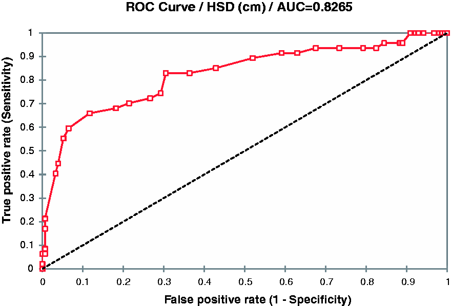


The cut-off value for HSD below which head engagement would be diagnosed was 2.8 cm, as it was the average HSD value corresponding to station 0. On the basis of this threshold, AUC, the sensitivity and specificity of ultrasound for diagnosing engaged fetal head were 0.8265, 74.5%, and 70.8%, respectively. The positive predictive value and negative predictive values for HSD were 44.0 and 90.0%.
The threshold for AoP above which head engagement would be diagnosed was 101°, as it was the average AoP value corresponding to station 0. On the basis of this threshold, AUC, the sensitivity and specificity of ultrasound for diagnosing engaged fetal head were 0.7729, 68.1%, and 68.2%, respectively. The positive predictive value and negative predictive value were 39.5 and 87.5%, respectively.
Discussion
This study investigated the diagnostic performance of sonographic parameters in detecting engaged fetal head. Earlier studies by Maticot-Baptista et al. 17 and Dimassi et al. 18 reported that a HPD of 5.5 cm or higher was predictive of fetal head non-engagement with high sensitivity and specificity, but provided no HPD predictive for an engaged fetal head. Given that the distance from the perineum to the maternal ischial spines is reported to be 5 cm apart,16,19 an HPD value for an engaged fetal head would be expected to be ≤5 cm since engagement occurs around station 0 or at a station further below. 14 However in the 62 parturients whose digital VE head station was 0, the HPD obtained was in the range of 3.4–3.9 cm (Table 1). Consequently, the average HPD of 3.6 cm was used as the cut-off value for high likelihood of predicting an engaged fetal head, just as a HPD of ≥5.5 cm was reported as being predictive of non-engagement. The 3.6 cm average HPD for station 0 was in perfect agreement with Tutschek et al. 20 who earlier reported that a HPD of 3.6 cm corresponded with station 0 in their study population. However, their study provided no information on the diagnostic performance in connection with fetal head engagement.
With regards to the AoP, this study noted that digital VE station 0 averagely corresponded with 101°. This AoP value obtained by this study in correlation with station 0 is apparently lower than the 123° reported by Chan et al. 21 in the Chinese study population and the 116° reported by Tutschek et al. 20 in the Norwegian study population. It is, however, closer to the 99° obtained by Barbera et al. 22 which compared the AoP to CT scan findings in a separate non-gravid population. In a related finding, this study also noticed that the 62 parturients with digital VE head station 0 had an AoP in the range of 98°–105°, and that using an AoP of ≥99° was a good predictive value for fetal head engagement as the findings indicate (Table 2). It however implies that these studies all agree on station 0 typically corresponding to an AoP above 99° and therefore highly probable to be indicative of an engaged fetal head as the AoP gets wider above 100°.
In addition, although not as well known as the HPD and AoP, the HSD was also investigated by this study because it is measured in the same plane as the AoP and could therefore serve as an additional sonographic parameter that could complement the HPD and the AoP for determining an engaged fetal head. This suggests that all three parameters may be measured to confirm agreement, as in the use of fetal biometry for the estimation of fetal growth where it is standard to measure more than one parameter for comparability and verification. It also indicates that a high agreement amongst the three may increase confidence in these intrapartum sonographic parameters if chosen as the method for determining fetal head engagement in a given parturient.
To the best of our knowledge, this is the first time the diagnostic performance of ultrasound in detecting engaged fetal head has been reported. Second, this study was performed in a black African population for the first time. The shape of the female pelvis might differ between populations. Thus, this article also adds important knowledge about using ultrasound in labour in other populations.
Footnotes
Acknowledgement
We are grateful to the midwives and obstetricians of Komfo Anokye Teaching Hospital, for their cooperation and assistance in the collection of data in this study.
Declaration of Conflicting Interests
The author(s) declared no potential conflicts of interest with respect to the research, authorship, and/or publication of this article.
Funding
The author(s) received no financial support for the research, authorship, and/or publication of this article.
Ethical approval
Local approval was obtained from the Committee for Human Research, Publication and Ethics of Komfo Anokye Teaching Hospital, with reference number, CHRPE/AP/251/15.
Guarantor
ATO.
Contributors
YAW, BW, and HV researched literature and conceived the study. ATO supervised data collection. YAW wrote the first draft of the manuscript. All authors reviewed and approved the final version of the manuscript.