




Abstract
Extracellular vesicles (EVs) are small, nonreplicating, lipid-encapsulated nanoparticles that carry protein and nucleic acid cargo derived from their tissue of origin. Due to their capacity to provide comparable insights to solid organ biopsy through a minimally invasive collection procedure, EVs provide an attractive biomarker source. This review will provide an insight, how EVs in circulation may provide a novel way to assess cholestasis and will address the possibility of getting a better understanding of the mechanisms of cholestasis of pregnancy through the use of serial hepatic-specific EVs as a window.
Keywords
Intrahepatic cholestasis of pregnancy (ICP)
ICP is the most common liver disease related to pregnancy, with per annum incidence in Australia of 0.6%–0.7%. 1 It is characterised by pruritus with increased serum bile acids and other liver function tests. 2 Intrahepatic cholestasis is associated with significant risk to the fetus including stillbirth, preterm birth and neonatal morbidity. 3
Women are usually diagnosed with ICP in the third trimester of pregnancy, however, early-onset ICP carries a higher incidence of preterm labour, fetal distress and lower birth weights compared to later-onset ICP. 4
The aetiology of the disease is poorly understood and is thought to be complicated and multifactorial.1,5 Genetic susceptibility, hormonal, and environmental factors have been proposed as possible mechanisms of the disease. 6
Although ICP resolves shortly after delivery, there is growing evidence of increasing risk of developing hepatobiliary cancer, autoimmune and cardiovascular disease later in life, highlighting the need for better understanding of the disease aetiology and subsequent management. 7
Extracellular vesicles (EVs)
EVs are a heterogenous population of small, non-replicating, membrane-encapsulated particles, that are released into biofluids including blood, saliva, and urine by virtually all cells.8,9 They contain biologically functional cargo, comprised of proteins, lipids, metabolites and nucleic acid cargo, including mRNA, miRNA, and non-coding RNAs, that mirrors protein and nucleic acid content of their parent cells, thus serving as cellular surrogates and are hypothesised to carry cellular markers from distant, even inaccessible anatomical locations (Figure 1).10–13
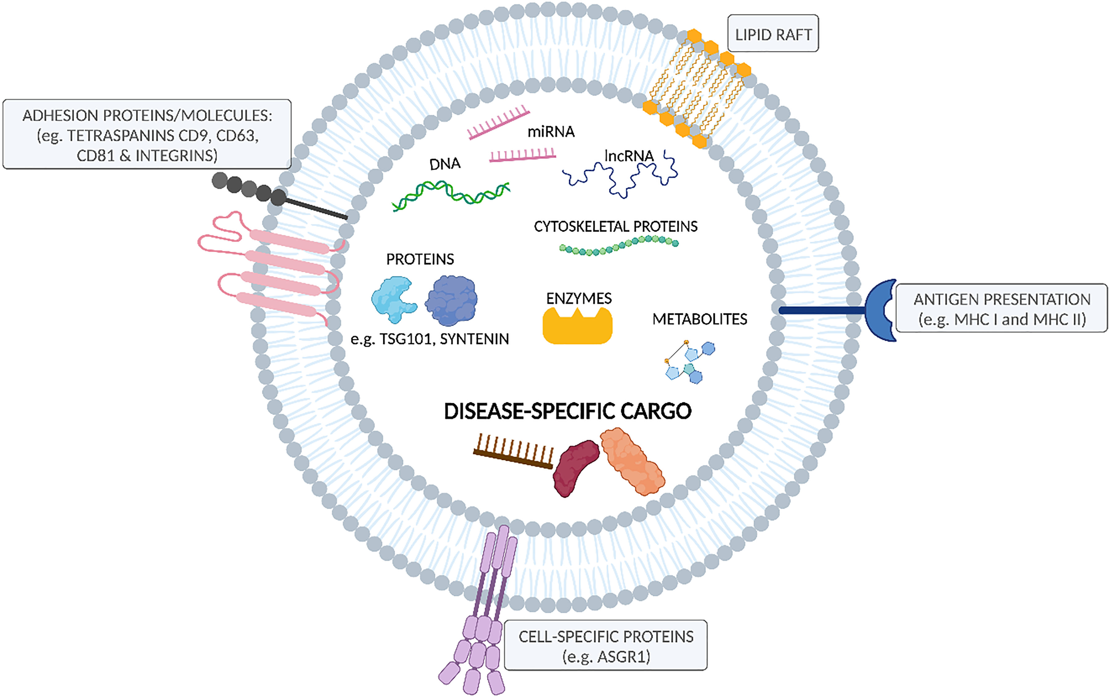
Schematic representation of extracellular vesicle structure and cargo. The phospholipid bilayer is integrated with membrane proteins such as tetraspanins (CD9, CD63, CD81), cell-specific proteins (ASGR1 – liver-specific protein), adhesion molecules (integrins), major histocompatibility complex (MHC I and MHC II) and lipid rafts. Membrane surrounds the content of nucleic acids (mRNA, miRNA, DNA, lncRNAs), proteins (TSG101, Syntenin), enzymes, metabolites and disease-specific cargo.
This capacity of EV-encapsulated cargo to reflect the state of their parent organ has driven the prominent rise to the studies investigating EVs as a minimally invasive liquid biopsies. 14 In fact, based on the promising data from numerous preclinical studies, more than 50 clinical trials have been registered (www.clinicaltrials.gov) in aim to evaluate the potential of EVs as biomarkers for disease diagnosis. 13
The key advantage of EVs for disease biomarker studies is the ability to efficiently diagnose and monitor disease progression or treatment response in near real time at the molecular level. 15 For biomarker studies, EVs can be isolated from routinely collected blood samples from randomised controlled trials or from freshly collected samples of patients attending their medical appointments. 16 Typically, global (crude) EVs are isolated from biological fluids using a wide range of techniques, that offer various degree of sample specificity and yield, however, global EV sample contains a pool of EVs originating from all cells in the body, providing a highly heterogeneous EV sample. 9 In order to address the challenge associated with EV sample heterogeneity in liver disease setting, liver-specific EV isolation strategy can be used. 15
Liver-specific EVs
A Global EV sample includes all EVs released in the body, therefore, traditional approaches to isolate global EVs do not distinguish between tissue of origin. Around 90% of EVs in human plasma originate from platelets and other haematopoietic or endothelial cells and EVs from every other organ in the body, with the majority of EVs present in the sample not being relevant to analysis and potentially masking disease-specific low abundant disease biomarkers by reducing assay sensitivity. 17
This is especially important in the diagnosis of early-stage patients when high assay sensitivity is required. The ability to The ability to efficiently isolate relevant subpopulations of single organ or disease-associated EVs containing candidate biomarkers represents an important step towards their clinical translation. 13
Since EVs inherit specific proteins from their parent cells, they can be selectively isolated by immunoprecipitation. 12 This technique is reliant on a protein marker specifically expressed or at least enriched in the tissue of interest, and that is localised to the EV plasma membrane and has an external domain, that can be targeted by a specific antibody. Liver-specific EVs can be selectively isolated by anti-asialoglycoprotein receptor 1 (ASGR1) immunoprecipitation, reducing the ‘noise’ produced by non-liver EVs and lipoproteins and increasing sensitivity of liver disease biomarker analysis (Figure 2). 17
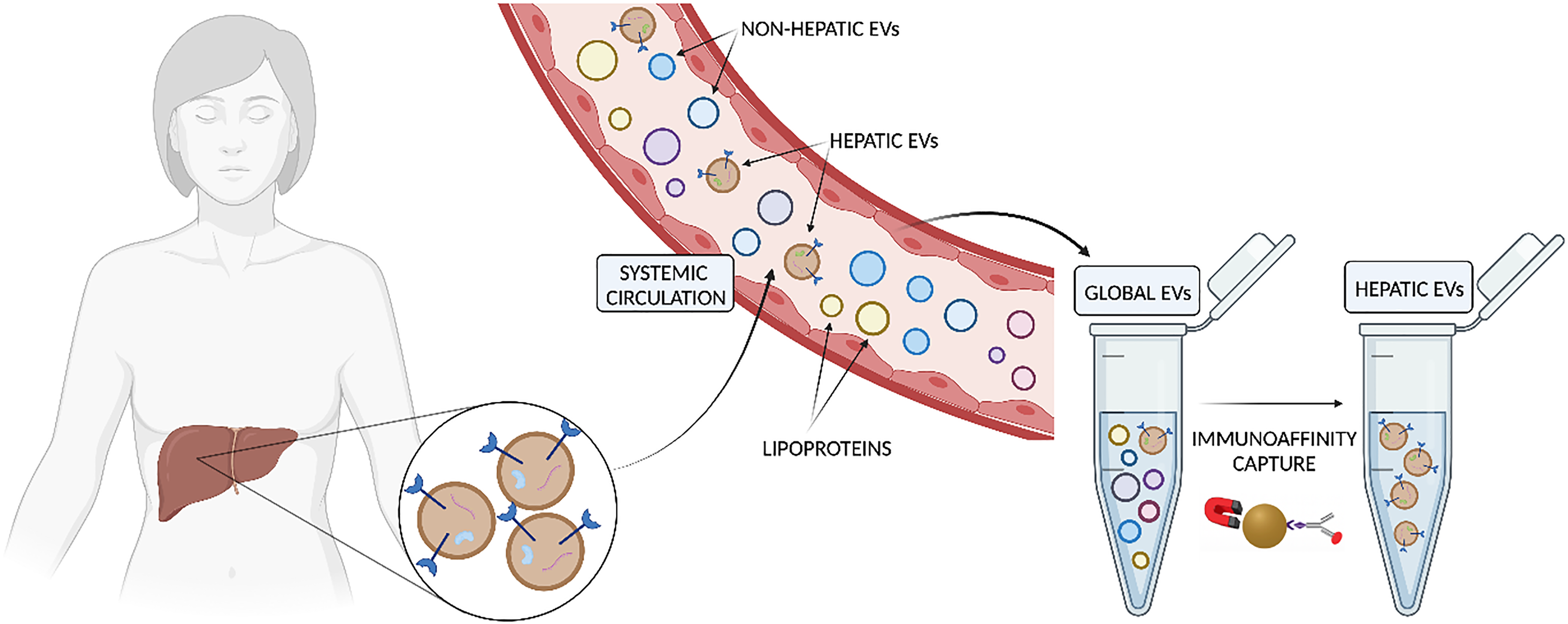
Schematic representation of immunobead-based capture for selective isolation of hepatocyte-derived EVs. Global EVs in circulation can be isolated by an array of conventional EV isolation methods. The addition of liver-derived EV isolation step by anti-asialoglycoprotein receptor 1 (ASGR1) immunoprecipitation increases sensitivity of liver disease biomarker analysis.
Recent evidence demonstrates high concordance between proteins detected in human liver tissue and EVs isolated from the same liver tissue, with proteome of EVs in the human liver tissue containing 58% of the proteome of the liver. 18 Furthermore, representation of proteins across hepatocyte cellular compartments is comparable between liver tissue and EVs originating from paired liver tissue. 18
When coupled with robust methods to selectively isolate hepato-derived EVs from blood, there is a promising potential for the use of circulating liver-derived EVs as a liquid biopsy for this organ.
Liver-specific EVs as liquid biopsy
The use of anti-ASGR1 immunoprecipitation for the selective isolation of liver-specific EVs was shown to benefit the analysis of EV-encapsulated miRNA biomarkers in non-alcoholic fatty liver disease (NAFLD) and was able to assist with staging of the disease.
Specifically, it was demonstrated that liver-specific EV-derived miR-122, -192, and -128-3p were significantly associated with disease severity, while disease severity could not be differentiated with either total cell-free or global EV-derived RNA. 17 This provides the first direct evidence that selective isolation of liver-derived EVs enhances assay sensitivity enabling detection of minor changes in miRNA expression between different stages of chronic liver disease.
In addition to liver disease biomarkers, there is an emerging body of evidence to support the use of liver EV-derived proteins as biomarkers of drug metabolism and disposition capacity. 19 Liver-specific EVs were used as a liquid biopsy to study induction of cytochromes P450 (CYP3A4, CYP3A5 and CYP2D6) and organic anion-transporting peptides (OATPB1 and OATP1B3) during pregnancy and after administration of rifampicin (RIF) to healthy male subjects. Proteomic analysis revealed induction of liver-derived EV CYP3A4 following RIF administration consistent with the mean oral midazolam area under the plasma concentration-time curve ratio in the same subjects. It was also possible to show that CYP2D6 was not induced by RIF. Further analysis of liver-derived EVs revealed significantly higher CYP3A4 and CYP2D6 protein expression in women at the third trimester of pregnancy compared to non-pregnant women, while expression of both OATPs in liver-derived EVs was unaltered by RIF administration and pregnancy. 19
Liver-specific EVs to gain a better understanding of the mechanisms of cholestasis in pregnancy
There are limited studies investigating the use of EVs in ICP in humans, however, easy access of EVs from minimally invasive routine blood sampling and promising data from chronic liver disease and drug metabolism and disposition studies offer a great opportunity to gain a better understanding of the mechanisms of ICP through the use of EVs as a window.
The abnormal synthesis, metabolism, secretion and excretion of bile acids may lead to ICP. 20 Aetiology contributing to the development of ICP disease is multifactorial and includes genetic predisposition.20,21 A number of ABC family transporter genes like ABCB4, ABCB11 and ABCC2 are known to have a functional role in the development of ICP, however, the role of other ABC transporter genes is less studied. 22 EVs carrying ABCB4, ABCB11 and ABCC2 have been studied as biomarkers in cancer and drug absorption, distribution, metabolism and excretion studies, indicating the feasibility of these markers to be detected in EVs.23–25
Recent studies identified mutations in ATP8B1 and ANO8 genes, which may also provide new insight into genetic mapping on ICP. 26 Interestingly, a recent EV study showed that ATP8B1 is a marker of vasculogenesis and angiogenesis, critical processes in fetal circulation and placental vasculature development, further suggesting that ICP-associated markers are carried by EVs. 27
The significance of epigenetic regulation on gene expression is becoming more evident in ICP. 28 In ICP patients, miR-148a has been shown to be upregulated in the placenta and the peripheral blood, and miR-148a is upregulated by oestradiol and may be involved in the induction of oestrogen. 29 In addition to miR-148, miR-590-3p was reported to regulate vascular cell adhesion protein 1 (VCAM1), while miR-21 and miR-29a were shown to directly target and inhibit the expression of intercellular adhesion molecule 1 (ICAM1).
Both VCAM1 and ICAM1 are already studied in non-ICP EV studies. A recent EV study demonstrated rapid neutrophil mobilisation by VCAM1-positive endothelial cell-derived EVs in acute myocardial infarction and EV-associated ICAM1 has been shown to be upregulated by interferon-γ and immune checkpoint blockade-based therapies.30,31
A promising EV study suggested that EV-derived miR-21, miR-29a and miR-590-3p can be used as potential biomarkers for the diagnosis of ICP. This study also has shown the urinary EVs collected from ICP patients displayed upregulated expression of miR-21, miR-29a and miR-590-3p, compared with the urinary EVs collected from healthy controls. 32 A number of studies show EV-derived miR-148a, miR-590-3p, miR-590-3p to be detectable and studies in pleural mesothelioma and colorectal cancer, further demonstrating the feasibility of ICP-associated miRNA markers to be further investigated in EVs.33,34
At the protein level, matrix metalloproteinases (MMPs) MMP-2 and MMP-9 were shown to be upregulated in ICP, suggesting potential new drug targets. 35
The dysfunction of the endoplasmic reticulum (ER) and mitochondria is also known to be associated with pathogenesis of ICP. 36 Elevated levels of glucose-regulated protein (GRP-78) were reported to be upregulated in trophoblasts of ICP patient placental tissues. 36 Elevated bile acid levels in ICP can activate mTOR signalling pathway causing ER stress and leading to a decrease in trophoblast viability. 37 Comparative proteomics analysis found placental proteome alterations in women in ICP, which include proteins involved in cytoskeleton activity, blood coagulation, platelet activation, heat shock proteins, calcium-binding and RNA-binding proteins and a number of enzymes, compared to women with a normal pregnancy. 38 Promisingly, recent EV studies show MMP2 and MMP2 detectable in EVs; EV-associated MMP2 abrogates intercellular hepatic miR-122 transfer to liver macrophages and curtails inflammation, and EV-associated MMP-9 has been shown to act as a prognostic marker for brain tumours.39,40 EV-associated GRP-78 has been shown to alter the migration and proliferation of cancer cells. 41
Genetic predisposition for ICP, tissue specificity (enrichment in the liver tissue) of the ICP-associated markers and the ability to study EV nucleic acid and protein cargo, may open a novel avenue to interrogate the pathway of the disease further, that may lead to earlier diagnosis, before women present with symptoms, and as a result improving maternal and perinatal outcomes.
Conclusion
In order to study and understand the pathway of liver disease, tissue biopsy is often required, followed by consecutive biopsies to evaluate hepatic changes in the course of the disease. In ICP, tissue biopsy is not required for the disease diagnosis and monitoring, therefore, studies interrogating the pathway of this disease are not well understood in humans. Easy access to liver-specific EVs in circulation and the ease of serial sample collection throughout the course of the disease, offers a novel opportunity to understand this disease, improving outcomes.
Footnotes
Acknowledgements
Figures were created using BioRender.com. ZU would like to thank Prof. Andrew Rowland, Prof. in Clinical Pharmacology at the College of Medicine and Public Health at Flinders University, for his guidance and support in writing this review.
Contributorship
ZU researched literature and wrote the review.
Declaration of conflicting interests
The author declared no potential conflicts of interest with respect to the research, authorship, and/or publication of this article.
Ethical approval
Not applicable.
Funding
The author received no financial support for the research, authorship, and/or publication of this article.
Guarantor
ZU.
Informed consent
Not applicable.

Trial registration
Not applicable.