
Abstract
Tissue-inducing biomaterials, which promote tissue regeneration without the addition of exogenous cells and/or bioactive factors, have recently attracted increasing interest in the repair of nonosseous tissues. As a key strategy for transforming data into actionable evidence, evidence-based biomaterials research plays a critical role in guiding material development. In this study, evidence mapping method was employed to systematically analyze and visualize animal study designs, material characteristics, outcome indicators, and evaluation methods, aiming to identify current research trends and emerging focal areas. The results revealed a wide diversity of experimental animal species, with a predominance of small animal models. Among the 19 types of nonosseous tissues investigated, skin, abdominal wall, cartilage, and blood vessels were the most frequently studied. Materials were mainly classified into bio-derived materials, polymers, and composites. Outcome indicators span from macroscopic to molecular levels, with tissue-level indicators being the most commonly applied. Histological analysis served as the primary method for validating inductive effects, supported by gross observation, imaging analysis, molecular biology assays, and biomechanical testing. Overall, tissue-inducing biomaterials show promising potential for nonosseous tissue regeneration. However, challenges remain, including limitations of animal models, short follow-up periods, and insufficient evaluation systems. Future studies should strengthen the alignment between functional validation and clinical needs to promote the translation of these materials from experimental research to clinical application.
Impact Statement
This study provides the first evidence map of in vivo animal studies on the application of tissue-inducing biomaterials for nonosseous tissue regeneration. By integrating data on material types, target tissues, experimental models, outcome measurements, and detection methods, the study offers a comprehensive overview of current research. These findings highlight the potential of tissue-inducing biomaterials beyond bone regeneration and current research trends, providing valuable evidence-based guidance for future experimental design and translational research in nonosseous tissue engineering.
Introduction
Biomaterials play a pivotal role in regenerative medicine and tissue engineering. Traditional inert biomaterials have primarily served as structural substitutes to support and restore the morphology of damaged tissues. 1 However, with the advancement of research, the emergence of tissue-inducing biomaterials has significantly expanded the scope of the biomaterials field.2–7 These materials are engineered to induce the regeneration of tissues or organs solely through optimized material design, without the need for exogenous living cells and/or bioactive factors. 8
Tissue-inducing biomaterials were first identified in the 1990s. Xingdong Zhang from China, 5 Ripamonti from South Africa 3 and Yamasaki from Japan,2,4 successively reported the phenomenon of ectopic bone formation induced by calcium phosphate ceramics without the incorporation of any cells or bioactive factors in various animal models. This groundbreaking discovery demonstrated that biomaterials alone, without the addition of exogenous cells or growth factors, could still induce bone formation. Since then, tissue-inducing biomaterials have achieved significant progress in bone tissue regeneration and have gradually gained international recognition.1,9–12
Studies on the phenomenon and mechanisms of osteoinduction have provided valuable insights for extending material-induced regeneration to other tissue types. Building on this foundation, researchers began to explore the application of tissue-inducing biomaterials in nonosseous tissues, evaluating their biological responses and inductive effects through animal experiments. For instance, in a 2002 study, Marijnissen et al. reported that implantation of E210 (a nonwoven fleece of polyglactin)-alginate composites into the subcutaneous tissue of nude mice induced localized cartilage formation. 13 In addition, tissue-inducing biomaterials have shown promising potential in the regeneration of various tissues, including ligaments, muscles, and blood vessels.14–17 However, despite the emergence of diverse animal studies, there is currently a lack of research that systematically organizes and comprehensively analyzes existing research on nonosseous tissue-inducing materials.
In recent years, the concept and methodology of evidence-based biomaterials research have been proposed to transform data and findings from material studies into valid scientific evidence. 18 Evidence mapping, as one of the key methods in evidence-based research, offers distinct advantages such as systematic panoramic scanning, visual representation, and the ability to clarify the current research landscape and identify knowledge gaps. 19 Therefore, this study aims to systematically review existing research on tissue-inducing biomaterials for nonosseous applications using an evidence mapping approach. The goal is to reveal their potential and current trends in nonosseous tissue repair and regeneration, and to provide evidence-based insights for future material optimization and clinical translation, ultimately promoting the advancement of tissue-inducing biomaterials in regenerative applications beyond bone.
Methods
Literature search strategy
We conducted a comprehensive search of Medline (via Ovid) for original animal studies related to tissue-inducing biomaterials. The final search was performed on November 20, 2023. The search strategy was based on the core keywords “tissue induction” and “materials,” and incorporated the updated and extended version of the SYRCLE animal filter to identify all relevant animal experiments. 20 No restrictions were applied regarding language or publication date. The complete search strategy is provided in Supplementary Data S1.
Inclusion and exclusion criteria
Nonosseous tissues were defined as all tissue types in the body excluding bone, including soft tissues, neural tissues, vascular tissues, and others. The specific tissue categories were not predetermined, but rather inductively identified based on the descriptions in each study regarding the anatomical target or primary site of regeneration. These tissue types were then systematically categorized and consistently represented throughout the evidence map sections. The inclusion criteria for this study were established using the PICOS framework, as detailed below: P (Participants): Animal models involving the repair, regeneration, or functional improvement of nonosseous tissues. I (Intervention): Use of tissue-inducing biomaterials, defined as materials that can induce tissue or organ regeneration through optimized material design alone, without the addition of living cells and/or bioactive factors. No restrictions were placed on the degradability, form, or application method. C (Comparator): No limitations on the type of control group. O (Outcome): Inclusion of at least one outcome indicator related to the remodeling or regeneration of nonosseous tissues induced by the material. S (Study Design): Animal studies. In addition, eligible studies had to meet the following condition: The full text must be available in either Chinese or English. Duplicate publications and studies without an accessible full text were excluded.
Literature screening
All search results were imported into EndNote 21, and duplicate records were removed. Two reviewers independently screened the titles and abstracts of all identified studies based on the inclusion criteria. A pilot screening was conducted prior to formal screening, in which 10% of the studies (randomly selected) were independently assessed by both reviewers. Interrater agreement was calculated using percentage agreement, and formal screening commenced only after agreement exceeded 75%. During screening, studies were categorized into three groups: included, excluded, and uncertain. Full texts of studies deemed potentially eligible or classified as uncertain were then independently reviewed by both reviewers to determine final inclusion or exclusion. Any disagreements were resolved through discussion or, if necessary, by consulting a third reviewer.
Data extraction
Two reviewers independently extracted data from the eligible animal studies using a predesigned data extraction form. The following information was collected: (1) basic study information, including first author and year of publication, type of control, animal model details, material name, material composition, material form, and specifications; (2) information on nonosseous tissue induction and regeneration, including the targeted nonosseous tissue regenerated by the material, outcome indicators, evaluation methods, and follow-up time points; and (3) data standardization. Given the differences in lifespan across animal species, we categorized the animals by species based on the age of sexual maturity and classified follow-up durations according to the average life expectancy of each species.21–23 The classification criteria for animal age and follow-up time for each species are detailed in Supplementary Data S2. Prior to analysis, all extracted data were cross-checked by the two reviewers. Any discrepancies were resolved through discussion or, if necessary, consultation with a third author.
Statistical analysis
All raw data used for figure and chart generation are available in Supplementary Data S3 and S4. We used data visualization combined with descriptive analysis to present the mapped characteristics of the included studies, covering aspects such as publication distribution, material characteristics, outcome indicators, and animal experiment design. Data analysis and visualization were performed using GraphPad Prism 10.2.2, VOSviewer, and Microsoft Excel.
Declaration of artificial intelligence technologies
During the preparation of this work, the authors used ChatGPT·4o·[04/06/2025] to improve language clarity and correct grammatical errors. After using this tool, the authors reviewed and edited the content and took full responsibility for the content of the publication.
Results
Literature search and screening results
Using the search strategy described in the Methods section, a total of 2,304 relevant records were identified. After removing duplicates, 2,201 studies were included for screening. Based on the predefined inclusion and exclusion criteria, 71 animal studies were ultimately included. The study selection process is illustrated in Figure 1.
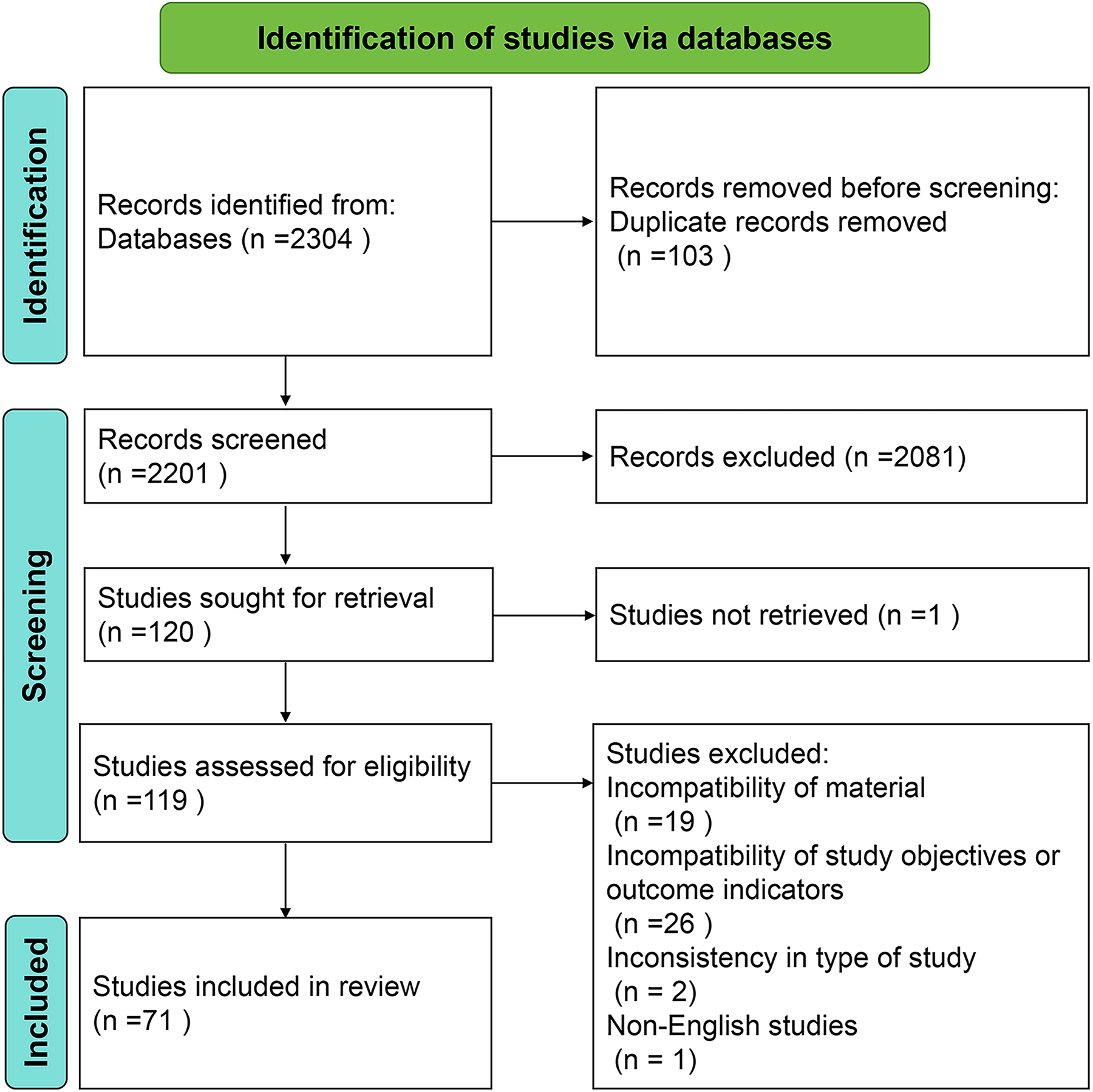
Flowchart of guidelines selection.
Characteristics of literature distribution
As shown in Figure 2a, the number of published animal studies has generally increased since 1990, with a marked rise in the past decade. Figure 2b presents a keyword clustering analysis of the included studies, highlighting the thematic distribution and current research hotspots. The major focus areas include biomaterial development, regulation of material structure and properties, cross-species animal validation systems, nonosseous tissue repair and regeneration, and clinical translation integrated with tissue engineering. Figure 2c illustrates the trends in animal species usage, revealing dynamic changes in species distribution over time. In total, the included studies involved 1,819 animals (excluding 16 studies that did not report animal sample sizes). Rats were the most frequently used species (917 animals), followed by rabbits (514 animals). Over time, species diversity increased, with a growing use of large animals such as miniature pigs, dogs, and sheep.

Characteristics of literature distribution.
Characteristics of material applications
As shown in Figure 3a, most materials used in the included studies were bio-derived materials (38%), followed by polymers (29%) and composite materials (29%). The use of metals (3%) and inorganic nonmetallic materials (1%) was relatively limited. Figure 3b further analyzes the commercialization status of the materials. Only 21% of the studies used commercially available products, while 79% utilized noncommercialized materials. Although the proportion of commercial products was relatively low, a total of 14 different products were reported, with Dual Mesh® (Gore-Tex®) being the most frequently used commercial material (3%).

Characteristics of material application.
We also summarized the correspondence between different nonosseous tissue types and material categories. As shown in Figure 4, a total of 19 types of nonosseous tissues were involved in the included studies. Skin-related tissues (including skin, subcutaneous tissues, and adipose tissues) were the most frequently studied, accounting for 26% of all studies. The materials used for these tissues primarily included bio-derived materials (46%), composite materials (36%), and polymers (18%). Surgical-related tissues (including abdominal wall, extrahepatic bile duct, and stomach wall) accounted for 24% of the studies, with polymers being the most used (58%), followed by composite materials (21%), bio-derived materials (16%), and inorganic nonmetallic materials (5%).

Categories of nonosseous tissues and corresponding material application characteristics in the included studies.
Studies involving cartilage, ligaments, and rotator cuff tissues accounted for 18% of the total, with materials primarily consisting of bio-derived materials (46%) and composite materials (39%), while polymers were used in a smaller proportion of studies (15%). Cardiovascular tissues (including blood vessels and ventricular walls) represented 15% of the studies and involved a wide range of material types, predominantly polymers (37%) and bio-derived materials (27%), along with metals (18%), composites (9%), and inorganic nonmetallic materials (9%). Spinal cord, brain tissue, and annulus fibrosus collectively accounted for 7%, with a strong reliance on bio-derived materials (60%). In addition, tissues such as the kidney, urethra, vocal cords, cornea, and periodontal tissue appeared less frequently in the included studies, with bio-derived and composite materials being the most used in these cases.
In summary, the included studies covered a wide range of nonosseous tissue types, and the materials used within each tissue category showed considerable diversity. For clarity of presentation, all material names in Figure 4 are shown in abbreviated form. The corresponding full names are provided in the material abbreviation table in Supplementary Data S3.
Characteristics of outcome indicators and evaluation methods
To analyze the application of outcome indicators in current studies, we categorized the indicators into five types: safety indicators (complications, adhesions), material-related indicators (degradation), tissue-level indicators (tissue integration, neovascularization, tissue growth, connective tissue, extracellular matrix deposition, fiber wrapping), cellular-level indicators (cellular infiltration), and molecular-level indicators (protein, cytokines, antibodies, enzymes, gene expression). Based on this classification, we calculated the usage frequency of each indicator type across different nonosseous tissue studies (Fig. 5a). Overall, tissue-level indicators were the most widely used, accounting for 56% of all indicators and dominating across nearly all tissue types. Cellular-level indicators (15%) and safety indicators (13%) were also relatively frequently reported and appeared across most nonosseous tissue studies. In contrast, material degradation indicators (8%) and molecular-level indicators (8%) were used less frequently.

Classification and proportion of outcome indicators in different nonosseous tissues.
Further detailed analyses were performed for the three indicator categories that included specific subitems: safety indicators, tissue-level indicators, and molecular-level indicators (Fig. 5b–d). Among the safety indicators, “complications” were reported in all relevant studies except those involving adipose tissue, stomach wall, ventricular wall, annulus fibrosus, and periodontal tissue. In contrast, “adhesion” was reported only in studies related to the abdominal wall, extrahepatic bile duct, and ligaments (Fig. 5b).
Tissue-level indicators were further divided into six subcategories. Among them, “tissue growth” was the most frequently used (25%) and was reported across nearly all tissue types. This was followed by “neovascularization” (23%), “extracellular matrix deposition” (21%), and “tissue integration” (17%), which were commonly applied in studies involving skin, blood vessels, abdominal wall, and cartilage (Fig. 5c). Molecular-level indicators were generally less utilized. “Gene expression” was the most frequently reported (33%), appearing in studies on various nonosseous tissues such as vocal cords, cornea, brain tissue, ventricular wall, and cartilage. “Antibodies” were the least used (2%), reported only in a few cartilage-related studies (Fig. 5d).
To further understand the methods used to evaluate the tissue-inductive properties of materials in current studies, we analyzed the assessment techniques applied across different nonosseous tissues (Fig. 6). The results showed that histological analysis was the most used method (76%), widely applied across all tissue types as a fundamental approach for confirming new tissue formation and integration. Gross observation (11%) was also frequently employed to record visually apparent healing outcomes, such as wound closure. Biomechanical testing (6%) was mainly used in models where restoration of mechanical or physiological function was critical, such as vocal cords, urethra, ligaments, and cartilage, but was less commonly applied in tissues like skin, where morphological healing is the primary focus. Imaging analysis (4%) was primarily used to assess the functional status of internal tissues, such as cardiovascular and tubular organs, and was commonly applied in studies involving the ventricular wall, blood vessels, annulus fibrosus, cartilage, and adipose tissue. Molecular biology assays (3%) were used in only a small number of studies, specifically those involving skin, cartilage, spinal cord, vocal cords, and cornea.
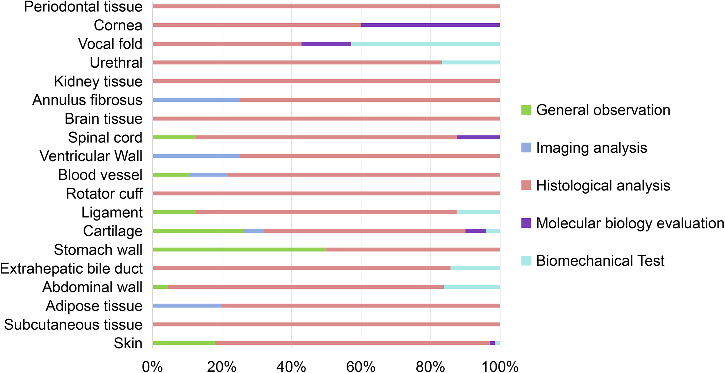
Distribution of evaluation methods for assessing tissue-inductive outcome indicators in different nonosseous tissues.
Characteristics of animal experimental design
We analyzed the overall experimental design of the included animal studies from three aspects: animal characteristics, modeling characteristics, and follow-up duration.
Regarding the choice of animal species, as shown in Figure 7a, rats were used in 41% of the studies, followed by rabbits (26%) and mice (15%). Other animal models included minipigs (5%), sheep (4%), pigs (4%), dogs (3%), guinea pigs (1%), and baboons (1%). Figure 7b illustrates the age characteristics of the animals. Many studies did not report animal age, but adult animals were used far more frequently than juvenile animals. In terms of animal sex, species-specific biases were observed. For example, rats and rabbits were predominantly male, whereas minipigs were more commonly female (Fig. 7c).
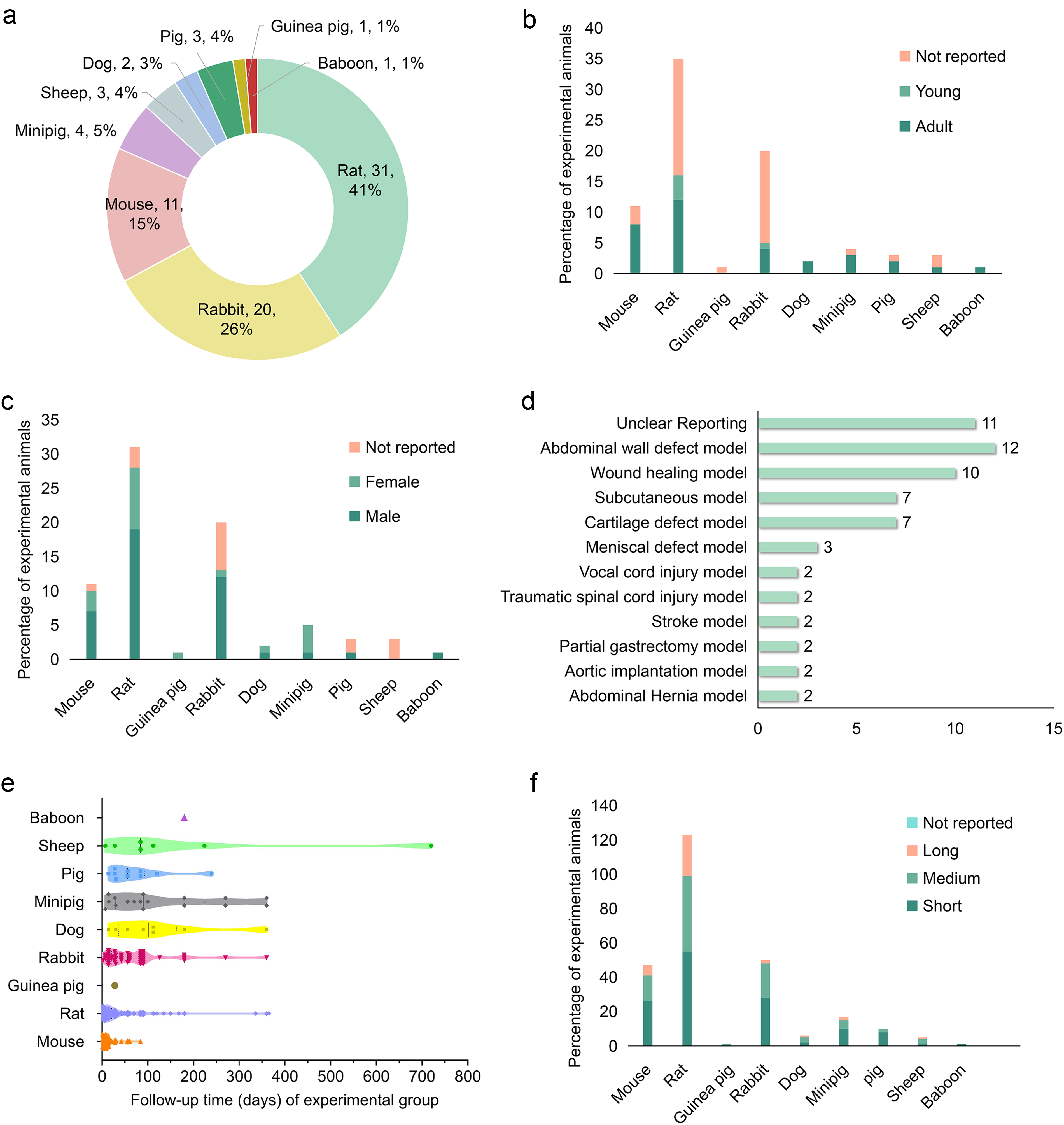
Experimental design characteristics of included animal studies.
Figure 7d shows the most frequently used animal models. Abdominal wall defect models (19%), wound healing models (16%), subcutaneous implantation models (11%), and cartilage defect models (11%) accounted for the largest proportions. Notably, a considerable portion of studies (18%) did not clearly report the specific type of animal model used. Detailed information on all animal models is available in Supplementary Data S4.
Figure 7e presents the follow-up durations in the included studies, which varied significantly across species. Long-term follow-up periods of up to 1 year were observed in studies involving rats, rabbits, dogs, minipigs, and sheep. The distribution of short-, medium-, and long-term follow-up durations for each species is shown in Figure 7f.
Discussion
Tissue-inducing biomaterials, as a cutting-edge technology in regenerative medicine, have gradually transitioned from passive substitution to active regulation. 24 This shift not only reflects advancements in material design and functionality but also signifies that, in future repair processes, biomaterials may actively participate in and guide the entire reconstruction of host tissues, rather than merely serving as structural scaffolds. 25 Based on this, the following discussion will focus on the research progress and application characteristics of representative tissues such as skin, cartilage, and blood vessels.
After reviewing the studies focusing on skin tissue engineering, we found that using naturally derived materials or their composites to promote skin regeneration in defect models is the major trend. Some studies further employed specialized disease models to simulate clinically relevant conditions. For instance, Sandeep Adem et al. 26 established a radiation-induced skin injury model to mimic damage caused by clinical cancer treatments. Similarly, Xiaoli Huang et al. 27 and Fang Lv et al. 28 conducted experiments in the context of chronic inflammatory wounds and diabetic ulcers, respectively. These specific models provide early-stage evidence for the translational application of materials under complex clinical scenarios.
In terms of evaluating the tissue-inductive capacity of biomaterials for skin, most studies employed conventional histological staining techniques such as Hematoxylin and Eosin (H&E) and Masson’s trichrome. However, several studies have begun to explore more specific or mechanism-oriented assessment methods. For example, one study investigated molecular-level markers to examine the mechanism by which bio-ceramic ion—loaded nanofiber scaffolds promoted skin regeneration through the EMT/EndMT pathway. 28 Another study used CD31 immunostaining to confirm early angiogenesis in diabetic wound models, achieving both structural and functional validation of the scaffold. 29 In addition, layered transplantation models were used to assess early vascularization capacity and to explore how quantitative microstructural regulation can accelerate tissue integration. 30 Biomechanical testing was applied to evaluate skin stiffness as a measure of functional recovery. 26 One study also identified newly formed hair follicle—like structures in the material implantation area using H&E staining. 31 In summary, future research should combine complex pathological models with multidimensional evaluation systems to more precisely target the ultimate goal of skin repair—not merely achieving wound closure, but restoring the full structural and functional integrity of the original tissue. 32
Cartilage regeneration is also one of the current research hotspots. Overall, cartilage repair still predominantly relies on small- and medium-sized animal models. Only one study utilized a sheep meniscus defect model with a follow-up period extending to 1 year, representing a rare combination of a large animal model and long-term dynamic observation in the field of cartilage regeneration. 33 In addition, this study evaluated the mechanical properties of the regenerated tissue induced by the material through tensile and compressive testing, providing a reference-worthy model framework for the clinical translation of cartilage repair materials. 33
Current evaluations of the inductive capacity of biomaterials for cartilage repair still primarily rely on histological observation and gross morphological analysis; however, the assessment systems are gradually becoming more diversified. qRT-PCR was employed in two studies to analyze the expression of cartilage-specific marker genes such as COL II, SOX9, and Aggrecan, revealing the mechanisms by which materials induce stem cell differentiation into a chondrogenic phenotype.34,35 Another study applied two histological scoring systems—the O’Driscoll scoring method 36 and a self-developed system—to compare the effects of type I and type II collagen, as well as their composites, on the types of newly formed tissues, highlighting the tunability of material composition in directing regenerative outcomes. 37 Notably, some studies placed more emphasis on improving the overall repair microenvironment. For instance, one study utilized TUNEL staining along with IL-1β and TNF-α expression analysis to evaluate the inflammatory response, focusing on cell survival and inflammation suppression following material implantation. 38 Another study constructed an IL-1β-induced inflammatory microenvironment model and verified the material’s regenerative performance under adverse conditions through immunological factor expression, thereby providing evidence for its applicability in clinical pathological settings. 39
Vascular regeneration also holds an important position in current research. The included studies involved animal models such as rats, mice, rabbits, and dogs. Cross-species models are beneficial for multidimensional verification of material performance; however, large animal models remain scarce in vascular research. Among the included studies, only one utilized a hybrid fiber construct with strong mechanical compatibility as a tissue substitute, applied in canine models of aortic and inferior vena cava repair. With a maximum follow-up period of 24 months, this study provides a valuable reference for long-term evaluation using large animal models. 40
In current vascular repair research, verification of tissue inductivity still primarily relies on traditional histological analysis. However, some studies have begun incorporating functional assessments to deepen the evaluation. One study proposed a “Guided Vascular Regeneration (GVR)” strategy, utilizing a nonporous ELP-RGD hydrogel scaffold to induce endogenous tissue growth by modulating the RGD sequence. In addition to histological analysis, vascular angiography was employed to assess scaffold patency, reflecting an integrated structure-function evaluation approach. 41 Similarly, another study modified the surface of LVAD (Left Ventricular Assist Device) cannulas with a titanium mesh structure to induce endomembrane formation and microvascularization. 42 And immunohistochemical staining of endothelial cells was used to verify the feasibility of functional regeneration mediated by the three-dimensional surface architecture of acellular materials, proposing a dual strategy of “antithrombosis and regeneration-guidance.”
In addition to skin, ligaments, and blood vessels, current research has also focused on the inductive regeneration of various other nonosseous tissues. Overall, the potential of tissue-inducing biomaterials has been preliminarily validated in the repair of multiple nonosseous tissues, including skin, vasculature, ligaments, abdominal wall, and vocal folds. At the same time, diverse explorations have been conducted regarding animal experiment design, material types, and their properties. However, common limitations persist across studies, such as limited use of large animal models, short follow-up durations, insufficient evaluation dimensions, and a lack of dynamic monitoring and functional assessment systems. These limitations may be related to the inherent biological characteristics of different types of tissue and the feasibility of studying them. For example, in skin tissue research, although the skin healing mechanisms of mice differ from those in humans, their rapid healing properties still make them a common model, which also leads to studies that often focus on short-term wound closure or histological repair outcomes.43,44 Large animal models, on the other hand, are limited in their application due to their high breeding costs, complex ethical approvals, and operational difficulties. 45 The lack of blood vessels and nerves in cartilage makes it difficult to observe significant functional improvements in the short term. This often leads to a focus on structural indicators in research, which ultimately limits the extension of follow-up periods and hinders the clinical translation. 46 Vascular tissue, due to its structural complexity, faces significant challenges in achieving long-term integration and functional maintenance in vivo. Consequently, it is difficult to monitor the parameters of new blood vessels such as the perfusion capacity, patency, and stability, especially the long-term functional equivalence. As a result, evaluation in this field still largely relies on the morphological observations. 47 Future research should aim to address these shared challenges. Animal models should be selected based on the functional characteristics of the target tissue, and incorporating disease or injury models may further enhance clinical relevance. Outcome evaluations should place greater emphasis on objective and long-term functional assessments, going beyond simple histological analysis. Through multidimensional optimization, the translational gap between animal studies and clinical needs can be narrowed, ultimately achieving the dual goals of functional reconstruction and clinical application of tissue-inductive biomaterials.
Conclusion
The findings of this evidence mapping study indicate a significant increase in research on tissue-inducing biomaterials for nonosseous tissue repair over the past decade. Animal models primarily involved rats, rabbits, and mice. The predominant types of tissue-inducing biomaterials used were bio-derived materials, polymers, and composites, with commercially available products accounting for less than one-quarter of cases. Skin, abdominal wall, cartilage, and blood vessels were the most frequently studied nonosseous tissues, with variations in material application across different tissue types. Outcome indicators span from microscopic to macroscopic levels, with a predominance of tissue-level assessments. Histological analysis was the most employed method to evaluate inductive capacity, while imaging techniques and molecular biology assessments were used less frequently. Most animal experiments had a duration of 28 to 90 days, with relatively few studies conducting long-term follow-up.
Authors’ Contributions
Y.L.: Conceptualization (supporting), methodology (equal), investigation (equal), data curation (equal), writing—original draft (lead), and visualization (equal). Q.W.: Conceptualization (supporting), methodology (equal), investigation (equal), data curation (equal), writing—original draft (supporting), and visualization (equal). W.Z.: Conceptualization (supporting), methodology (equal), investigation (equal), data curation (equal), writing—original draft (supporting), and visualization (equal). X.S.: Investigation (supporting), and data curation (supporting). F.D.: Investigation (supporting) and data curation (supporting). W.D.: Software (lead) and formal analysis (lead). M.N.: Formal analysis (supporting). Y.Z.: Validation (lead). B.Y.: Supervision (equal), project administration (supporting), and writing—review and editing (equal). H.L.: Funding acquisition (lead), supervision (equal), project administration (lead), writing—review & editing (equal). K.Z.: Funding acquisition (lead) and Writing—review and editing (supporting). B.M.: conceptualization (lead), supervision (equal), and writing—review and editing (equal).
Footnotes
Author Disclosure Statement
No competing financial interests exist.
Funding Information
This work was supported by the National Key Research and Development Program of China (Grant number 2022YFC2409705) and the National Key Research and Development Program of China (Grant number 2022YFC2409803).