
Abstract
BACKGROUND:
With inherent flexibility, high electroconductivity, excellent thermal conductivity, easy printability and biosafety, Ga-based functional liquid metals (LMs) have been extensively evaluated for biomedical applications. When implanted in the biological environment, the safety of the LMs is a major concern for future application.
METHODS:
In this study, we conducted several biocompatibility assessments through immersion experiments, in vitro cytotoxicity experiments and in vivo embedding experiments.
RESULTS:
The results showed that both the Al-assisted self-driven LM and the LM per se own good biocompatibility and retrievable properties when contacted with living organisms for a relatively long period of time.
CONCLUSION:
This study provides preliminary evidence about the biocompatibility of the functional LM materials, such as LM-based soft machine, which would promote and inspire other research to address other tough biomedical issues.
Introduction
Biomaterials provide new insight into efficiently treating tough diseases in modern medicine. They are supposed to interact with human cells, tissue or organs to elicit their effects [1]. Recently, liquid metal (LM) as promising materials have attracted great attention in biomedical field [2]. LM is a particular family of biomaterials that exhibits both metallic and fluidic properties simultaneously [3], in contrast to traditional metals, LM has favorable fluidity, sufficient radiopacity, low viscosity and plasticity [4]. Typical materials in the area, such as gallium, gallium–indium eutectic alloys (EGaIn) and gallium–indium–tin alloys (Galinstan), which have low melting points, have been widely used in printed circuits, soft electronics, and other biomedical areas. Accordingly, liquid metal gives rise to a revolution of conventional medical concepts and provides some novel treatments for tough diseases. Some biomedical application of LMs includes tumor embolotherapy [5], contrast agent used in human body [4], nerve connection [4], biosensing and health monitoring with high conformability, excellent conductivity and better signal stability [6], etc. However, the basic criteria for these clinical applications are safety and good biocompatibility, which means that the safety and stability of biomaterials in human body need to be ensured. The LM-related medical devices that work in human body will interact with microenvironment of organism, consequently having an impact on the function and viability of cells or tissues. Hence, there are long-term contacts between liquid metal and human cells or tissues, indicating the necessity of assessing the changes of the vivo environments and tissues caused by liquid metal.
The metal ions released from corrodible alloys to the surrounding cells and tissues, may cause biological responses in short or long period, consequently altering their normal morphology and viability. On the other hand, human will elicit an immune rejection to foreign body reaction (FBR) when the liquid metal device is introduced into organism, which may greatly affect the therapeutic effects of treatments. The previous studies have discussed and proved the biocompatibilities of some specific liquid metals, and researchers have cultured the cell with the immersion solution of the gallium and EGaIn alloy. The results have shown that the proliferation status of cells were normal, indicating the good biocompatibility of liquid metal [7]. Moreover, scientists have cultured hippocampus neurons with medium containing EGaIn alloy. The cell viability after 12 days was greater than 65% and the intrinsic calcium channels remained normal, suggesting its non-cytotoxic property [8]. Researchers have also analyzed the changes of blood ingredients and the impairments of key organs when EGaIn nano-particles were injected into mice, the results demonstrated its nontoxicity in vivo [9]. However, for some novel applications of liquid metal used in biomedical fields, the LMs are usually interacted with aluminum for self-driven motion in medical devices. As a consequence, in this manuscript we aim to evaluate the biocompatibility of Al-assisted self-driven liquid metal machine. The experiments were divided into 3 categories: firstly, the toxicity of a metallic material is governed not only by its composition and toxicity of the component elements but also by its corrosion and wear resistance [10]. Hence, the stability of LM in physiological solution need to be ensured that their biocorrosion products not deleterious to the surrounding tissues. The amounts of metal ions released from LMs in Hank’s solution were measured to prove their stability in vivo; secondly, the morphology and growth status of cells surrounding the LMs were evaluated; lastly, taking into account the complicated microenvironments in vivo, so it is very necessary to further carry out embedding experiments to observe the growth status and tissues morphological changes of organisms after introducing the liquid metal machine. Proliferation rate and morphology of cells cultured in the presence of designed LMs were monitored after 4 h, 24 h and 48 h of growth of propagation.
Materials and methods
Liquid metal ion release experiments
In general, the ingredients of Hank’s solution are similar to that of biological body fluid, therefore, we regard it as a simulated body fluid to evaluate the amount of metallic elements released from different LM alloys. 6-well plates were numbered from 1 to 6 and added 8 ml Hank’s solution into each well plate by pipette, followed by the injection of 50 μL Ga, Ga-Al alloy, EGaIn alloy and EGaIn-Al alloy into 1–4 wells by sterile injectors, respectively. The 6-well plate was put in the incubator (Sanyo MCO-18AIC, Japan) under 5% CO2 concentration and 37 °C temperature which mimic the physiological environment in vivo. 6 ml soaking solution was transferred from number 1–4 wells to 4 centrifuge tubes after 48 h, inductively coupled plasma-mass spectrometry (ICP-MS, Thermo Fisher Scientific, Thermo X Series II, Germany) was employed to measure the concentrations of Ga, In and Al which had dissolved from the alloy plates. An average of three measurements was taken for each group.
Liquid metal cytotoxicity experiments in vitro
In this study, EMT6 cells and NIH3T3 cells were chosen as objects under investigation and divided into four groups: the test groups treated with LMs; and the control group cultured simply with culture medium. The EMT6 cells and NIH3T3 cells took out from liquid nitrogen reservoir for cell resuscitation, transferred them into centrifuge tubes for centrifugation, the supernatant was discarded. Continuously the cells were cultured in the DMEM (Dulbecco’s modified Eagle’s medium) supplemented with 10% FBS (fetal bovine serum) and 1% P/S (penicillin/streptomycin) in a humidified atmosphere of 5% CO2. Cell was passaged per 48 h with the replacement of culture medium per day to maintain the normal growth of cells, the cells could be utilized for LM cytotoxicity test after 2 passages.
Numbered the 8 culture plates, adding 6ml fresh culture solution (DMEM supplemented with 10% FBS and 1% P/S) into each culture plate. The EMT6 cells were seeded into number 1–4 culture plates, the NIH3T3 cells were seeded in number 5–8 culture plates, both at a density of 10,000 cells∕cm2. The LMs used in this experimental group include Ga, Ga-Al alloy and EGaIn alloy. To ensure the fluidity of LM, the temperature under 32 °C is necessary before the experiments. Due to the chemical reaction of Ga-Al alloy in solution is a dynamic process, the Ga-Al alloy had to be freshly made through adding 50 μL Ga and a piece of 4 mm × 4 mm Al foil into NaOH solution of 0.2 mol∕L concentration. Holded the Al foil with tweezers and made it corrosion when contacting with the Ga. After about half a minute, the Al foil accelerated the corrosion into the LM and quickly released the gas. Furthermore, to avoid the effect of alkaline solution on cell growth, we washed the freshly-made Ga-Al alloy by phosphate buffered saline (PBS) solution for several times, then the Ga-Al alloy was put into number 3 and 7 culture plates. Meanwhile the 50 μL Ga was put into number 2 and 6 culture plates, the 50 μL EGaIn alloy was put into number 4 and 8 culture plates. And the rest of number 1 and 5 culture plates were set as control groups. These culture plates were placed in a constant temperature incubator to allow the cells to grow adherent to the wall. After 4 h, 24 h and 48 h of growth, the cells were taken out and observed under the microscope (Nikon TS100-f, Japan).
When recording the cells growth status, the results of the central location of the culture plates, namely site 3 in Fig. 1, were first recorded as the indicators of the impacts of LM on the whole cells growth environment. The results of midpoint from the center to the LM, known as site 2 in Fig. 1, were then recorded to assess the effects of the presence of the LM on cells growth as it moved closer to the LM. Finally, cells growth near the edge of the LM were recorded, namely site 1 in Fig. 1, to observe the effect of the LM on cells growth when cells near the LM in the local microenvironment.
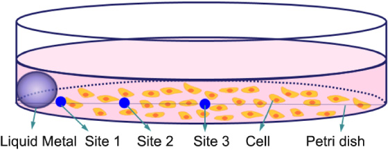
Schematic diagram of experimental observation position in a dish.
To further demonstrate the biocompatibility of LM alloy, in vivo experiments performed with BALB/c mice were carried out. Five 8-week-old male mice were employed for subcutaneous implantation. Before the animal experiments, the anesthetic we used is 1% sodium pentobarbital of the 50 mg∕kg dose, injected around 0.11 mL into the abdominal cavity for each BALB/c male mouse, the mice were basically under general anesthesia after 1–2 min. Disposable needles were used to make a defect measuring 1.2 mm in diameter on dorsal skin partly shaved and sterilized with 75% alcohol. LMs were exposed to ultra-violet radiation for 20 min and then embedded subcutaneously through the injection. Five mice were numbered. Mice no. 1 was set as the control group and was injected with 0.9% saline 50 μL in the middle of the back (as shown in Fig. 2). Mice 2–5 were injected with 50 μL of LMs Ga, Ga-Al, EGaIn and EGaIn-Al, respectively. Taking the chemical reaction of aluminum into account, when Al was dissolved into LM in NaOH solution, immediately washed twice with PBS solution to wash away the lye to avoid the bias on the experiments. The reacting LM alloy containing Al was then inhaled with a syringe and injected subcutaneously into the mice’s back. After that, the mice were raised normally and their activity and hair status were observed regularly. After 3 weeks, two mice injected with EGaIn alloy and EGaIn-Al alloy were sacrificed for anatomical observation, and the intercostal muscle tissue directly below the liquid metal was obtained. Meanwhile, the corresponding part of the contralateral side of the same mouse was taken as the control sample. All 4 samples were cut into small pieces of 0.5 cm × 1 cm and placed in 4% formaldehyde solution for 24 h tissue fixation. Followed by HE staining for histological analysis, the slicing direction was longitudinal, and three slices were cut for each sample. The current animal tests have been approved by the Ethics Committee of Tsinghua University, Beijing, China under contract no. SYXK (Jing) 2009-0022.
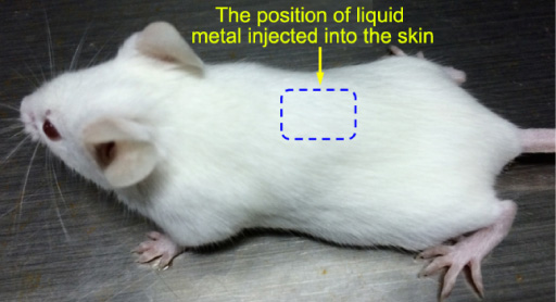
The position of liquid metal injected into the skin of mice.
Results of the liquid metal ion release experiments
To assess the stability of elements in liquid metal under the physiological conditions, the concentration of the three metal elements were analyzed by inductively coupled plasma-mass spectrometry (ICP-MS, Thermo Fisher Scientific, Thermo X Series II, Germany, power:1300 W), among which the concentration of Ga and Al were in the order of microgram, and that of In was 1.309 ng∕mL. The concentration of Ga and Al in solution were both below the limit value [10]. Therefore, Ga and In in liquid metal are relatively stable in simulated physiological solution. As for the Al, its concentration in the immersion solution of Ga-Al alloy and EGaIn-Al alloy were not high, as shown in the histogram of Fig. 3.
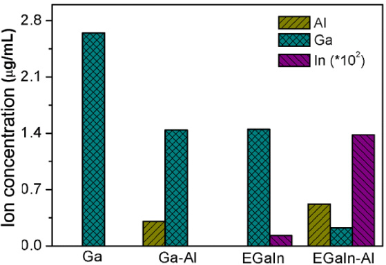
Concentration of related metal ions in Hank’s solution after 48 h immersion.
The growth status of EMT6 cells
The growth status of two cell lines were visualized using optical microscope, specifically the morphology and proliferation rate of cells define the growth status, which acting as indicators of LM cytotoxicity. As shown in Fig. 4, namely site 1, all EMT6 cells had significantly increased after 24 h, and there was no significant difference in the number of cells between the control group and experimental group. After 48 h, the dense growth status of cells in both the experimental group and control group were observed, indicating that EMT6 cells could survive and reproduce normally in the environment surrounding liquid metal.

The growth status of site 1 of EMT6 cells after 24 h and 48 h: (a) control group; (b) medium with 50 μL Ga; (c) medium with 50 μL Ga-Al; (d) medium with 50 μL EGaIn. (i), (ii) represents the growth status of the cells after 24 h and 48 h, respectively.
When it comes to site 2, as expected, although the growth of cells after 48 h was not as dense as that of site 1, the proliferation and morphology of the cells were normal within 24 h and 48 h, as shown in Fig. 5, suggesting that the presence of liquid metal did not seriously impact the cell survival.

The growth status of site 2 of EMT6 cells after 24 h and 48 h: (a) medium with 50 μL Ga; (b) medium with 50 μL Ga-Al; (c) medium with 50 μL EGaIn. (i), (ii) represents the growth status of the cells after 24 h and 48 h, respectively.
As shown in Fig. 6, observation can be found at site 3, after 24 h and 48 h cell inoculation the LM edge cells had a regular growth situations, and less dense than the cells of sites 1 and 2, but their normal morphology and proliferation rate could be investigated.
The growth status of site 3 of EMT6 cells after 24 h and 48 h: (a) medium with 50 μL Ga; (b) medium with 50 μL Ga-Al; (c) medium with 50 μL EGaIn. (i), (ii) represents the growth status of the cells after 24 h and 48 h, respectively. The dark shadow in the figure is where the liquid metal is under the light microscope.
Specifically, for the medium containing Ga, the existence of the Ga basically wasn’t cytotoxic to the cells proliferation. As for the Ga-Al alloy, after inoculation for 24 h, the growth status of cells didn’t significantly differ from that of the cells surrounding Ga. While the density of cell growth was just lower than that of Ga after 48 h, but still exhibited the normal proliferation, indicating that the presence of Ga-Al alloy would not seriously affect the growth and proliferation of cells. In the medium containing EGaIn alloy, the density of cells at 24 h and 48 h after inoculation were significantly lower than that in the medium containing Ga and Ga-Al alloy, thus, EGaIn alloy might abrupt the growth situation of cells to some extent, determining its biocompatibility was not as moderate as Ga. However, as shown in Fig. 6, cells contacting with the EGaIn alloy still existed, compared to the cells in the medium containing Ga and Ga-Al alloy at the observation sites 1 and 2, their growth status were in the relatively same level, meaning that the biocompatibility of EGaIn alloy was relatively good in some case.
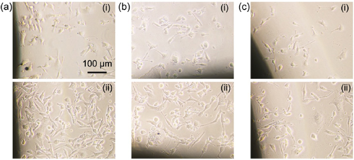
NIH3T3 cells behaved similarly to EMT6 cells in the cytotoxicity test. The density levels of NIH3T3 cells at sites 1 and 2 were not significantly different among these groups, while at site 3 the cell density of the control group was higher than medium with Ga. The amount of cells are relatively great near the edge of Ga. Conversely, the cells near the edge of EGaIn alloy were sparse, some still survived. It can also be concluded that the biocompatibility of Ga is more moderate than EGaIn alloy, as shown in Fig. 7.

Under 40-fold objective lens, the growth status of NIH3T3 cells for 24 h. (a) Control group; (b) medium with 50 μL Ga; (c) medium with 50 μL EGaIn. (i), (ii), (iii) represent the site 1, site 2 and site 3, respectively. The dark shadow in the figure is where the liquid metal is under the light microscope.
As is known, the chemical reactions of LM reacted with Al will release H2 and Al3+, which might influence the physiological growth process of cells. Surprisingly, under the 10-fold objective lens, the living cells could be roughly investigated, but some visual fields are obscured by flocculent deposits. Under the 40-fold objective lens, there were living cells at all three sites, indicating that the introduced reaction did not seriously poison the cells, as shown in Fig. 8.

The growth status of NIH3T3 cells cultured in medium containing Al-interacted liquid metal for 24 h: (a) 10-fold objective lens, medium with 50 μL Ga-Al; (b) 40-fold objective lens, medium with 50 μL Ga-Al; (c) 10-fold objective lens, medium with 50 μL EGaIn-Al; (d) 40-fold objective lens, medium with 50 μL EGaIn-Al.
Along with the remarkably excellent biocompatibility of the LM, in vivo experiments were performed by embedding LM alloy in BALB/c mice back subcutaneously, the inflammatory responses and capsule formation of the LM were investigated in a mouse model [11]. The results revealed the injection of LM had a moderate biocompatibility on the normal life of the mice. As expected, the morphology of mice no. 1 of control group wasn’t different compared to the normal mice (as shown in Fig. 9(a)), and there was a small bump on one side of the back of mice nos. 2–5 with good activity state and hair growth which were identical to that of mice no. 1. Besides, the morphology of mice nos. 2 and 4 look similar (injected with liquid metal without Al) that small bumps were appeared on their back (as shown in Fig. 9(b)), and there were no significant difference in the morphology of mice nos. 3 and 5 (injected with liquid metal containing Al), but with larger bumps on their back (as shown in Fig. 9(c)).

The growth status of BALB/c mouse after 3 weeks injection test: (a) the control group mice injected with saline; (b) mice injected with EGaIn alloy; (c) mice injected with EGaIn-AI alloy.
In addition, neither the fester nor the discoloration were observed in the dissected muscle tissue underneath the LM by naked eyes, also shown normal growth of tissue characterized by smooth and intact structures. Specifically, each mouse had only one bump that the LMs were encapsulated together by a kind of mucous membrane, while the volume of bump was larger for mice that injected with LM containing Al. The bump of LM containing Al can be seen filling with gas, after cut by surgical scissors, the gas in the bump was discharged and the volume of the bump became smaller. To investigate the conformational change of the muscle tissue contacting with the LM, the pathological changes of the tissues could be observed by HE staining, directly determining the impact of LM on tissues. The HE staining slides of EGaIn alloy injected mouse has shown the morphology of muscle tissue and the contralateral corresponding muscle tissue remained normal, and the myofilament fibers were arranged in an orderly manner. Also the normal morphology of the muscle cells were clearly observed, without obvious inflammatory cells (Fig. 10).
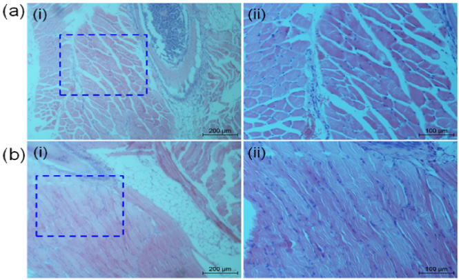
HE staining slides after 3 weeks injection of EGaIn alloy into mice: (a) sections of intercostal muscle tissue in the area closely contacting with liquid metal; (b) section of the intercostal muscle tissue of the corresponding area of the contralateral side. (i) is under the 10-fold objective lens, (ii) is under the 20-fold objective lens. The dotted box indicates the corresponding position of (ii) in (i).
Considering the HE staining slides of EGaIn-Al alloy injected mouse, similar to EGaIn alloy injected mouse, both the morphology of bilateral muscle tissue and the myofilament fibers performed normally. Also the regular morphology of the muscle cells was clearly visible. The main difference was the presence of lymphocytes infiltration in muscle tissue contacting with EGaIn-Al alloy, shown as dark dots in Fig. 11(a), indicating the elicitation of inflammatory response in the local area rather a systemic inflammatory response due to the absence of lymphocytes infiltration in the contralateral corresponding muscle tissue, as shown in Fig. 11(b). However, after a long time of observation, this inflammation will not cause local tissue decay or necrosis, thus, the material is relatively safe. Besides, the morphology of adipose cells was normal, and there was no obvious deformation or necrosis, indicating that the inflammatory reaction had no serious effect on the tissue.
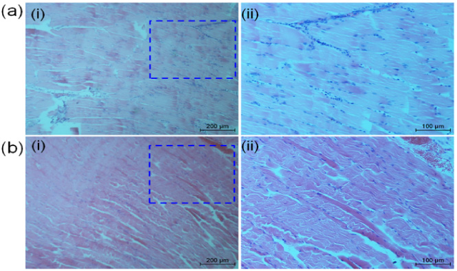
HE staining slides after 3 weeks injection of EGaIn-Al alloy into mice: (a) sections of intercostal muscle tissue in the area closely contacting with liquid metal; (b) section of the intercostal muscle tissue of the corresponding area of the contralateral side. (i) is under the 10-fold objective lens, (ii) is under the 20-fold objective lens. The dotted box indicates the corresponding position of (ii) in (i).
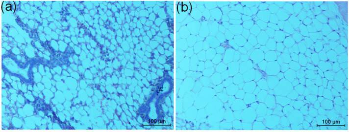
The adipose tissue in HE staining slides after 3 weeks injection of EGaIn-Al alloy under 20-fold: (a) adipose tissue sections in close contact with EGaIn-Al alloy; (b) adipose tissue sections of the corresponding area on the contralateral side.
Researchers regard Hank’s solution as a simulated body fluid to assess the metal corrosion rate by soaking liquid metal materials [12–14]. Hank’s solution can be used to simulate the blood environment when studying the corrosion of metal stent implanted in the blood vessels [15]. As shown in the histogram of ion concentrations (Fig. 3), the concentration of Ga was highest in the pure Ga, followed by Ga-Al alloy and EGaIn alloy, and the EGaIn-Al alloy obtained the lowest concentration. These phenomena could be explained by the formation of primary battery system between Ga and Al, In, due to the chemical activities of Al and In are higher than Ga, which act as anode to protect Ga. And in the EGaIn-Al alloy both the Al and In protected Ga so that achieved the lowest concentration. The histogram also revealed the tiny release of In, the main reason is that the mass fraction ratio of In and Ga is 1:3, so the metal surface of EGaIn alloy was mostly occupied by Ga, which took a great advantages in chemical reactions so the electrochemical corrosion rate of the In was slow. While in EGaIn-Al alloy, the activated Al constantly reacted with the solution, causing continuous flow on the liquid metal surface. This dynamic flow on the liquid metal surface accelerated the electrochemical corrosion of In. Therefore, in the presence of Al, the ion concentration of In will increase correspondingly. Surprisingly, the concentration of Al was low either in Ga-Al alloy or EGaIn-Al alloy. The reason why this phenomenon occurred was that Al will be involved in the chemical dissolution in the neutral Hank’s solution, generating the Al(OH)3 deposit and H2, consequently diminishing its concentration in Hank’s solution. Besides, the dosage of Al is lower than Ga and In, resulted in its low concentration as well. Accordingly, the concentration of 3 metal ions in the physiological solution is maintained at a relatively low level, and the introduced Al could protect the core ingredient of LM-Ga. The In similarly protected Ga whose concentration was 1.309 ng∕mL in the EGaIn alloy group that is under the limit value of 3 μg∕L [10,11].
To clarify the overall performance of LMs, we have conducted the cytotoxicity tests to demonstrate its biocompatibility. In many studies, the cytotoxicity of implanted metal materials was studied by using the immersion solution to culture the cells after soaking the metal materials in cell culture medium [7,10]. This method is widely applied because it is convenient and quick for the metal materials with special sizes or shapes. However, it is an indirect cytotoxicity test rather than assessing the effect of local environmental changes on cell growth after contacting with the metal material directly. As expected, the LM will stay at a certain position on the side wall of the medium (as shown in Fig. 1). Moreover, there is a concentration gradient of metal ions in the medium characterized by the closer to the LM the higher concentration will be, as the metal ions take time for diffusion. Thus, we set 3 observation sites and evaluated the growth status of cells in all 3 sites. From the representative results (Figs 4–6), although the amounts of cells in the site 3 were less dense than that of cells at sites 1 and 2 after 24 h and 48 h inoculation, the morphology and proliferation status of EMT6 cell maintained normally at all 3 sites. These phenomenon could be explain by the initial amount of cells at site 3 was less than sites 1 and 2, which was observed after 4 h inoculation, because the cells are more likely to adhere and grow at the center of medium. Besides, at site 3 the EGaIn alloy might abrupt the growth situation of cells to some extent, determining its biocompatibility was not as moderate as Ga. But compared to the cells in the medium containing Ga and Ga-Al alloy at the observation sites 1 and 2, the cells cultured with EGaIn alloy was in the relatively same level. And the density of cell growth in medium injected with Ga-Al alloy was just lower than that of Ga after 48 h, indicating the presence of Ga-Al alloy would not seriously affect the growth and proliferation of cells. The NIH3T3 cell performed similarly to EMT6 cell, proliferated normally at sites 1 and 2, while at site 3 the amount of cells near the edge of Ga was more than that of the EGaIn alloy. However, the immersion test have revealed the released amount of metal ions of EGaIn alloy was lower than that of pure Ga, theoretically the amount of cells near the edge of EGaIn alloy was more than that of the pure Ga. Thus, we assume the effect of In on cells is more severe than that of Ga on cells, especially the area close to the LM. But due to the tiny release of In, it has little impact on cells far away from the liquid metal. Therefore, it can also be concluded that the biocompatibility of Ga is more moderate than that of EGaIn alloy. As we known, the chemical reactions of LM reacted with Al will release H2 and Al3+ for self-driven, which might influence the physiological growth process of cells [16,17]. Surprisingly, under the 10-fold objective lens, the living cells could be roughly investigated, but some visual fields are obscured by flocculent deposits. Under the 40-fold objective lens, there were living cells at all three sites, indicating that the introduced reaction did not seriously poison the cells (shown in Fig. 8). In a word, the cytotoxicity test demonstrates that both the Al-assisted LM and fundamental LMs have very low or even no toxicity to the NIH3T3 cell and EMT6 cell. These results indicate that this material may be quite beneficial to be used in vivo.
In regard to the embedding experiments of LM in BALB/c mice in vivo, daily observation shown their activity and diet remained normal after injection. While the mice nos. 3 and 5 who injected with Al containing LMs had a larger bump on their back compared to the mice nos. 2 and 4 who injected with LMs without Al (Figs 9(b) and 9(c)). The main reason is the release of H2 after chemical reaction involved in Al enlarged the volume of bump. Furthermore, after being implanted for 3 weeks, it was found that the implants were located with stability in situ through dissection, but the locations slightly differed from the initial injected points. This phenomenon can be attributed to the fluidity of LM and initial injected points were on the spinal cord which cannot maintain the same location of LM. In addition, we observed the LM was encapsulated by mucous membrane, because the LM provoked the immunological responses of body, known as foreign body responses (FBR), forming a collagenous capsule outside the LM [16,18]. The collagenous capsule didn’t adhere to the back skin or the underlying muscles, so it can be completely separated from the back with surgical scissors. Also neither the fester nor the discoloration were observed in tissue, conversely the intact and smooth structures still maintained. Accordingly, liquid metal is relatively safe in vivo in a long period of time and will not have a serious impact on the tissue. And we concluded the liquid metal is retrievable when utilized in the body as a result of its high surface tension and insolubility. As shown, the minor changes of muscle tissue by HE staining, both the morphology of bilateral muscle tissue and the myofilament fibers, performed normally in the EGaIn-Al alloy injected mouse and EGaIn alloy injected mouse (Figs 10, 11). The single difference between two mice was that the increased infiltration of inflammatory cells appeared in the surrounding tissues of EGaIn-Al alloy injected mouse rather in the contralateral corresponding tissue (Fig. 11(a)), suggesting the inflammatory response is locally elicited instead of systematic inflammatory response. The reason lying behind the difference is that the chemical reaction involved in Al caused the surrounding environment became alkaline [19], consequently stimulated the tissue and further elicited the inflammatory response. It can be found that the inflammatory cells caused by liquid EGaIn-Al alloy are mainly concentrated in the myofilament fiber gap (Fig. 11(a)). In detail, as shown in Fig. 12, the inflammatory cells can be observed mainly around adipose tissue, the number of inflammatory cells infiltrating in the adipose tissue which closely contacting with the EGaIn-Al alloy was more than that in the muscle tissue, no inflammatory cells were observed in the adipose tissue in the contralateral corresponding adipose tissue, which further proves that this inflammation is a local inflammatory response. Therefore, the biocompatibility of EGaIn alloy is more moderate than EGaIn-Al alloy. And with a long time observation, this inflammation does not cause local tissue decay or necrosis, so the material takes a good biocompatibility with tissues in a relatively long period of time. In summary, no obvious evidence showed the occurrence of severe inflammation reaction when the LM was subcutaneously implanted in mice. These in vitro and in vivo experiments are both consistent and show that the LMs are biocompatible to a great extent.
Conclusions
In summary, we have conducted the biocompatibility assessments among the immersion test, LMs cytotoxicity test in vitro and LMs embedding test in vivo. The results have been proven that both the Al-assisted self-driven LMs and Ga-based LMs take good biocompatibility with living organisms in a relatively long period of time, with retrievable and non-toxic properties. The LMs will undoubtedly further applied in medical field to address some tough issues, and especially the Al-assisted self-driven LMs [20] opens great promise as self-driven machine with many improved properties which are expected to change the basic concept of current biomedical material.
Footnotes
Acknowledgements
This work was supported by the National Key Research and Development Program of China under grant no. 2020YFC0122301, the National Natural Science Foundation of China under grant nos. 51890893 and 91748206.
Conflict of interest
None to report.