
Abstract
Cartilage repair is a common problem in the clinic. Owing to the absence of vascular and lymphatic systems, cartilage exhibits a very limited capacity for self-repair, which complicates related research. The decellularized extracellular matrix (dECM), obtained by removing cellular components, preserves the natural structure and bioactive molecules of native ECM. This offers a biocompatible and bioactive environment for cell growth, making it a suitable and effective biomimetic scaffold material. In recent years, many studies have shown that the dECM has good effects on cartilage regeneration. However, there are no studies on the cartilage regeneration of decellularized matrix from different tissue sources, especially the related mechanisms. This article reviews the preparation methods for dECM and research on decellularized matrix derived from cartilage, fat, synovium, and dermis with respect to cartilage repair and regeneration, and further explores the application value and broad prospects of acellular ECM as a new tissue engineering biomimetic scaffold material. With further progress in dECM research and 3D bioprinting, their combination can better replicate native tissue architecture and function. This approach enables precise control of cells and materials, improves the regenerative niche, and may speed the clinical translation of biomimetic ECM for tissue repair.
Impact Statement
In this review, we systematically compared dECM for cartilage repair from various tissues and proposed a more targeted breakthrough direction for cartilage repair treatment by clarifying how component differences (collagen, glycosaminoglycan [GAG], growth factors) affect regeneration, which can accelerate the clinical transformation of dECM-based treatment. More importantly, it positions the integration of dECM-3D bioprinting as a new construction method of biomimetic ECM for tissue engineering, which will become a new mode of cartilage tissue engineering.
Introduction
Cartilage defects, such as the repair of various types of articular cartilage, nasal cartilage, and other injuries, are common problems in the clinic. At present, its treatment relies mainly on surgical and tissue engineering methods. 1 The cell tissue engineering method is a promising therapeutic method, while it is greatly hampered by the lack of sufficient cell sources and inappropriate biomaterials. 2
As a promising biomaterial, decellularized extracellular matrix (dECM) preserves the native structure and bioactive molecules of the ECM through the removal of cellular components. In cartilage regeneration, dECM has demonstrated significant potential. With their inherent multipotent capacity, adipose-derived mesenchymal stem cells (ADSCs) can be induced to differentiate into osteoblasts, adipocytes, and chondrocytes, establishing them as a widely utilized cellular resource in tissue engineering.3,4 This article summarizes the research on cartilage repair and regeneration induced by dECM from different tissue sources, focusing on its mechanism of inducing ADSC chondrogenic differentiation. Meanwhile, we review the new methods and application prospects of constructing biomimetic ECM to promote tissue repair by combining dECM with 3D printing and discuss the clinical needs and tissue engineering experimental research, providing ideas and references for more efficient and safe cartilage cell tissue engineering.
Decellularized Extracellular Matrix
Decellularization refers to the use of appropriate methods to remove cells and DNA from tissues, retain their structure and regulatory proteins, and form a suitable biological scaffold to decellularize and form target cell (single) sheets, tissues, or organs. 5 The ECM provides an essential niche that regulates cell growth, differentiation, and tissue homeostasis. In tissue engineering, an effective biomimetic scaffold should replicate these functions to approximate the properties of natural ECM. Among various candidates, dECM is regarded as a promising material for reproducing the intricate structure of native ECM. When cellular components are removed, the dECM can retain most ECM-related components. 6 In addition, dECM from different species has low immunogenicity and can be well tolerated even by xenoreceptors. 7
Studies have demonstrated that dECM can support the mobilization and differentiation of mesenchymal stem cells, regulate macrophage polarization, promote tissue repair, and exhibit excellent biocompatibility without biological toxicity. 8 It provides the intricate composition of native tissues, which is challenging to reproduce with conventional biomaterials. Therefore, dECM is regarded as a promising biomaterial 9 and can serve as a scaffold in diverse applications of tissue engineering and regenerative medicine. 5 (The process used to obtain nasal cartilage dECM is outlined in Fig. 1).
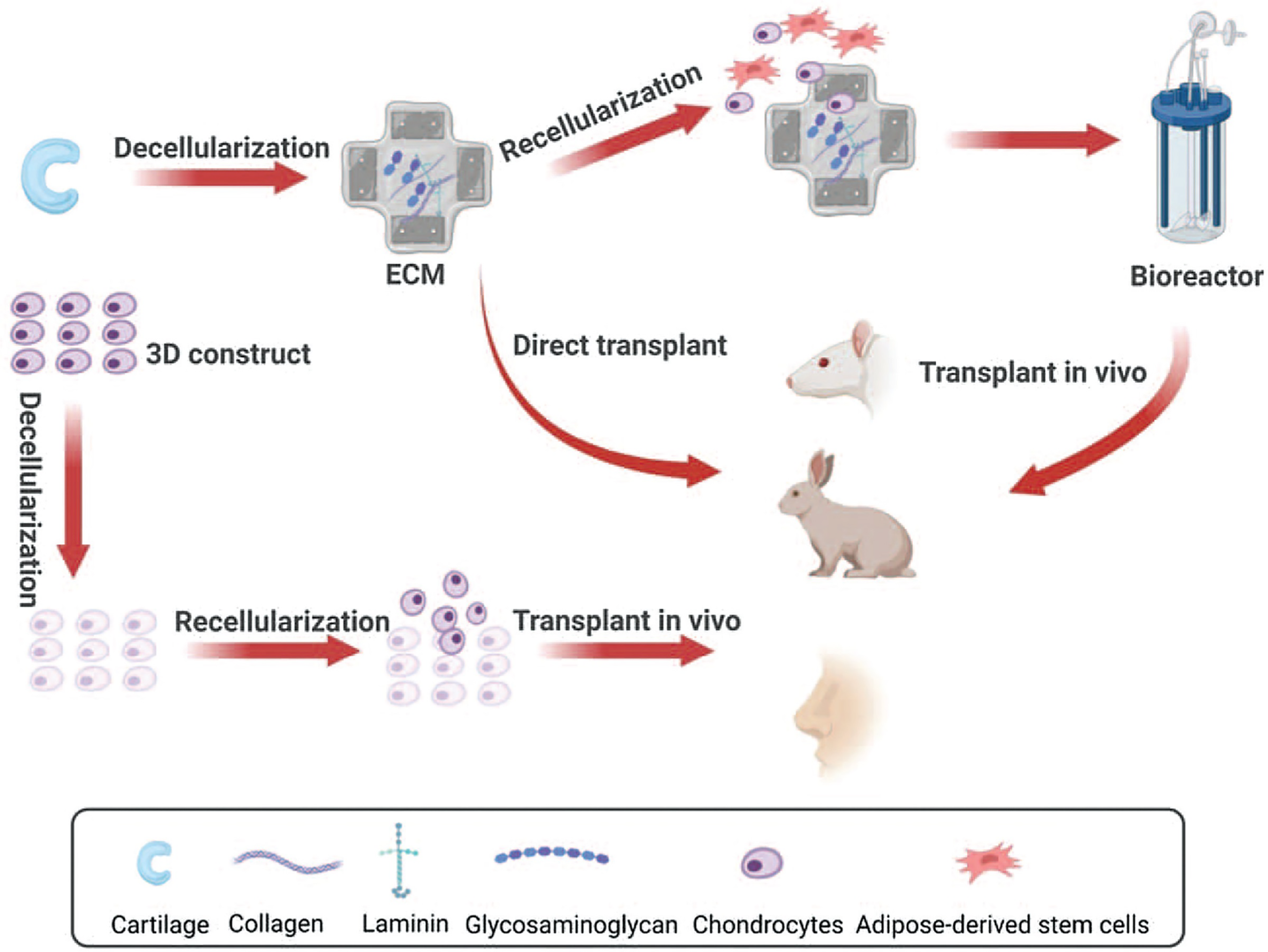
Rationale of nose decellularization. Reused from Ref. 5 with permission from Wolters Kluwer Health, Inc., copyright 2023.
Source of the dECM
The dECMs commonly used for tissue engineering can be divided into human-derived, animal-derived, and plant-derived dECMs. 10 Human-derived dECM can be prepared from different tissues and parts, such as the heart, upper limb, ovary, pancreas, ear, liver, skin, teeth, lung, and other tissues and organs. 11 Animal-derived dECM can be sourced from pigs, cattle, goats, rats, and other animals. The tissue sources include bone, cartilage, joint synovium, dermis, fat, heart, and liver.
Preparation methods for the dECM
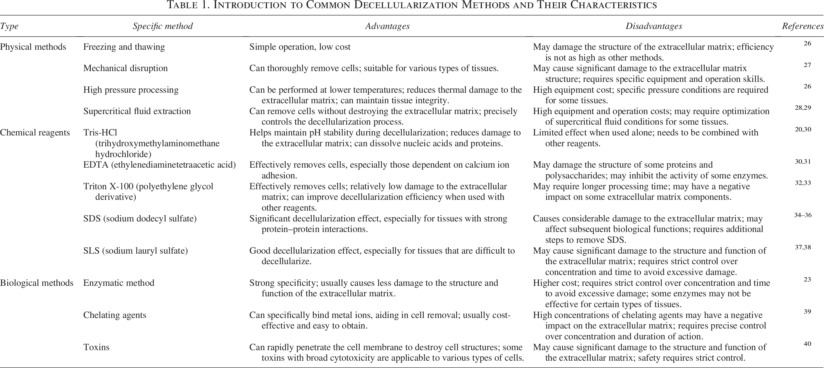
The preparation of dECM involves physical, chemical, and biological approaches. Physical methods primarily include freeze–thaw cycles, high hydrostatic pressure, and supercritical fluid techniques.11,12 The freeze–thaw cycle method involves repeatedly freezing tissues at low temperatures and then thawing them at room or physiological temperatures, causing cell lysis through the formation and melting of ice crystals. During rapid freezing, intracellular ice crystals are generated, resulting in membrane disruption and subsequent cellular necrosis. 13 To ensure sufficient decellularization, this approach generally relies on several successive freeze–thaw cycles. 14 The prevalent technique of high hydrostatic pressure processing utilizes pressures above 600 MPa to efficiently eradicate cells from biological tissues. Cell debris can be removed by washing to obtain decellularized tissue. 15 This method can effectively retain the ECM ultrastructure. 16 The supercritical fluid method refers to the removal of biological components via the fluid generated when the gas changes under critical temperature and pressure while retaining most glycosaminoglycans (GAGs) and collagen. 17 Physical methods can effectively protect the ECM structure but generally cannot effectively remove cell debris. 18
The chemical method involves the use of detergent, acid, alkali, and other reagents for decellularization. Common acidic reagents include deoxycholic acid, hydrochloric acid, peracetic acid, sulfuric acid, and acetic acid, 19 while common basic reagents include sodium hydroxide, sodium sulfide, ammonium hydroxide, and calcium hydroxide. 20 Cleaners mainly include ionic, nonionic, and zwitterionic types, which can decellularize and treat cells by permeating and dissolving cell membranes. 21 In addition, hypotonic and hypertonic saline, alcohols, and tributyl phosphate have also been used for decellularization treatment. 22
The biological enzyme method utilizes enzymes such as proteases, nucleases, and esterases to achieve decellularization. Following cell lysis, nucleic acid residues are eliminated, and intercellular as well as extracellular connections are cleaved. 23 Proteases are able to cleave peptide bonds between arginine and lysine. 24 Nucleases hydrolyze DNA and RNA chains. When used individually, these enzymes are often insufficient, so they are typically combined to achieve more complete decellularization.
At present, to reduce the adverse effects of traditional single decellularization methods on dECM, the combined application of the above decellularization methods is preferred when decellularizing tissues and organs. 25 (The principal techniques for decellularization and their key attributes are summarized in Table 1).
Introduction to Common Decellularization Methods and Their Characteristics
Different Kinds of Tissue-Derived dECM for Cartilage Repair
Cartilage-derived dECM
At present, research on the mechanism of ADSC chondrogenic differentiation induced by the cartilage ECM has focused on the role of various cytokines. Zhang 41 and others have shown that chondrocytes can induce the chondrogenic differentiation of ADSCs in a paracrine manner. Yin’s results 42 demonstrated that scaffold integration with TGF-β1-loaded microspheres effectively enhanced both the proliferation and differentiation of chondrocytes. Related studies 13 have also confirmed that rabbit ADSCs can enhance their cartilage differentiation and formation, the production of cartilage constructs in vitro, and the regeneration of hyaline cartilage in vivo by expressing TGF-β3/bone morphogenetic protein (BMP)-6. Alexander 43 investigated the chondrogenic differentiation of ADSCs following implantation of poly(lactic acid–hydroxyacetate) scaffolds in nude mice. The study reported that TGF-β1 enhanced ADSC adhesion to ECM components, thus inducing chondrogenic differentiation—a result corroborated by qPCR verification. (As depicted in Fig. 2, exposure to TGF-β1 significantly elevated the transcriptional levels of ACAN and COL2A1). Furthermore, reported findings 44 indicate that N-cadherin and neural cell adhesion molecule-mediated cell–cell adhesion is essential for early chondrogenic differentiation, while later stages rely on integrin-based adhesion signals from ECM proteins to regulate the proliferation, differentiation, and hypertrophy of mesenchymal precursor cells. Cell attachment mediated by integrins is not only crucial for migration 45 but also significantly influences proliferation. Key ECM components—such as laminin, fibronectin, vitronectin, and collagen—interact with specific integrins; for example, fibronectin engages integrin α5β1. 46 These interactions between integrins and ECM components can activate several signaling cascades, such as MAPK, Akt, ERK1/2, and PI3K, 47 and can even directly trigger growth factor receptors through adhesion receptor engagement. 48 It has also been shown that dECM can enhance the proliferation of stem cells and chondrocytes by reducing intracellular reactive oxygen species (ROS) levels, thereby creating an enhanced environment that supports cell growth and function.49–53
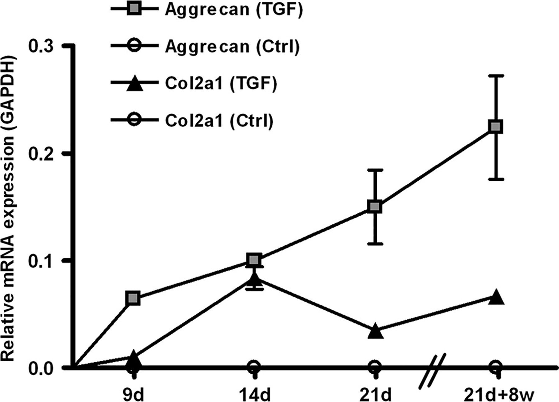
Aggrecan and collagen type II (COL2A1) messenger RNA (mRNA) expression was detected only in samples treated with transforming growth factor beta 1 (TGF-β1) in vitro (9, 14, and 21 days) and in samples pretreated with TGF-1 in vivo (21 days and 8 weeks). mRNA expression of samples treated with TGF-β (controls) was measured using quantitative real-time polymerase chain reaction at 9, 14, and 21 days, and after transplantation in vivo at 21 days and 8 weeks (n = 4 each). mRNA expression was displayed relative to the housekeeping gene glyceraldehyde 3-phosphate dehydrogenase. Values are reported as means ± standard deviations of the mean. 103 × 71 mm. Reused from Ref. 43 with permission from Mary Ann Liebert, Inc., copyright 2009.
Mechanical stimulation is also an important condition. Mahsa Sani 54 reported that insulin-like growth factor (IGF)-1 contributes to the chondrogenic differentiation of ADSCs, although its effect is less pronounced than that of mechanical stimulation. Furthermore, the combination of compression and IGF-1 synergistically elevates COL2A1 levels while showing minimal impact on other markers. Chen 12 reported that biological mechanical stimulation promoted cartilage differentiation through the Yap receptor, a known mechanical stimulation receptor. (The immunofluorescence staining results for Yap and cartilage-specific markers across different groups are presented in Figs. 3 and 4). The elevated level of chondrogenesis promoted by the dECM can be attributed to the upregulation of YAP caused by periodic tensile changes in the local microenvironment.55,56 (The qPCR verification results and immunofluorescence staining results of YAP pathway are shown in Fig. 5). YAP, 57 an essential regulator of the Hippo signaling pathway, acts as a critical mechanotransduction factor that conveys mechanical signals to cells, thereby regulating and promoting subsequent cellular activities. Knockdown of the YAP gene inhibits cartilage formation in vitro and in vivo. 58 Mechanosensitive stretch stimuli, along with various external modifications, can increase cell numbers, with synergistic effects and parallel signaling pathways collectively amplifying cellular responses.
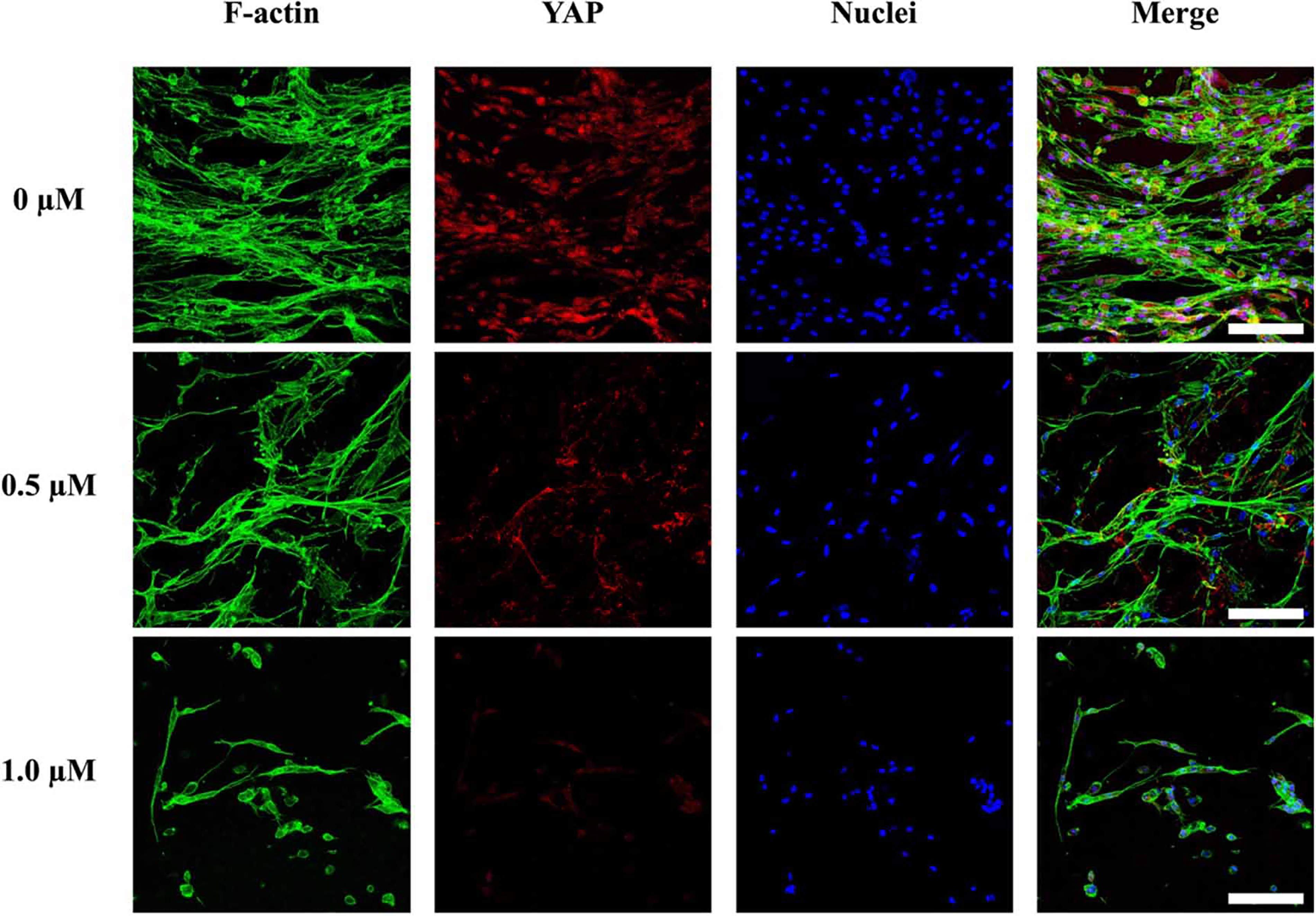
Immunofluorescence staining of YAP in the presence of veteprofin at 0, 0.5, and 1 μm. The scale bar is 150 μm. Upon exposure to mechanical stimulation, there was homogenous expression of YAP in the dECM/FGelMa auxetic scaffolds, as observed in the control group without the YAP inhibitor. Exposure to the YAP inhibitor resulted in a dose-dependent decrease in YAP expression, along with a dose-dependent decrease in cell proliferation. Reused from Ref. 12 with permission from lOP Publishing, copyright 2023.
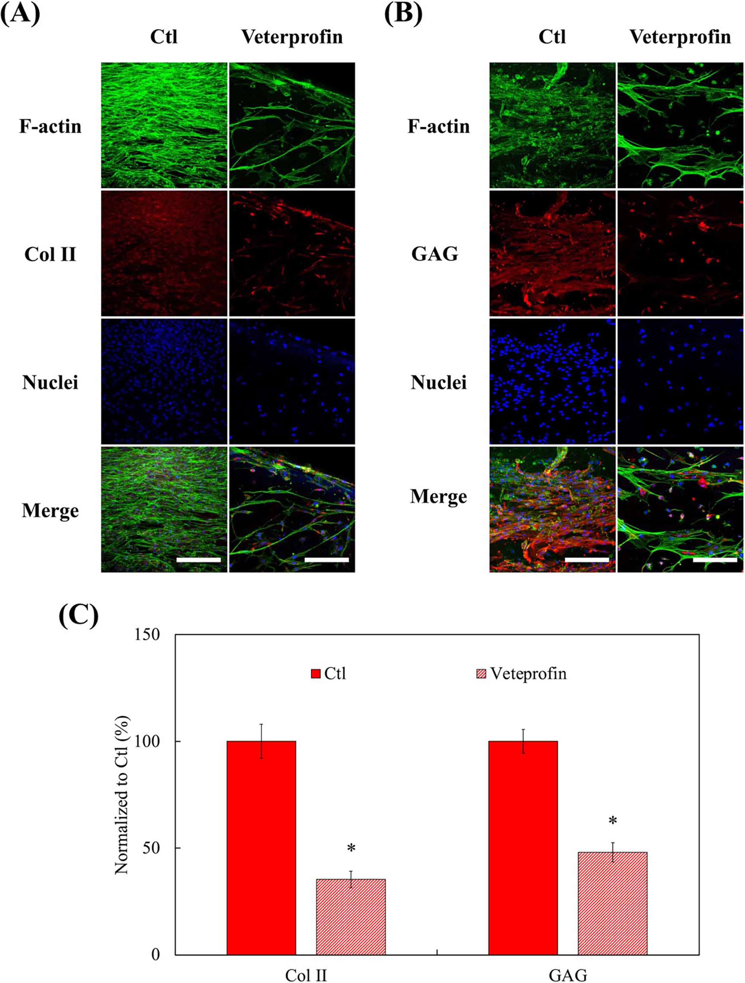
Immunofluorescence staining of chondrogenesis-related markers in the presence of veteprofin at 0 and 0.5 μm.
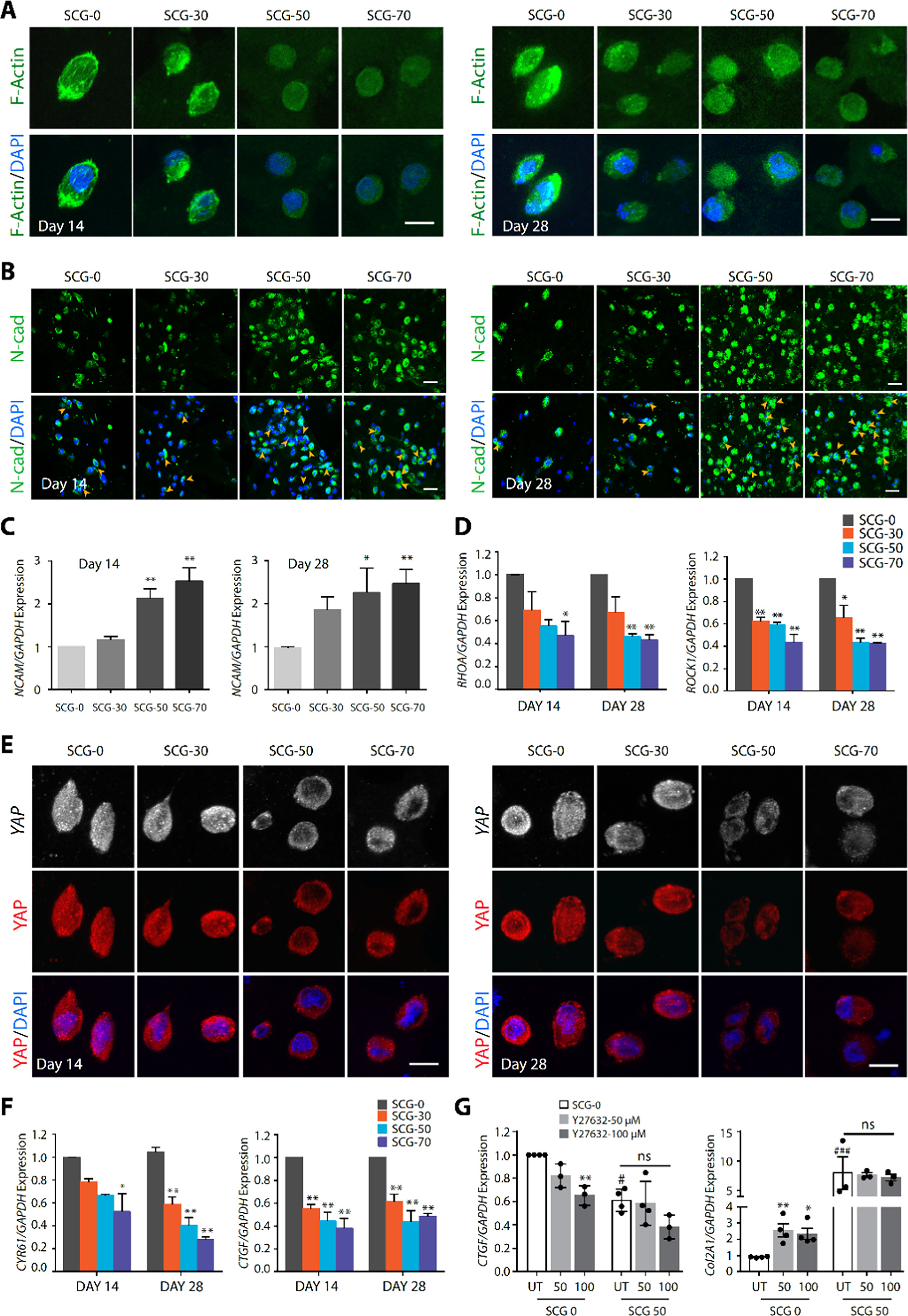
Effect of hydrogel stiffness and contraction on cellular shape, condensation, and YAP signaling during in vitro chondrogenesis.
Adipose-derived dECM
As a common material in plastic surgery, fat is usually obtained from human tissue sources. Over the past few years, notable developments have occurred in adipose tissue engineering, 59 and soft tissue reconstruction approaches utilizing adipose-derived dECM have shown promising potential. 60 Ji Suk Choi 61 mixed an ECM scaffold isolated from adipose tissue with ADSCs and cultured them in cartilage culture medium. They reported that ADSCs presented cartilage differentiation characteristics. The research on using ADSCs to induce cartilage regeneration and repair has also been carried out. Nidia et al. 62 developed an implant utilizing ADSCs (characterized by CD105, CD90, CD73, CD14, and CD34 immunophenotypes) transduced with IGF1.The implant was incorporated into a composite scaffold containing dECM, and the findings suggested that the scaffold exhibited structural and functional characteristics comparable with those of natural knee cartilage. However, the current research mainly focuses on adipogenic differentiation and regeneration of adipose dECM, and the specific application of adipose dECM scaffolds in inducing cartilage differentiation at the cellular and tissue levels is still rare. This issue needs further discussion.
Synovial-derived dECM
Jinku reported that synovial dECM can promote chondrocyte regeneration through the SIRT1 pathway. RT-qPCR analysis revealed that chondrocytes expanded on dECM exhibited notably higher SIRT1 mRNA levels, showing a 1.1-fold increase in chondrogenic medium, 79.7% higher with IL-1β (1 ng/mL), and 1.5-fold higher with tumor necrosis factor (TNF)-α (1 ng/mL) compared with controls. Western blotting confirmed elevated SIRT1 protein expression in the dECM group, with increases of 73.6% and 57.0% under IL-1β and TNF-α treatment, respectively. Additionally, SOX9 protein levels were significantly enhanced in dECM-expanded chondrocytes, with rises of 29.6% in chondrogenic medium, 57.1% with IL-1β, and 48.5% with TNF-α. 63 SIRT1 is a class III deacetylase dependent on NAD+ that removes acetyl groups from both histone and nonhistone proteins, 64 suppresses p53 activation, and influences cell survival, 65 differentiation, 66 and oxidative stress. 67 SIRT1 can effectively protect chondrocytes in an inflammatory environment. 68 When exposed to TNF-α, activating SIRT1 markedly promotes chondrocyte survival, while silencing SIRT1 through siRNA triggers apoptosis in these cells. 69 SIRT1 activation facilitates the deacetylation-driven nuclear translocation of SOX9, which in turn upregulates aggrecan and COL2A1 expression in chondrocytes. 70 (The diagram of the mechanism of the SIRT1 pathway under inflammatory and healthy conditions is shown in Fig. 6).
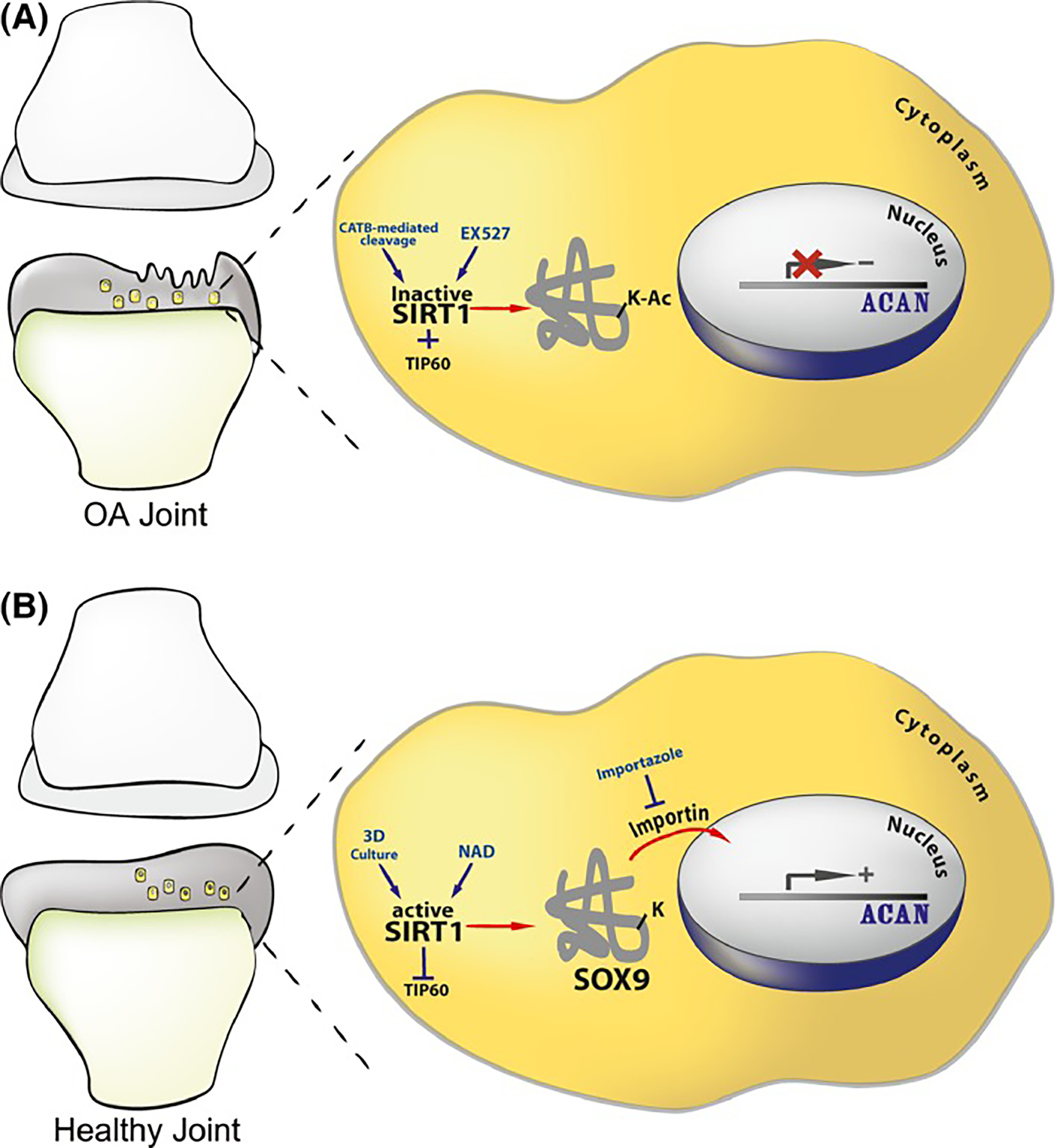
Scheme illustrating the mechanism of SOX9 nuclear entry. The illustration shows how acetylation state could affect SOX9 nuclear entry and ACAN expression in intact and osteoarthritis (OA) cartilage.
SIRT1 modulates cartilage matrix gene expression by interacting with the histone methyltransferase Set7/9 to form a protein complex that associates with the COL2A1 promoter, thereby promoting its transcription. 71 In addition, the enhanced antioxidant capacity of the dECM helps increase the resistance of chondrocytes to proinflammatory cytokines. In chondrocytes expanded on dECM, upregulation of SIRT1 facilitates the nuclear translocation of the transcription factor FOXO3a, leading to increased expression of antioxidant enzymes such as superoxide dismutase (SOD)2 and catalase. 72 During redifferentiation, dECM expansion also mitigates cartilage matrix degradation triggered by interleukin-1β and TNF-α, indicating that elevated SIRT1 may play a key role in suppressing matrix-degrading enzymes. 63 In human chondrocytes, overexpressing SIRT1 inhibits IL-1β-induced production of matrix metalloproteinases MMP-1, MMP-2, MMP-9, and MMP-13, whereas SIRT1 knockdown via siRNA enhances their expression. 73 SIRT1, as an important pathway signal for synovial dECM to promote cartilage regeneration and repair, also suggests the application of targeted gene editing and active factors, which would provide more possibilities for improving cartilage regeneration efficiency.
Dermis-derived dECM
Wei Gao’s study 74 revealed that dermal dECM scaffolds were superior to cartilage dECM scaffolds (the staining results of chondrocytes seeded in cartilage and dermal dECM scaffolds are shown in Fig. 7), which could result in a higher cell adhesion rate and greater potential for cartilage regeneration. Two-dimensional dECM scaffolds derived from the dermis were converted into three-dimensional constructs, exhibiting favorable physicochemical characteristics, excellent biocompatibility, and strong potential to support cartilage regeneration both in vitro and in vivo. These scaffolds facilitated the presentation of adhesion molecules and bioactive factors, enhanced the expansion and specialization of chondrocytes, and stimulated cartilage matrix synthesis. 75 Moreover, in rabbit models, dermis-derived dECM scaffolds effectively repaired cartilage defects, highlighting their strong capacity for cartilage regeneration both in vitro and in vivo.
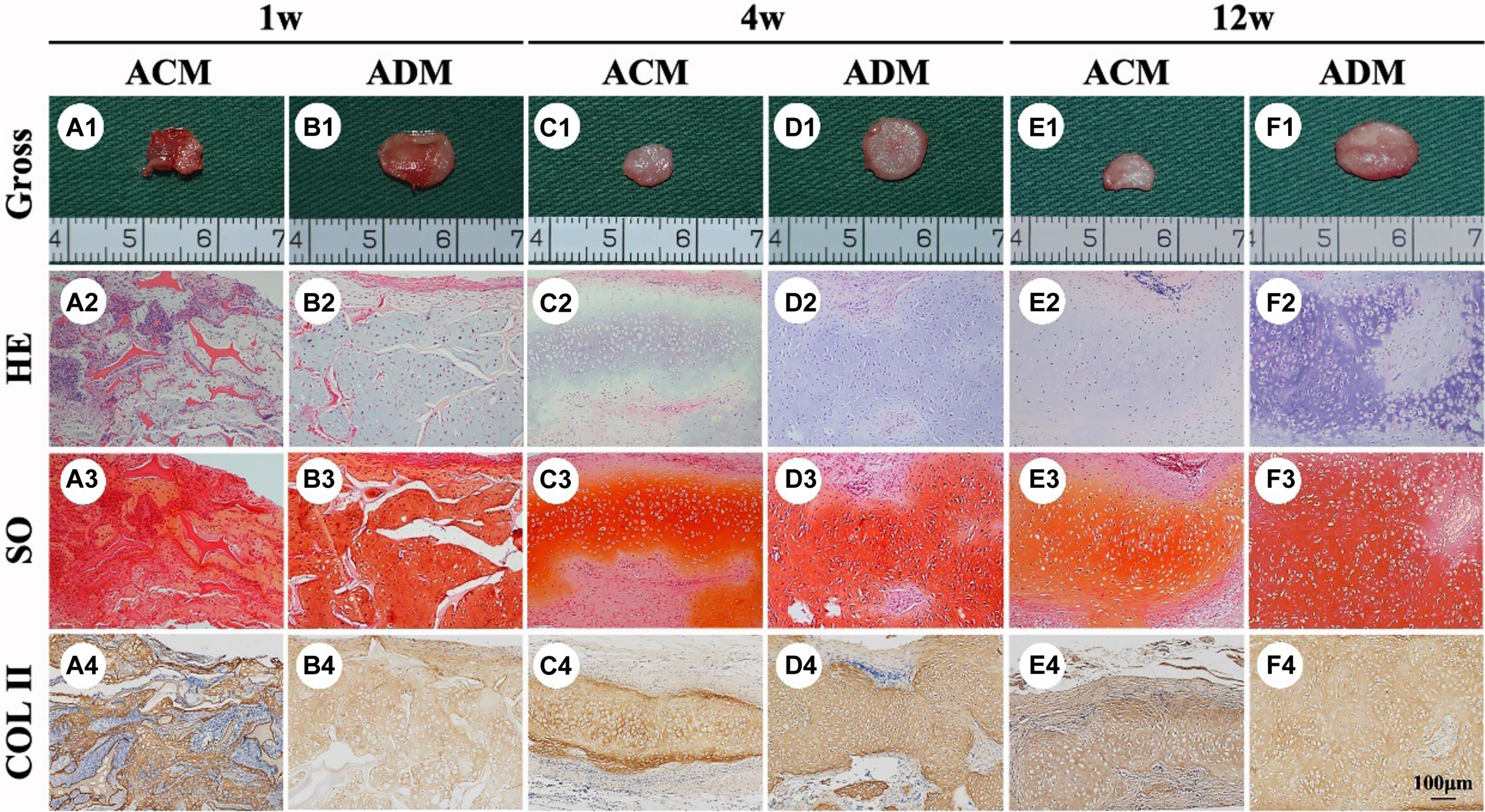
In vivo ECs formed by chondrocytes seeded in ACM and ADM scaffolds. Gross view, HE, safranin-O, and collagen II immunohistochemical staining of ACM A1–A4 and ADM B1–B4 scaffolds after in vivo implantation for 1 week. Gross view, HE, Safranin-O, and collagen II immunohistochemical staining of ACM C1–C4 and ADM D1–D4 scaffolds after 4 weeks of in vivo implantation. Gross view, HE, Safranin-O, and collagen II immunohistochemical staining of ACM E1–E4 and ADM F1–F4 scaffolds after 12 weeks of in vivo implantation. Reused from Ref. 74 with permission from Frontiers, copyright 2020.
The characteristics and application mechanisms of dECM derived from four organizational sources refer to Table 2.
Comparison of dECMs from Different Tissue Sources in Cartilage Repair
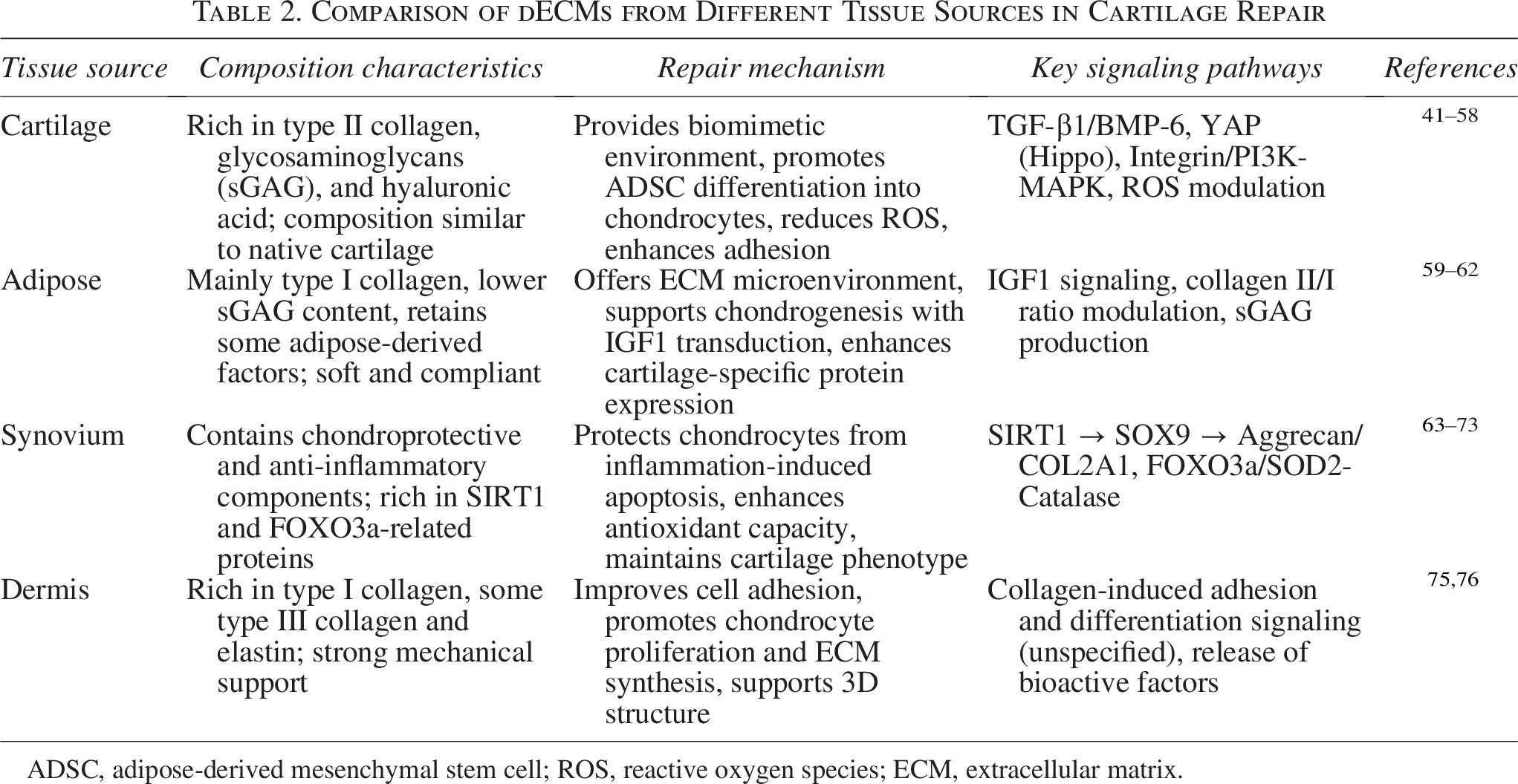
ADSC, adipose-derived mesenchymal stem cell; ROS, reactive oxygen species; ECM, extracellular matrix.
Prospects of Biomimetic ECM Materials for Cartilage Repair Combining dECM with 3D Bioprinting Technology
Cell-based therapy for cartilage defects remains a major challenge in cartilage tissue engineering, requiring an ideal biomimetic scaffold that closely replicates the structure of natural cartilage ECM. The ECM functions as a dynamic framework within the tissue microenvironment, directly regulating cell adhesion, migration, differentiation, and functional activity.77–80 The functional importance of ECM is further emphasized by its role in guiding cellular behavior, 81 dynamic responsiveness, and regenerative potential. 82 Therefore, the complexity of ECM requires biomimetic scaffold materials to achieve multi-scale simulation at the molecular, structural, and functional levels. Constructing biomimetic ECM for tissue repair often requires the integration of multiple materials. Cheng et al. 83 reported that a sugar-to-sulfate ratio of 2.5:1 critically influences whether bone marrow mesenchymal stem cells (BMSCs) undergo osteogenic or chondrogenic differentiation. These studies highlight the critical effect of component ratios on cell fate regulation, suggesting that biomimetic ECM materials need to finely regulate the proportion of each component to mimic the dynamic equilibrium of native ECM.
3D bioprinting facilitates the fabrication of tissue and organ analogues by precisely arranging living cells and biomaterials in a layer-by-layer manner. 84 Leveraging the ability of dECM to retain key features of native ECM, 85 dECM-based bioinks provide a supportive microenvironment that promotes the formation and maturation of 3D tissue constructs. 86 For example, Lin et al. 87 reported that ECM scaffolds substantially improved the recovery of subchondral bone–cartilage lesions, and Cunniffe et al. 88 found that biphasic ECM-derived scaffolds effectively guided stem cells toward tissue-specific differentiation. Therefore, biomimetic ECM scaffolds combined with 3D bioprinting have become a promising manufacturing strategy for cartilage repair. 45 At present, the construction and application of various biomimetic ECM materials have also been studied. We summarize the application mode of biomimetic ECM into three steps: “construction → functionalization → mechanism exploration.” (Three-tier strategy of biomimetic ECM combining dECM with 3D bioprinting is shown in Fig. 8). The following is the detailed introduction.
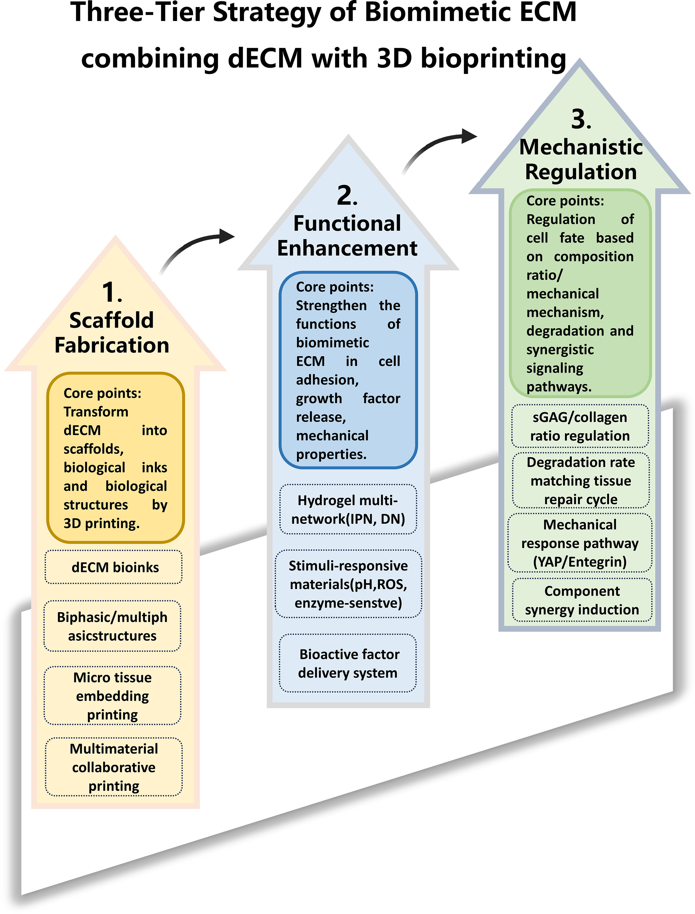
Three-tier strategy of biomimetic ECM combining dECM with 3D bioprinting.
Construction and application of various biomimetic ECM
The applications of diverse hydrogels
Hydrogel has become the core carrier of biomimetic construction because of its high moisture content and structural similarity to ECM, including natural polymer-based hydrogels and synthetic hybrid hydrogels. Chitosan combined with collagen to form a multilayer osteochondral scaffold 89 enhances both the mechanical strength and chondrogenic potential of chitosan-based scaffolds. 90 A double-network hydrogel of hyaluronic acid (HA) and gellan gum has been observed to substantially facilitate cartilage regeneration in rabbit defect models. 79 Moreover, a 3D-printable bioink composed of HA, gelatin, and chondroitin sulfate integrated with graphene oxide nanoparticles can drive hMSC chondrogenic differentiation without the need for exogenous factors. 91 Silk fibroin (SF), when combined with chitosan nanoparticles, can significantly improve angiogenesis while mimicking the porous architecture and chemical composition of native ECM. 92 In addition, Qu et al. developed a three-component hydrogel consisting of gellan gum, SF, and chondroitin sulfate. After optimizing the component ratio (0.5% GG/3.5% SF/CS), cell proliferation and cartilage-specific gene expression were increased by 2 times. 93
Hybrid hydrogels combining synthetic and natural components focus on modifying existing ECM-inspired materials via various crosslinking strategies to generate multifunctional hydrogel scaffolds. Cheng et al. 83 demonstrated that adjusting the sugar-to-sulfate ratio to 2.5:1 in a chondroitin sulfate analogue hydrogel, produced through light-induced crosslinking, precisely regulates the balance between osteogenic and chondrogenic differentiation of bone marrow-derived mesenchymal stem cells (BMSCs). Interpenetrating network hydrogels have also been engineered to support vascularized bone regeneration. 94 Furthermore, a chitosan/mesoporous silica nanoparticle (mSiO2 NPs) composite hydrogel developed by Cui et al. 95 promotes cartilage ECM deposition by delivering dehydrated icariin anhydroicaritin via the nanoparticles.
Construction and application of inks for various 3D printing materials
The construction of ink from biological tissue is still a common material for many studies. The researchers functionalized the porcine cartilage ECM into alginate ink, combined it with PCL fiber network, and can print out the composite scaffold matching the compression modulus of natural cartilage (∼2 MPa). 96 In addition, Xie et al. also developed biological ink based on microtissue, encapsulated ear chondrocytes and ECM particles in GelMA, and successfully printed the complex structure with stem cell shelter function through digital light processing (DLP). 97 These bioinks formed under different conditions are important achievements of 3D bioprinting in the field of ECM bionics research. The collaborative preparation of multimaterials and bioink also plays an important role. Melo’s research used a dual-material system (hard polyethylene glycol alginate and soft fibrin) to simulate the mechanical heterogeneity of cartilage and finally proves that this dual-material system can maintain high strength in the macro and provide a soft environment in the micro to promote cartilage differentiation. 98 In addition, through the spatial modification of DNA network hydrogel and aptamer, BMSCs were recruited and vascular endothelial growth factor was dynamically released to achieve vascularized bone regeneration. 81 Rathan et al. used microtissue biological ink combined with DLP printing technology to achieve high-precision regeneration of ear cartilage. 96
Although dECM use in tissue engineering and 3D printing has advanced markedly, it is still necessary to systematically distinguish dECM biological ink from ordinary biological material ink in the research of ECM bionics. The former emphasizes the specific regulation of dECM active components (such as GAGs and growth factors) on cell behavior, 99 which is also its unique advantage. Some researchers pointed out that the preparation of dECM ink needs to balance the decellularization efficiency and component retention to avoid immune rejection caused by DNA residue (<50 ng/mg). 100 Wang et al. found that young dECM can best promote the differentiation of BMSCs cartilage and inhibit calcification. 101 Cao et al. proved that children’s cartilage dECM can enhance subchondral bone repair through unidirectional collagen dECM stent combined with microfracture surgery. 102 The intervertebral dECM hydrogel developed by Borrelli et al. successfully increased the synthesis of sGAG in nasal chondrocytes by 40% by regulating the degradation rate through chondroitin sulfate functionalization, 103 which also revealed the cross-tissue repair potential of the intervertebral ECM.
Multi-scale and intelligent construction of biomimetic ECM
The multi-scale bionic design of ECM is beneficial to better simulate the structure and microenvironment of tissues. The fiber diameter, porosity, and degradation rate of NF MCS can be regulated by orthogonal design. 104 Ding et al. proposed the trinity strategy of “composition-structure-function”, which effectively simulated the multilevel structure of ECM through the gradient composite of SF nanofibers and chitosan. 92 Zhang et al. used microfluidic technology to prepare PrP-loaded microcarriers (IGMs), combined with dECM hydrogel to construct a composite system for hair follicle regeneration, and achieved high-throughput optimization of the composition ratio. 105 Biomimetic integration of multiple tissues can promote the repair effect. Collagen I maintains hepatocyte function in soft hydrogel (elastic modulus∼1 kPa), which also provides enlightenment for liver–cartilage composite tissue engineering. 106
In recent years, the design of various intelligent materials has been gradually applied to the construction of biomimetic ECM. Sophora bean gum MA hydrogel can dynamically regulate degradation and cartilage differentiation through photocrosslinking. 107 HA/Alg-RGD hydrogel utilizes the reversibility of ionic crosslinking to achieve in situ remodeling of vascular channels after printing. 108 New bionic design combined with machine learning design is also being explored. Optimizing the proportion of components 83 and developing 4D printing response materials 98 will become new breakthroughs. Studies have analyzed the component–function correlation of cartilage ECM at different ages through big data, which has effectively guided the design of personalized biomimetic ECM scaffolds. 101 In addition, GelMA/DNA composite hydrogel 81 can dynamically regulate cell recruitment through aptamers, which also shows the potential of smart materials.
Functionalization of biomimetic ECM
After the basic components of biomimetic ECM are constructed, it is necessary to carry out biomimetic functionalization to adapt to the function of the natural microenvironment. For the optimization of mechanical properties, we can design covalent/noncovalent double-network hydrogels and use the synergy of HA-SH and self-assembled short peptides to increase the expression of collagen Ⅱ/ACAN and maintain the phenotype of hyaline cartilage. 109 Other studies have reproduced the anisotropic mechanical properties of the meniscus (compression modulus ∼15 MPa) through 3D printing of PCL/SF scaffolds, achieving effective protection of articular cartilage. 110 The application of active factor delivery system can effectively protect tissue and promote tissue repair. Zhao et al. developed fucoidan self-assembled peptide hydrogel, which can effectively inhibit chondrocyte hypertrophy by removing ROS and activating NRF2 pathway. 111 Liu et al. combined with nanofiber poly-L-lactic acid scaffold and Matrilin-3 (MATN3), inhibited cartilage osteogenesis in BMSCs, and maintained the stability of cartilage phenotype. 112
Dynamic crosslinking technology has also been gradually applied. For example, the photocrosslinked silk glue gel (SMH) developed by Qi et al. uses ultraviolet light to trigger in situ molding, which is synchronized with cartilage regeneration, providing a new treatment strategy for cartilage regeneration. 113 Some researchers also used tyrosinase to crosslink sulfated alginate to achieve strong adhesion of hydrogel to natural cartilage (shear strength >50 kPa) and effectively promote collagen II deposition. 114 The integration of vascularization and neuralization, as a difficult point in the functionalization of various types of tissue regeneration, has attracted much attention in recent years. Yang et al. constructed chitosan-fibrin hydrogel and effectively promoted the formation of tubular structures of human umbilical vein endothelial cells (length >200 μm). 115 The HA/Alg-RGD interpenetrating network hydrogel developed by Liu et al. successfully constructed a vascularized microfluidic channel through RGD peptide to enhance cell adhesion and 3D printing technology. 108 These functionalized methods and functions provide a more appropriate reference for the further refined application of dECM in the future.
Regulation mechanism of biomimetic ECM
At present, the research on the regulation mechanism of biomimetic ECM on tissue repair mainly includes the proportion of components, synergistic effect, mechanical mechanism, degradation regulation, and so on. Studies have explored the effect of the balance between GAG and collagen on differentiation. The proportion of sugar to sulfate groups in a chondroitin sulfate analogue hydrogel (2.5:1) has been shown to optimize the production of COL2 and GAGs in BMSCs. 83 Similarly, maintaining an appropriate ratio of sulfated GAGs to collagen (1:3) in young cartilage-derived dECM effectively suppresses calcification while enhancing differentiation, reducing the calcified area by approximately 60%. 101 Synergistic effects among multiple components are also critical. For instance, a ternary hydrogel composed of gellan gum, SF, and chondroitin sulfate (0.5% GG/3.5% SF/CS) increases hydrogel water absorption by 50% and doubles cell proliferation. 93 In addition, a chitosan/collagen II/nano-hydroxyapatite (nHA) composite hydrogel, with a chitosan-to-collagen II ratio of 70:30, induces hypertrophic differentiation of ATDC5 cells, offering a novel strategy for regenerating the bone–cartilage interface. 116
The mechanism of mechanical regulation also deserves attention. 117 With the increase in mechanical load, the collagen/proteoglycan ratio of the lower periodontal ligament ECM decreases, suggesting that the dynamic changes of different components and their effects on cell behavior should be considered. 82 The degradation rate also plays a key role in regeneration regulation. Studies have found that chondroitin sulfate-functionalized dECM hydrogel can match the degradation cycle (6–8 weeks) with intervertebral disc regeneration by adjusting the crosslinking density, 103 while lightly crosslinked SMH can adjust the degradation rate through the degree of methacrylation to achieve “on-demand degradation.” 113 These mechanisms of cell growth and proliferation also suggest the key breakthrough point of dECM in biomimetic application in the future, which is expected to implement more precise regulation on cartilage tissue repair.
Conclusion
In conclusion, dECM has a broad application prospect in cartilage regeneration. Its core potential is to promote the functional recovery of chondrocytes by maintaining the biological activity of ECM and providing a growth microenvironment. Although it has great potential, its clinical transformation still faces challenges. First, the risk of immunogenicity has not been completely eliminated. Second, the biological characteristics of dECM depend on the species, age, and health status of the donor, which makes it difficult to ensure the consistency and reliability of products in mass production. Therefore, there is still no standardized production process for obtaining dECM from different organizations, hindering the subsequent wider commercialization. In addition to the effective implantation and fixation of stents in the face of complex defects, it also needs the accurate bionic natural structure, which needs the support of more clinical patient imaging data. Moreover, it is worth noting that dECM products are usually classified as composite products with strict approval requirements. At present, only a few products (such as for skin repair) have been approved by FDA, while most of the products suitable for cartilage regeneration are in the research stage. The cooperation of researchers, clinicians, and regulatory agencies is essential for the establishment of clear guidelines.
In addition to the barriers to transformation, many basic scientific problems and technological bottlenecks have not been solved. For example, the research on cartilage repair mainly focuses on the application of cartilage dECM scaffold, while fat and dermal dECM need to be further explored and developed. At the same time, the impact of donor age and health on the biological activity and effectiveness of dECM requires further investigation. Technical challenges also include achieving high-precision bioprinting to replicate the layered architecture of native cartilage and enhancing subchondral bone vascularization without compromising the integrity of the cartilage layer. Previous studies 118 have initially achieved stratification through stem cell differentiation, but there is still a gap between personalized printing and large-scale production.
It can be anticipated that dECM will continue to advance through interdisciplinary applications in the coming years. For example, using artificial intelligence to analyze big data on material properties and biological outcomes to design the optimal biomimetic scaffold and predict its in vivo performance. The synergy of bioengineering, computational science, and developmental biology will also enhance the application value of dECM. In the future, the solution of these problems will also promote dECM to become the “gold material” of cartilage tissue engineering, provide very encouraging strategies for cartilage injury repair, and drive new advances in tissue engineering.
Authors’ Contributions
W.L.: Conception and design, article writing; G.W.: Conception and design; M.J.: Collection and assembly of figures; Z.L.: Design of tables; R.A.: Collection and article writing; H.W.: Collection and article writing; C.S.: Collection and article writing; Y.A.: Final approval of article.
Footnotes
Availability of Data and Materials
All the data included in this study are available upon request by contacting the corresponding author.