
Abstract
Low back pain, linked to nucleus pulposus degeneration and annulus fibrosus (AF) defects, is a significant cause of global disability. The AF’s multilayered structure supports the spine, but its limited self-repair capacity makes treating AF injuries and herniation difficult. Traditional surgical interventions like discectomy often fail to restore AF integrity, resulting in high reherniation rates. In response, biomaterial-based strategies have emerged as promising alternatives, aiming to replicate AF structure, deliver bioactive factors, and promote tissue regeneration. This review evaluates the composition, biomechanics, and pathophysiology of the AF, emphasizing the fundamental properties of available biomaterials, integration with host tissues, mechanical properties, and biomimetic microenvironments in AF tissue remodeling and repair. We examine recent advances in AF repair biomaterials, including natural and synthetic hydrogels, decellularized extracellular matrix, electrospun scaffolds, and emerging technologies like three-dimensional bioprinting. These materials provide mechanical reinforcement, enhance cell adhesion, and modulate the degenerative microenvironment through controlled drug or growth factor release, offering a comprehensive approach to address the challenges of AF repair.
Impact Statement
This review systematically expounds on the revolutionary role of innovative strategies based on biomaterials in the field of AF repair. By applying advanced materials such as smart hydrogels, electrospun scaffolds, decellularized matrices, three-dimensional bioprinted structures, and smart microneedles, the limitations of traditional surgical intervention have been overcome. These biomaterials possess excellent biological properties, enabling them not only to reconstruct biomechanical integrity through biomimetic microstructures but also to actively regulate the degenerative microenvironment through delivery. Such cutting-edge tissue engineering technologies significantly reduce the risk of reherniation, promote endogenous regeneration, and pioneer new technologies for functional and long-lasting AF repair. The synergy between materials science and tissue engineering marks a new frontier in spine medicine, offering scalable solutions to alleviate dysfunction caused by disc degeneration worldwide.
Introduction
Low back pain, affecting 60 − 80% of the global population during their lifetimes, represents a major socioeconomic burden, and intervertebral disc (IVD) herniation is one of its primary etiologies. 1 The incidence of IVD is about 2–5% annually, and most of the cases occur in adults aged 30–50 years old. 2 Due to the IVD degeneration (IDD), or genetic, injury, environmental, and other driving factors, NP tissue can extravasate into the annulus fibrosus (AF) or beyond its confines, which may involve the metabolic responses of nucleus pulposus (NP) cells.3,4 The progression of pathological events leads to more AF fissures, thus altering spinal biomechanics, which exacerbates IDD. This degenerative cascade manifests clinically as characteristic symptoms, including radicular pain, numbness, and severe disability. Discectomy remains the gold standard surgical intervention for refractory cases unresponsive to conservative methods.5,6 During this procedure, the extruded NP tissue and part of the AF tissue are removed to alleviate the symptoms caused by nerve compression. 7 However, as a palliative treatment, it does not reverse the initial herniation process or repair the persistent defects of AF. 8 In addition, the self-repair and regeneration capacity of AF is limited, and its natural lamellar structure and mechanical properties cannot be reestablished, so that the residual NP tissue may continue to herniate.5,9 The clinical reports have indicated 5 − 25% reherniation rates and 40% reoperation rates following primary discectomy.10,11 Consequently, the unresolved challenge of AF defects after IVD herniation represents a critical therapeutic gap in spinal surgery.
Many attempts have been made to develop AF repair strategies. The AnchorKnot system, an implantable suture-based system, has been designed to seal AF defects through sutures for preventing NP reherniation, but has yet to receive regulatory approval. 12 Wang et al. developed a novel microendoscopic suture device for AF defect repair. Compared with conventional discectomy, this full-endoscopic AF suture effectively reduced reprotrusion and reoperation rates. However, these findings require validation through studies with longer follow-up periods and larger sample sizes. 13 More importantly, the advancements in tissue engineering and regenerative medicine have given new perspectives for AF restoration. Bioengineered constructs incorporating hydrogels, electrospun fibers, decellularized extracellular matrices (dECMs), and microneedles (MNs) have shown promising repair efficacy in disease models, especially when loaded with bioactive molecules and drugs. These biomaterials demonstrate superior biological performance in terms of matrix recapitulation and mechanical strength, with a growing focus on AF repair.
This review begins with a concise overview of the AF’s organization and biomechanical properties, establishing the fundamental design criteria for effective repair strategies. It then proceeds to present a systematic classification and a comprehensive evaluation of the recent advances in biomaterials and technologies developed for AF repair.
Structure and Mechanobiology of AF
The AF is a multilayered composite ring surrounding the NP at the center of the IVD. It interconnects adjacent vertebral bodies along the spinal, playing an indispensable role in maintaining spinal stability and flexibility. The AF is organized into 15–25 unique concentric lamellae exhibiting a characteristic “angular ply” microstructure. Each lamella consists of an extracellular matrix (ECM) with highly aligned collagen fibers arranged at varying angles, contributing to the anisotropic, nonlinear, and multiphasic fibrous structure of the AF.14–17 These collagen fibers exhibit a gradient transition from predominantly type I collagen in the outer AF (oAF) to type II collagen in the inner AF (iAF), with alternating orientations of ±28°–43° angles to the horizontal axis. 18 Adjacent lamellae are connected by thin interlamellar matrix (ILM) layers, composed of nonfibrous proteoglycan, elastic fibers, cells, water, and lipid. Furthermore, there are partition boundaries (PBs) that provide structural integration between neighboring ILM. 19 It is believed that the translamellar bridging network (TLBN), primarily comprising radially oriented elastic fibers, which span over multiple lamellae, forms an orthotropic network with ±45° and 0° orientations relative to collagen fibers. This elastomeric TLBN facilitates elastic recoil and restoration of the AF’s native crimped configuration after loading.20,21
AF cells represent an integral component for maintaining the structural and functional integrity of AF. These cells originate from the sclerotome during embryonic development and are responsible for synthesizing and maintaining the ECM, primarily collagen fibers and proteoglycans, while being highly sensitive to mechanical and biochemical stimuli.22,23 Previous studies have shown the differences in cell phenotype and performance of AF cell populations. Specific markers have been confirmed for AF subpopulations: oAF cells highly express collagen I (Col1a1) and lumican (Lum), while iAF and NP-border cells express more fibromodulin (Fmod), collagen II (Col2a1), and aggrecan (Acan).24,25 One recent single-cell RNA sequencing study further clarified the heterogeneity of AF cell populations and novel progenitor populations such as Saa2-High and Grem1-High mesenchymal stem cells (MSCs), which have potential for differentiation into AF-like lineages. Additionally, AF-derived mesenchymal stem cells express stemness markers such as CD44, Sca1, and mKi67, and may exhibit regional specificity with markers like Saa2 and Grem1, which are implicated in inflammatory response and bone morphogenetic protein antagonism, respectively. 26
The injury and apoptosis of AF cells significantly contribute to IDD, often involving dysregulated matrix metalloproteinase expression that accelerates matrix breakdown. 27 In response to acute injury, AF cells undergo transcriptomic shifts, with key populations such as oAF cells, neutrophils, Saa2-High MSCs, macrophages, and Krt18+ NP cells driving the differential gene expression. These changes are associated with upregulated pathways in angiogenesis and T cell recruitment, while wound healing and ECM regulation are suppressed. Notably, AF cells in injured discs exhibit advanced differentiation states and increased expression of senescence-associated secretory phenotype factors such as p53, p21, and Kras, indicating a compromised regenerative microenvironment. 26 In terms of structure, progressing radially from the oAF to iAF, lamellar thickness decreases alongside the reduction in collagen type I and elastic fiber concentrations. 28 Concurrently, glycosaminoglycan (GAG) content increases from 3% to 8% per wet weight, elevating hydration and permeability in the iAF, thereby blurring its morphological distinction from the NP. 29 Consequently, the iAF demonstrates more effective compressive load resistance, while the stiffer oAF effectively counteracts radial pressure and cranial–caudal stretch from the NP. 14 In addition, the number of cells attached to the AF gradually decreases from the outer layer to the inner layer, while the cell morphology and phenotype also change.30,31 oAF cells display fibroblast-like morphology aligned with collagen fibers, whereas iAF cells have rounded, chondrocytic phenotypes resembling NP cells. 32 Significantly higher expression of F-actin and β-actin in cells has been reported in the oAF, while more vimentin expression in the iAF.33,34 Further studies have described cell heterogeneity as a result of different fiber orientations and mechanical loads. 35
The unique composition and structure of AF confer great mechanical properties to withstand complex spinal loads. During vertebral compression, the hydrostatic pressure of NP induces radial and axial tensile stresses in the AF, generating a combination of shear, compressive, and tensile hoop stresses. In this load-bearing process, circumferential tension is the key tension of the AF resisting external loads.14,16,36 The structural and functional synergy between the AF and NP underscores the vital importance of AF integrity for the IVD’s load-sharing capacity. Meanwhile, the AF exhibits a tensile strength ranging from 10 to 90 MPa and a compressive modulus of 0.4–3 MPa, reflecting its anisotropic nature stemming from the highly organized collagen fibers and proteoglycan-rich matrix. 37 These mechanical properties, which vary depending on testing direction and tissue region, are fundamental for understanding disc biomechanics and developing effective biomaterial-based repair strategies.
Biomaterials in AF Tissue Repair
With the in-depth understanding of the structure and function of AF, strategies for addressing AF defects have expanded beyond surgical interventions. Biomaterial-based repair approaches are gaining attention, primarily categorized into three types: bioadhesive sealing strategies, bioactive molecule-loaded scaffolds to promote AF regeneration, and hybrid strategies combining both. Regardless of the approach, fundamental requirements for biomaterials include excellent biocompatibility, biodegradability, appropriate mechanical properties, and tissue adhesion.9,15,38–41 To meet these criteria, synthetic materials such as thermoplastic polyurethane (TPU) and polycaprolactone (PCL) have been utilized with three-dimensional (3D) printing technology to replicate AF’s unique “angle-ply” microarchitecture, thereby enhancing regenerative outcomes.9,42 Furthermore, the inflammatory microenvironment in AF degeneration is characterized by elevated proinflammatory cytokines (IL-1β, TNF-α), which upregulate matrix metalloproteinases (MMPs) and degrade ECM. This process is further exacerbated by abnormal mechanical loading through mechanosensitive pathways involving Caveolin-1 and Integrinβ, which amplify NF-κB activation. 43 Concurrently, increased reactive oxygen species (ROS) contribute to oxidative stress, creating a degenerative cycle that compromises tissue integrity and highlights potential therapeutic targets for biomaterial-based interventions. 44 Given the complex inflammatory microenvironment caused by AF defects, advanced materials like hydrogels and MNs—through chemical modifications or sustained release of bioactive molecules—exhibit intelligent response to remodel pathological structures (Table 1).29,41,48,51 These features demonstrate their remarkable efficacy in AF regeneration and repair, highlighting significant clinical potential (Fig. 1).
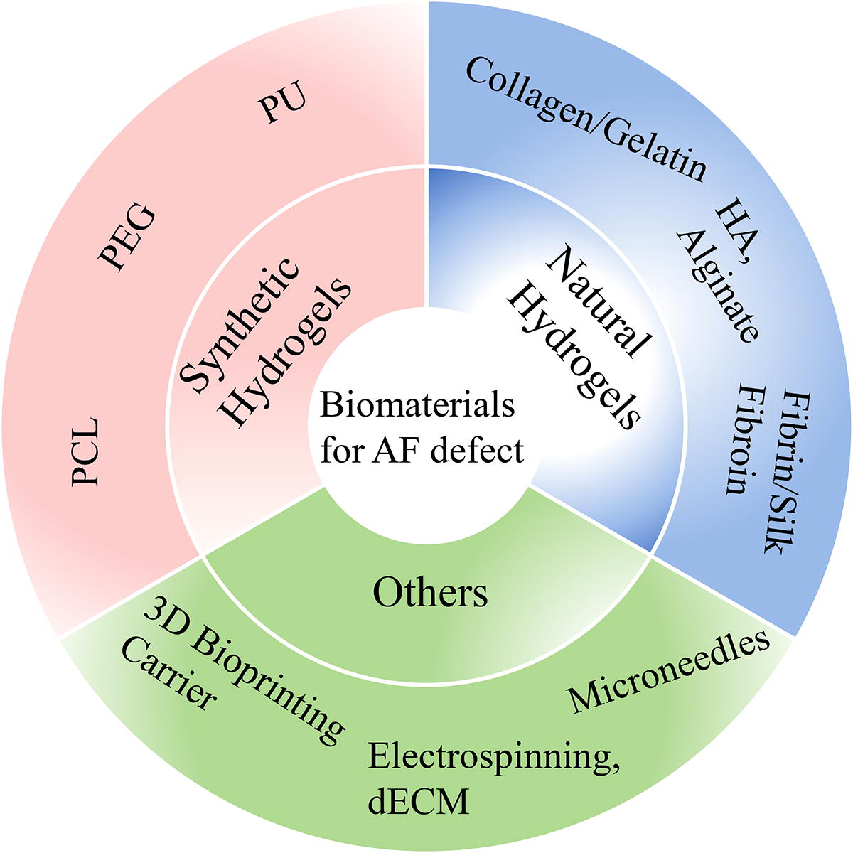
Biomaterials used in annulus fibrosus regeneration tissue engineering. dECM, decellularized extracellular matrix; HA, hyaluronic acid; PCL, polycaprolactone; PEG, polyethylene glycol; PU, polyurethane.
Other Biomaterials Evaluated In Situ for AF Repair
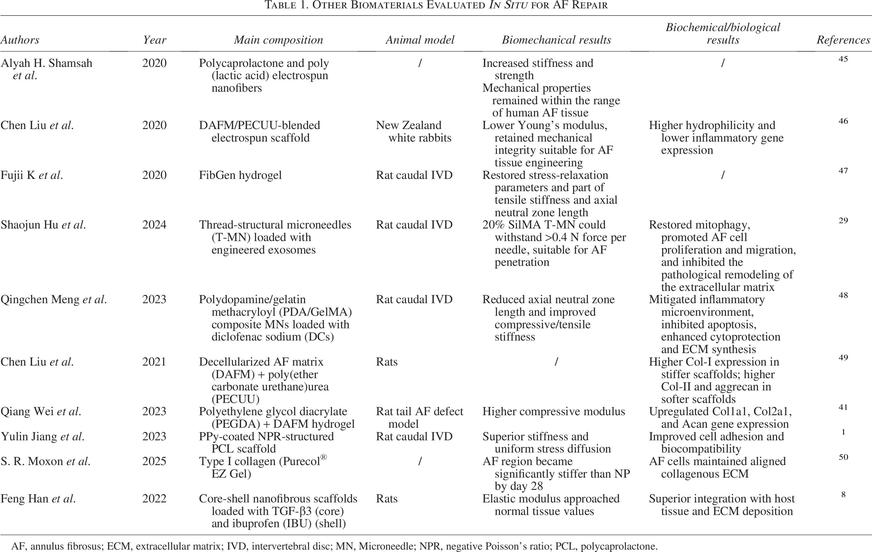
AF, annulus fibrosus; ECM, extracellular matrix; IVD, intervertebral disc; MN, Microneedle; NPR, negative Poisson’s ratio; PCL, polycaprolactone.
Hydrogels
Hydrogels, characterized by their highly hydrated three-dimensional cross-linked polymeric networks, exhibit exceptional liquid absorption, water retention, biocompatibility, and tissue-like properties, thus becoming prominent scaffold materials in tissue engineering and biomedicine.52,53 Recent advancements in regenerative medicine demonstrate their dual therapeutic potential for AF repair to effectively prevent recurrent herniation: bioadhesive or drug-eluting scaffolds. Strategic optimization of hydrogels via chemical modifiers and synthetic engineering has yielded implantable hydrogel sealing systems with enhanced mechanical robustness and tissue adherence while maintaining cytocompatibility.54,55 The development of smart hydrogels responsive to specific microenvironmental cues enables sustained controlled release of therapeutic agents and bioactive molecules, significantly advancing in situ AF tissue repair. 40 Current hydrogel fabrication utilizes natural polymers including hyaluronic acid (HA), alginate, and collagen, alongside synthetic materials such as polyurethane (PU), polyethylene glycol (PEG), and PCL (Table 2).62,63
Hydrogels Evaluated In Situ for AF Repair
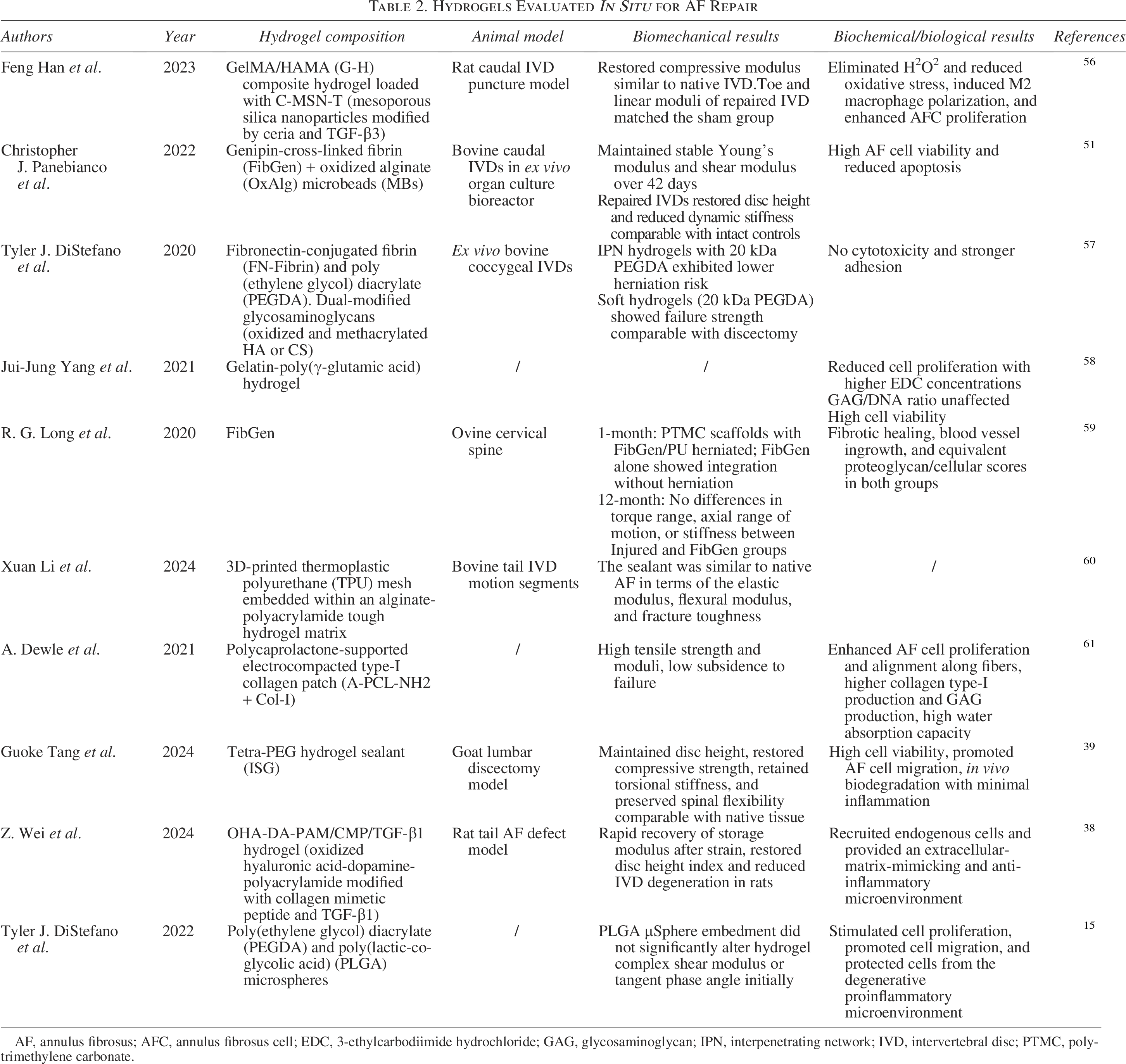
AF, annulus fibrosus; AFC, annulus fibrosus cell; EDC, 3-ethylcarbodiimide hydrochloride; GAG, glycosaminoglycan; IPN, interpenetrating network; IVD, intervertebral disc; PTMC, polytrimethylene carbonate.
Natural hydrogels
Hyaluronic acid
HA, identified as GAG, is ubiquitously distributed in vertebrate tissues and the ECM. 64 Renowned for its unique biocompatibility, controllable degradability, and nonimmunogenicity, HA actively participates in critical cellular metabolic and physiological processes. 65 Currently, the chemical modification of functional groups, such as establishing bonds with water molecules by its carboxyl and acetamido groups, addition or condensation reactions of thiol 66 and dihydrazide, 67 has become a common strategy to engineer structurally stable and functionally adaptable hydrogel scaffolds.
Nie et al. developed a combined AF repair strategy using a PLDLLA (poly-
Application of natural hydrogels in AF tissue engineering.
Alginate
Alginate with great biocompatibility and biodegradability is classified as a linear anionic polymer called polysaccharide extracted from brown algae and some bacteria. 64 Its gelation process, which is facile and operable under physiological pH conditions, is primarily attributed to the preferential bond of divalent cations (e.g., Ca2+) with α-l-guluronic acid (G), while the α-d-mannuronic acid (M) governs the digestibility. 70 Moreover, alginate can be coformulated with other biopolymers (e.g., HA) to fabricate multilayer capsules or improve properties for drug delivery. 71 Reasonably, encapsulation within alginate hydrogels not only protects bioactive/living species from external stressors (e.g., UV) but also achieves precise, sustained release by modulating structure, pH, temperature, humidity, and ionic strength. 72
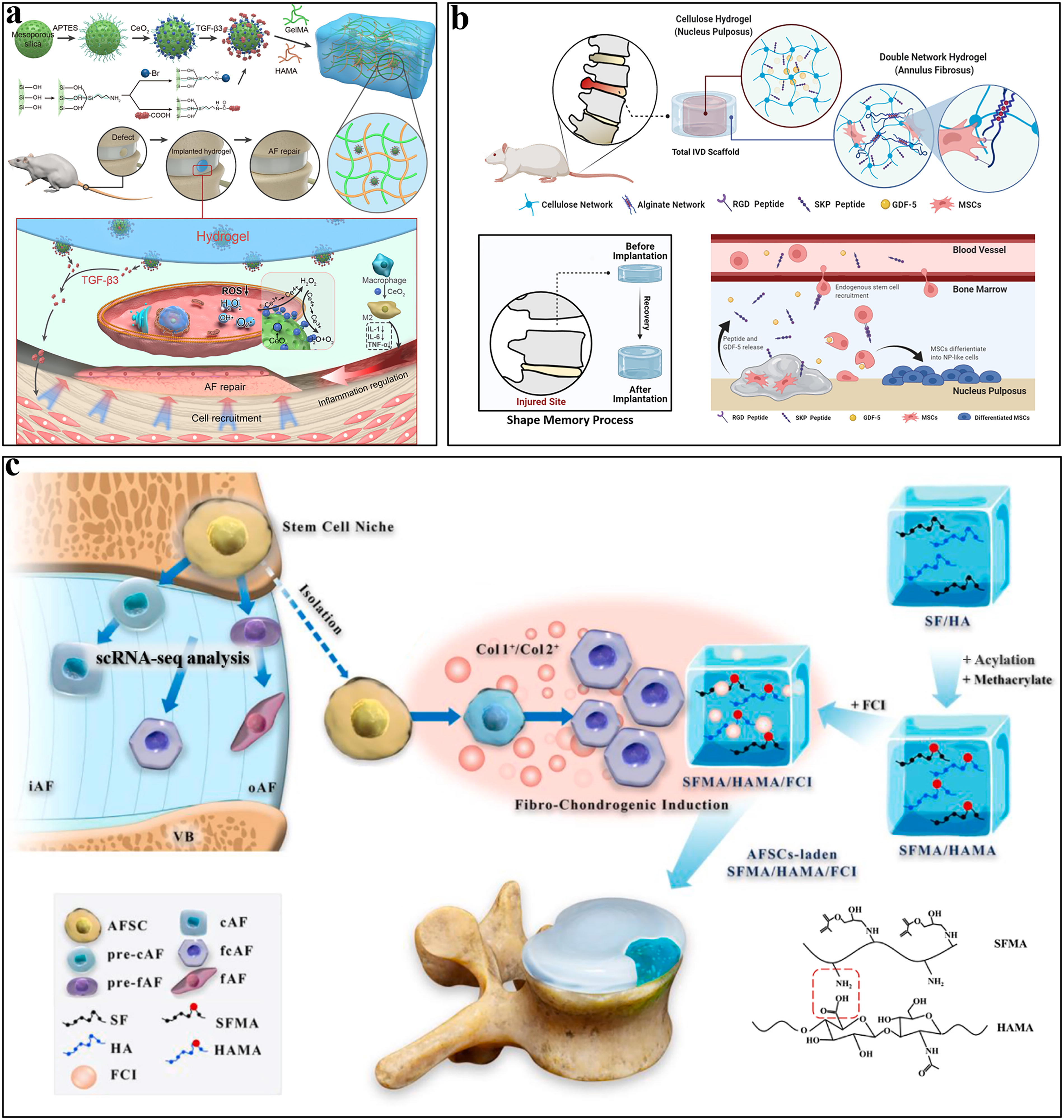
Mirit et al. introduced a biomimetic strategy by embedding long collagen fibers with different orientations into alginate-based hydrogels to mimic the mechanical behavior of native AF. The hydrogel was fabricated by embedding oriented collagen fiber arrays into a sodium alginate solution, followed by cross-linking in calcium chloride. Mechanical performance was evaluated through tensile tests and compared with human, bovine, and ovine AF samples. A finite element model of a lumbar spinal segment was used to simulate physiological loading. The findings demonstrated that both longitudinal and angle-plied biocomposites showed outstanding compatibility with the entire stress-strain. Moreover, further biomechanical testing involving extension, flexion, and lateral bending revealed comparable performance between the calibrated functional spinal unit model and the biocomposite model. This novel composite system exhibits significant potential to mimic native human AF under different orientations as a promising candidate for AF replacement. 73 Chia-Yu et al. developed a cellulose-alginate double-network hydrogel scaffold for AF repair. Experiments were conducted using in vitro models with rat MSCs and an in vivo rat caudal disc degeneration model. By adding chelating agents, the hydrogel acquired shape memory property to recover the disc height after being compressed. Additionally, MSC homing peptides (SKP peptides) and cell-adhesive peptides (RGD peptides) were grafted onto the cellulose and alginate polymer chains to enhance endogenous MSC recruitment, cell survival, and adhesion (Fig. 2b). 69
Collagen and gelatin
Collagen, a kind of critical structural polymer and the most abundant protein in the ECM of connective tissues, is widespread present in skin, tendon, bones, teeth, and cornea. 74 The collagen molecules exhibit triple-helical structures formed of three intertwined polypeptide chains and perform their roles by forming collagen fibrils. 75 Gelatin, which is a water-soluble polypeptide derived from the hydrolytic degradation of collagen, possesses abundant surface functional groups that render it easy to modify and functionalize. 76 The incorporation of functional nanostructures can modify the gelatin and endow it with enhanced water solubility, superior biocompatibility, and reduced immunogenicity. 77 However, due to its intrinsic limitations such as poor mechanical strength and limited water resistance, gelatin-based micro/nanostructures often require cross-linked by agents, which concurrently prolongs drug release in physiochemical environments. 58 Consequently, gelatin/collagen-based hydrogel systems have demonstrated remarkable progress in tissue engineering involving bone and cartilage regeneration, wound healing, dental, or cardiovascular tissue repair. 78
Yon et al. developed a coiled hydrogel microfiber designed to replicate the native AF tissue microstructure for bone marrow stromal cell (BMSC) encapsulation, incorporating both collagen and HA. Specifically, the hydrogels were fabricated by extruding alginate-cell suspensions into a calcium chloride solution containing type I collagen and/or HA, followed by coiling around a needle to mimic the multilamellar AF structure. Their study demonstrated that the synergistic combination of HA and collagen effectively modulated AF-specific gene markers and protease activity, highlighting its critical role in AF tissue engineering applications. 79 Yang et al. devised a gelatin-based hydrogel reinforced with gelatin-poly(γ-PGA). Under the action of the cross-linking agent 1-(3-dimethylaminopropyl)−ethylcarbodiimide hydrochloride (EDC), the carboxyl groups of γ-PGA react with the amino groups in gelatin to form amide bonds, thereby enhancing the binding strength. In parallel, the study evaluated the cytotoxicity of varying EDC concentrations during hydrogel fabrication. These favorable outcomes underscore these materials’ potential as a sealant for AF defects. 58
Fibrin and silk fibroin
Fibrin, a linear protein formed by thrombin-mediated aggregation of fibrinogen, consists of three interconnected polypeptide chains. 80 As a natural plasma protein, its exceptional hemostatic efficacy has been clinically utilized in various surgeries. Additionally, the properties of high cell adhesion and ECM-mimetic architecture allow it to stand out in tissue engineering. 81 However, in order to address its degradation rate and improve mechanical properties, modifications (e.g., methacryloyl reaction) and composite formulations (e.g., cross-linking agents) are often performed to adapt to diverse application. 29 Rose et al. introduced a repair strategy about fibrin hydrogel cross-linked with genipin (FibGen) in an ovine cervical spine model. In a 1-month screening study, FibGen alone integrated with surrounding tissue without herniation, whereas composite strategies with polytrimethylene carbonate (PTMC) scaffolds and PU membranes showed displacement. Consequently, a 12-month long-term study focused on FibGen alone. Results demonstrated that FibGen-treated defects exhibited fibrous healing, biomaterial resorption, and no hydrogel-related complications. Their study demonstrated using only FibGen integrated well with the AF tissue without inducing herniation. Furthermore, long-term evaluations revealed complete fibrous tissue healing, biomaterial absorption, and no significant hydrogel-related complications. 59
Silk fibroin (SF), a natural biopolymer composed of 18 amino acids, is typically derived from Bombyx mori and has been extensively utilized as surgical sutures in humans for decades. Owing to continuous advancements in processing techniques and its inherent biocompatibility, sterilizability, and biodegradability, SF has been widely utilized as a scaffold material in bone, skin, and nerve repair.6,19 In addition, SF demonstrates robust mechanical properties as a biomaterial, attributed to its unique secondary structures (silk I and silk II). 82 Wang et al. adopted a composite hydrogel by the acylation of methacrylated silk fibroin with HAMA, which was prepared by adding fibrochondrocyte-inducing supplement and a photoinitiator, and cross-linking under 405 nm blue light. This hydrogel, loaded with AF-derived stem cells (AFSCs), was tested in a mouse tail AF defect model. The results demonstrated that this novel hydrogel facilitated annulus AF defect repair, restoring chondroid matrices content to 31.7%. Furthermore, later implantation revealed favorable interaction between the hydrogel and surrounding tissues, establishing a suitable microenvironment conducive to anatomically precise AF reconstruction (Fig. 2c). 13
Synthetic hydrogels
Polyethylene glycol
PEG, which is a polymer with a variable structure composed of ethylene oxide and water, primarily exists in linear, four-arm, six-arm, and eight-arm configurations. 83 The functionalizability of the arms enables PEG to achieve improved performance through being block copolymerized with other polymers or physical integration with nanomaterials and natural hydrogels. 84 PEG is soluble in both water and most organic solvents with controlled biodegradability. Its degradation products are safely excreted via kidneys without bioaccumulation, ensuring minimal systemic toxicity. 85 Because of its inherent noncytotoxicity, biocompatibility, and antiprotein adsorption, PEG-based hydrogels have emerged as prominent candidates in tissue engineering. However, there is a lack of large-scale clinical studies to prove the efficacy and safety of PEG-based hydrogels, and the future studies should focus on improving the physical and chemical properties of PEG-based hydrogels, so as to achieve a more intelligent drug delivery system for bone repair.86,87
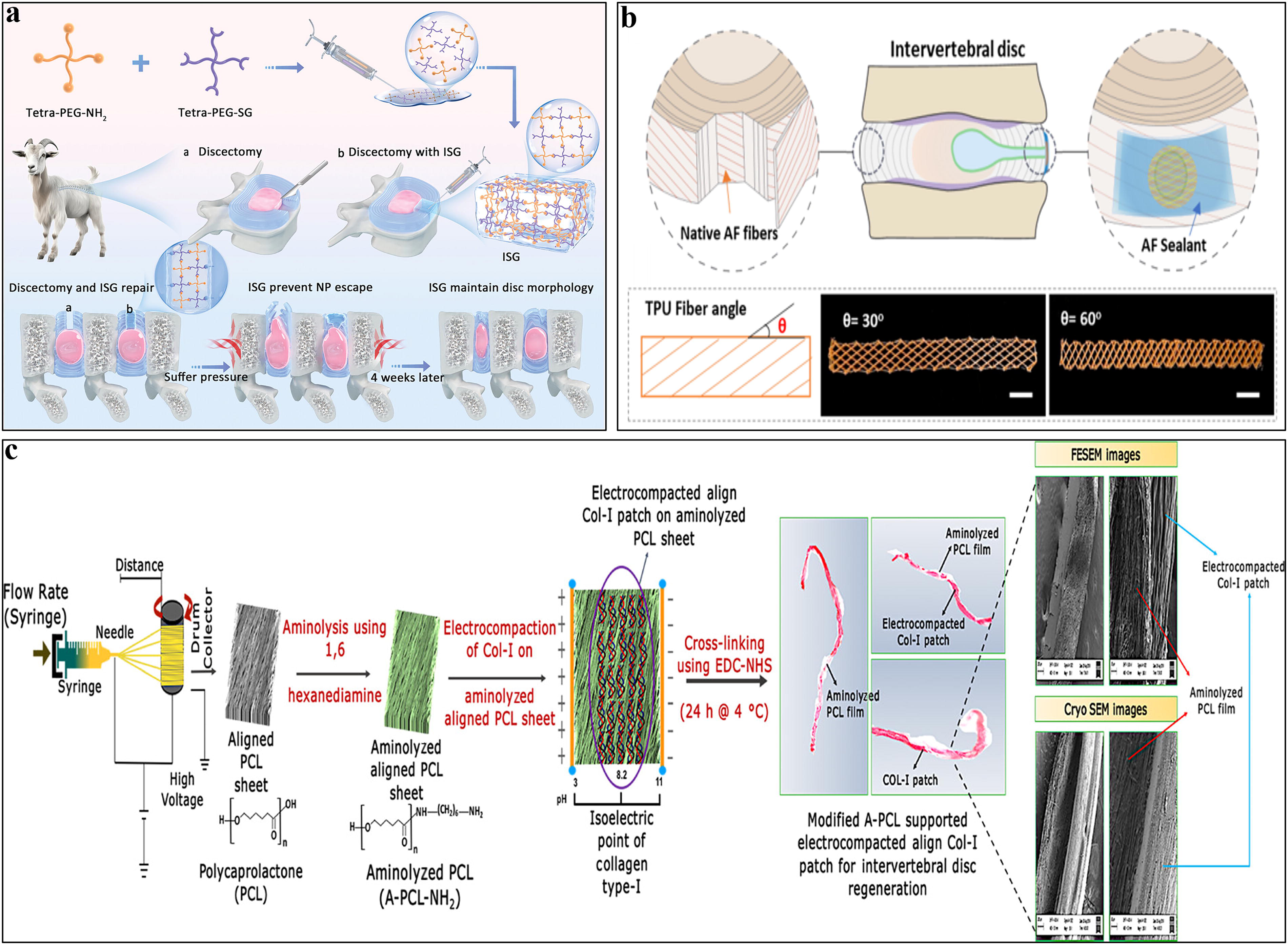
Tang et al. reported an injectable bioadhesive composed of FDA-approved tetra-armed poly (ethylene glycol) amine (tetra-PEG-NH2) and tetra-armed poly (ethylene glycol) succinimidyl glutarate (tetra-PEG-SG). Rapid formation of an in situ stabilization network was achieved through quick ammonolysis reaction with superior durable compliance, tissue adhesion, and efficient waterproofness. In vivo experiments confirmed the ability to maintain IVD height and mitigate disc degeneration (Fig. 3a). 39 Tyler et al. developed an interpenetrating network (IPN) hydrogel system comprising dual-modified (oxidized and methacrylated) GAGs and poly (ethylene glycol) diacrylate (PEGDA). The HAMA aldehyde covalently bonds the hydrogel to AF tissue, enhancing adhesion strength. The hydrogel’s mechanical properties were tuned by varying PEGDA molecular weight, with softer hydrogels (20 kDa PEGDA) showing lower herniation risk. When applied to an ex vivo bovine discectomy model, this strategy revealed that PEGDA molecular weight critically modulates hydrogel mechanical properties and influences herniation risk. This approach offers strong tissue integration, cytocompatibility, and clinical feasibility, outperforming traditional methods in sealing AF defects. 57
Application of synthetic hydrogels in AF tissue engineering.
Polyurethane
PU, a polymer material characterized by a microphase separation structure, is derived from renewable biomass resources such as starch, vegetable oil, and lignins. 50 The chemical structure of PU comprises two distinct phases: alternated hard segments and soft segments. 89 And, phase separation arises from the different polarity and chemical structure of these segments, endowing PU with excellent mechanical properties, biocompatibility, corrosion resistance, and many other properties. 90 Consequently, bio-based PUs have a good application prospect in tissue engineering and regenerative medicine, and as a result of their extensive properties and structural diversity, they are widely utilized in medical fields where biological and hemocompatibility are critical. 91 Furthermore, PU exhibits remarkable formulation flexibility and can be designed for a variety of mechanical properties—whether soft or rigid. Meanwhile, PU is recognized as a classical shape-memory polymers with the ability of shape recovery, retraction temperature, inherent soft/hard segments, softening modulation, adjustable physical properties, and a wide-ranging transition temperature. This all depends on the composition of soft and hard segments. 92 In tissue engineering, PU-based strategies are advancing effectively in scaffolds, cell growth, and tissue repair, involving a variety of technologies, such as electrospinning, 93 three-dimensional printing, 56 solvent casting/salt leaching, 94 and so on.
Du et al. reported a combination of PU-based scaffold and type I collagen hydrogel supplemented with TGF-β1 and human AF cells. The hydrogel was prepared from rat-tail-derived collagen I and used to encapsulate TGF-β1-pretreated human AF cells within a porous PU scaffold. The constructs were evaluated using in vitro 3D culture and an ex vivo bovine caudal IVD organ culture model under physiological mechanical loading. This hydrogel system effectively served as a cell carrier to maintain the functional phenotype of AF cells while promoting cell proliferation and matrix production in vitro and ex vivo. These findings highlight the therapeutic potential of combining cells, biomaterials, and bioactive agents for the repair of AF tissue damage (Fig. 3b). 88 Li et al. introduced a composite hydrogel sealant comprising a 3D-printed TPU mesh and hard hydrogel. The fiber orientation and volume fraction of the specific TPU mesh were engineered to mimic the “angle-ply” architecture and biomechanical properties of native AF tissue. Thus, the hydrogel sealant resembled the elastic modulus, flexural modulus, and fracture toughness of natural AF tissue, demonstrating significant promise for applications in tissue regeneration. 60
Polycaprolactone
PCL is a linear aliphatic polyester primarily synthesized by ring-opening polymerization of ε-caprolactone monomers. This polymerization can proceed through diverse mechanisms, including cationic, anionic, coordination, or radical pathways. PCL with different molecular weights and polydispersity index can be synthesized by precisely controlling several factors, such as polymerization duration, temperature, and solution composition. 95 Currently, PCL is widely used because of its bioabsorbability, biocompatible, and biodegradable, which makes it one of the main candidate materials in the medical field, especially for repairing damaged bone tissue. 96 The end-groups (i.e., hydroxyl groups) of PCL chains tend to be more stable than carboxylic acids commonly found in other polymers, hence enhancing the chemical stability of polymers and providing mechanical support for some load-bearing tissues. 38 Despite these advantages, PCL’s hydrophobicity and slow degradation rate (2–3 years) limit the performance in vivo, and may lead to reducing cell attachment, affinity, and proliferation. 97 Strategically blending PCL with other polymers or modifying its surface groups, can improve its performance in specific applications, thereby making up for the inherent limitations. 98
Dewle et al. developed a PCL-supported electrocompacted collagen type-I patch (A-PCL-NH2 + Col-I) for AF repair. Aligned PCL nanofibers were aminolyzed to enhance hydrophilicity, then cross-linked with electrocompacted collagen using EDAC-NHS. The composite scaffold exhibited high tensile strength (4.21 MPa), modulus (24.5 MPa), and water absorption, closely mimicking native AF’s anisotropic structure. In vitro, it supported aligned AF cell proliferation and significantly upregulated collagen I (+32-fold) and GAG (+2.3-fold) production. The scaffold’s biomechanical robustness, biodegradability, and bioactivity present a promising strategy for sealing AF defects and promoting regeneration in IVD repair (Fig. 3c). 61 Zhao et al. utilized high-resolution electrohydrodynamic 3D printing to fabricate PCL scaffolds with angle-ply architecture mimicking native AF tissue. The multilayered ±45° fiber arrangement, validated by finite element analysis, enhanced mechanical resilience and load distribution. In vitro, the scaffolds supported AF cell adhesion, alignment, and matrix gene expression. In vivo rat disc replacement demonstrated maintained disc height, reduced nucleus pulposus dehydration, and tissue integration. Advantages include precise biomimetic microarchitecture, improved biomechanical performance, and compatibility with bioactive hydrogels, offering a promising strategy for AF repair and IVD regeneration. 49
Genipin
Genipin (GNP) is described as a kind of natural iridoid glycoside compound extracted from the fruits of Genipa americana and Gardenia jasminoides. It has attracted substantial attention as a biocompatible, natural cross-linking agent. 99 GNP covalently cross-links biocompatible materials such as chitosan, gelatin, and fibrin to enhance their mechanical properties. Compared with conventional cross-linkers (e.g., glutaraldehyde, formaldehyde, and epoxides), GNP stands out due to its remarkably low cytotoxicity and superior biocompatibility, which permit it to be directly introduced into living tissues. Therefore, it has been used to prepare hydrogels for drug delivery and tissue regeneration in the field of bone system tissue engineering. 100
Christopher et al. engineered an injectable, high-modulus cell delivery hydrogel. Degradable microbeads (MBs) were employed to transiently shield microencapsulated AF cells from potentially cytotoxic hydrogel cross-linking reactions, followed by controlled degradation to release AF cells and cause ECM remodeling. GNP, serving as a cross-linker, covalently cross-linked fibrin molecules to reinforce the hydrogel’s mechanical strength. This composite system represents a novel strategy with immediate mechanical stabilization and long-term regeneration potential for AF repair. 51 Kengo et al. added genipin-cross-linked fibrin hydrogel (FibGen) into an ex vivo rat caudal IVD herniation model and assessed its biomechanical properties. Their study demonstrated that FibGen effectively sealed the IVD defect, restored effective hydraulic permeability and stress-relaxation parameters, and also exhibited a trend toward improved tensile stiffness and axial neutral zone length. 47
Decellularized ECM
dECM scaffolds belong to a kind of biomaterials derived from cells, tissues, and organs, and have attracted much attention in regenerative medicine. 38 The unique biomimetic properties enable them to provide a distinctive microenvironment that promotes cell proliferation, migration, adhesion, and differentiation regulation. 29 During decellularization protocols, the cells and immunogenic components—such as DNA, α-galactosidase (α-Gal) epitopes, and major histocompatibility complexes—are removed, while the native ECM’s original structure and functional components are largely preserved.84,101 The dECM-based biomaterials can provide a network structure for cell growth and adhesion in the presence of bioactive components and signaling cues, thereby able to regulate cellular behaviors. 56 Specifically engineered dECM platforms, through the integration with polymers or incorporation of bioactive factors, closely resemble native tissue microenvironments and hold great promise in tissue repair and regeneration. 102 Hopefully, challenges such as unsuitable mechanical stiffness, rapid biodegradability, and inevitable immunogenicity have gradually been addressed.103,104
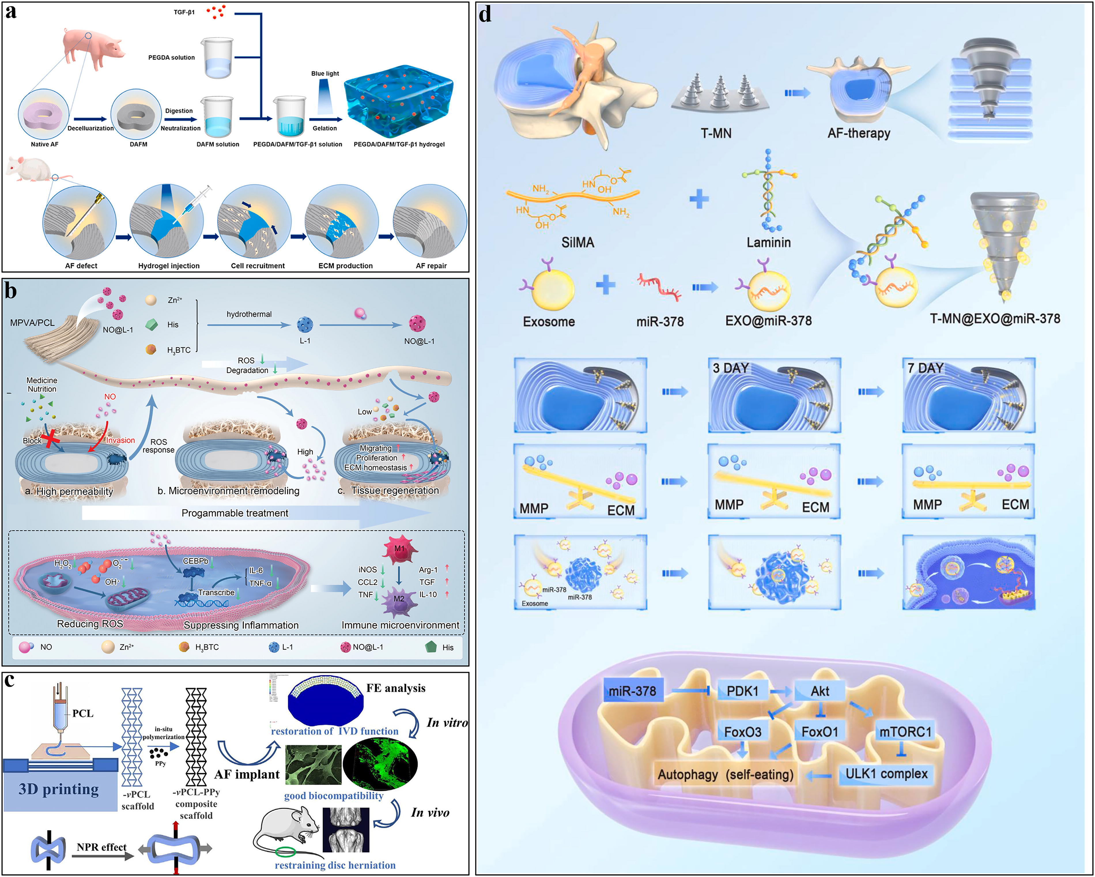
Liu et al. utilized coaxial electrospinning techniques to fabricate decellularized AF matrix (DAFM)/PECUU-blended fiber scaffolds, which exhibited similar elasticity to native inner and oAF tissue. This study demonstrated that the biomimetic AF scaffold could regulate the differentiation and related gene expression of AFSCs. In vivo evaluation confirmed that the DAFM/PECUU-blended fiber scaffolds effectively repaired AF tissue defects. 49 Wei et al. introduced an injectable, photocurable hydrogel for AF repair by combining PEGDA and DAFM with the incorporation of transforming growth factor-β1 (TGF-β1). Results indicated that the PEGDA/DAFM/TGF-β1 hydrogel, which could continuously release TGF-β1, supported AF cell adhesion, proliferation, and enhanced ECM secretion. In animal models, the hydrogel integrated well with adjacent AF tissues, result in effectively sealing AF defects, preventing nucleus pulposus collapse, and partially restoring disc biomechanical properties (Fig. 4a). 41
Application of other biomaterials in AF tissue engineering.
Future perspectives of biomaterial strategies
Recent advances in hydrogel-based AF repair focus on composite and bioadhesive systems that balance biomechanical integrity with biological functionality. 60 The cutting-edge hydrogel system realizes injectable, self-healing, shape memory, and intelligent response functions through physical cross-linking (hydrogen bonding, ionic bonding, hydrophobicity), chemical cross-linking (Schiff base, Michael addition, photoinduced polymerization, enzyme catalysis), or dual-network synergy mechanisms.82,106 Dual-modified GAGs (e.g., oxidized and methacrylated HA or CS) are used to prime tissue surfaces, enabling covalent bonding of injectable IPN hydrogels composed of PEGDA and fibrin-fibronectin. These systems are delivered via dual-barrel syringes for in situ gelation, offering seamless defect filling and strong tissue integration. 57 Looking forward, the integration of hydrogels with other biomaterials holds significant promise for enhancing AF repair outcomes. Combining hydrogels with genipin-cross-linked materials could improve mechanical stability while maintaining biocompatibility. 40 Incorporating dECM components into hydrogel matrices may better replicate the native tissue microenvironment and promote cell-mediated regeneration. 41 Furthermore, the strategic combination of natural and synthetic polymers could optimize both biological functionality and mechanical performance. 107 However, challenges remain in balancing high modulus with cytocompatibility, ensuring long-term interfacial stability under physiological loading, and achieving functional integration without eliciting immune responses. There is also a need for standardized large-animal validation and clinical translation pathways.108,109 Future directions should emphasize smart hydrogel designs with dynamic bonds, multifunctional cues, and stimuli-responsive degradation to match healing timelines.77,110 Combining microengineering (e.g., MBs, electrospraying) with biofabrication and in vivo models will accelerate the development of next-generation AF repair solutions. 51
Fabrication Technologies in AF Tissue Repair
The field of AF tissue engineering has witnessed remarkable advancements through innovative technologies such as electrospinning, 3D bioprinting, and MN systems. Each approach presents distinct advantages—from creating biomimetic microenvironments to enabling minimally invasive delivery—while facing unique challenges in the replication of native tissue complexity and mechanical properties.6,111 Electrospinning and three-dimensional bioprinting allow precise construction of scaffolds with complex architectures, therefore offering unique advantages in replicating the anisotropic structure of the AF. 112 In addition, as a minimally invasive treatment platform, microneedling systems can achieve local precision drug delivery, and the further development of stimulus-responsive intelligent microneedling provides important prospects for achieving on-demand precision treatment. 29
Electrospinning
Electrospinning is a technique for producing fibers from polymer solution or melt, 113 and these fibers typically in the range from tens to hundreds are usually used to engineer scaffolds. 114 The scaffolds feature a high specific surface area and a wide gap between fibers, which can significantly promote tissue regeneration when loaded with drugs or bioactive molecules. 99 By applying different processing parameters, such as the collector, voltage, temperature, and humidity, the nanofiber diameter and pore size can be adjusted to create loose and porous tissue engineering scaffolds. 15 Compared with traditional 2D culture systems, 3D culture scaffolds avoid flat or spindle-shaped cell morphologies and provide necessary biochemical cues for cell growth and proliferation. 115 In addition, the aligned nanofibers can be deposited on the collector by introducing conductive or dielectric regions for specific biomimetic tissue strategies. 116 The proper integration of aligned scaffolds and surrounding healthy tissue not only promotes cell phenotypic changes and gene expression but also improves mechanical properties. 117
Different materials have different effects on AF scaffolds. Scaffolds made by composite materials such as poly(
3D bioprinting carrier
3D bioprinting has emerged as a pivotal approach in bone tissue engineering and offers significant advantages for AF repair due to its precise and reproducible manufacturing capabilities. 121 There are various 3D printing technologies like gas foaming, fused deposition modeling, and bioprinting, extensively utilized to fabricate biological scaffolds with highly accurate biomimetic constructs. 122 The commonly used 3D-printed thermoplastic polymers have excellent biocompatibility, biodegradability, and biofunctionality, and when combined with bioactive molecules or drugs, they could enable enhanced performance for wide applications. 123
Jiang et al. designed a polypyrrole (PPy)-coated PCL scaffold with a negative Poisson’s ratio (NPR) structure through 3D printing technology to treat lumbar IVD herniation. They found that the NPR composite in the role of AF implant could withstand spinal load, resist the expansion of NP, and even exert a reacting force to inhibit NP herniation. This study confirmed that 3D-printed biomaterial scaffolds have good mechanical properties and biocompatibility, demonstrating potential to alleviate IDD (Fig. 4c). 1 Moxon et al. successfully developed an artificial IVD construct with precise AF architecture and NP-AF interfaces using suspended hydrogel bioprinting system (SLAM). This model naturally recapitulated the microstructural features of AF tissue regions and facilitated the incorporation of NP and AF cells, revealing the potential of SLAM bioprinting to control stem cell phenotype and morphology by modulating the extracellular environment’s structure. These findings provide a novel idea for studying the formation mechanism of IVD-like tissue, and it is expected to expand the application of IVD bioprinting to a wider range of tissue engineering and regenerative medicine. 50
Microneedles
MNs, a new type of localized and minimally invasive device for drug delivery, consist of a substrate (area from mm2 to cm2) and micron-sized projections with a height of 100–1000 µm in length.124,125 MNs can minimally penetrate the tissue barriers, avoiding pain or inflammatory responses, which cannot be achieved by traditional drug delivery methods, thereby enhancing drug bioavailability and patient compliance. 120 In the field of regenerative medicine, MNs can be designed for transdermal delivery of bioactive agents or as scaffolds for in situ tissue regeneration to improve the healing of damaged tissues. 72 Furthermore, the MNs’ devices can be engineered to respond to endogenous or exogenous stimulation, including pH, ROS, enzymes, light, temperature, or mechanical forces, allowing controlled release of active compounds within the epidermis and dermis. 40
Hu et al. reported a thread-structured microneedle (T-MN) loaded with BMSC-derived exosomes. The thread-like structure was designed to biologically match the hierarchical AF structure, thus providing stronger bonding. This composite MN system effectively adhered to the AF and slowly released therapeutic exosomes, which can prevent the progression of IVDD by promoting AF cell proliferation and migration and inhibiting pathological remodeling of ECM (Fig. 4d). 29 Meng et al. developed high-strength smart MNs with “offensive and defensive” effects for AF repair. These MNs can minimally penetrate AF tissue through localized delivery and achieve remote-controlled of the drug accelerated release and photothermal treatment under Near Infrared. The composite MNs, composed of polydopamine/gelatin methacryloyl (PDA/GelMA) and loaded with diclofenac sodium, are designed to “offend” the inflammatory microenvironment extracellularly (by alleviating cellular damage) and “defend” intracellularly (by upregulating cytoprotective heat shock proteins). This study demonstrated that the MNs effectively reduced inflammation, promoted ECM deposition, suppressed apoptosis, and restored the IVD biomechanical properties . This new “offensive and defensive” strategy provides a new view for repairing AF tissue and preventing recurrent IVD herniation. 48
Others
In addition to the common biomaterials mentioned above, there are a number of other materials that are used to repair AF. Zhou et al. evaluated CCL5-loaded fibrin gel for AF repair. In vitro, CCL5 exhibited dose-dependent chemotaxis on AF cells without altering catabolic gene expression. Fibrin gel enabled sustained CCL5 release, but organ culture (bovine discs) and sheep models showed no AF cell migration or repair improvement. While fibrin gel offered biocompatibility and controlled delivery, its dense structure likely hindered cell integration. Findings highlight the need for biomaterials enhancing cell motility alongside chemotactic agents for effective AF regeneration. 126 Ryan et al. developed an angle-ply collagen scaffold using decellularized porcine pericardium to mimic the native AF microarchitecture. The scaffold supported adipose-derived mesenchymal stromal cells (AD-MSCs) and AF cells, promoting viability, elongation, and alignment along collagen fibers. Gene expression of AF-specific markers (e.g., collagen types 1, 5, 12) was upregulated, and biaxial mechanical properties resembled native AF tissue. Histology confirmed new collagen deposition. Advantages include biomimetic structure, cell guidance, and mechanical reinforcement, offering potential for AF repair and prevention of disc reherniation. 18 Mohammad et al. utilized an absorbable poly(glycolic acid)-hyaluronan (PGA-HA) scaffold to repair ovine AF defects. Through partial AF resection in sheep lumbar discs (n = 10), experimental groups received PGA-HA implantation fixed via quadrant-suture technique, while controls remained untreated. Histological analysis at 3 months revealed the PGA-HA group demonstrated superior tissue regeneration with enhanced proteoglycan deposition (p < 0.01), organized lamellar structure (p < 0.05), and absence of degenerative features (cysts/necrosis) seen in controls. The scaffold’s complete biodegradability eliminates implant migration risks, while HA-mediated bioactivity promotes endogenous cell recruitment and matrix restoration, showing potential for preventing postdiscectomy degeneration. 127
Conclusion
The AF is a multilayered collagenous structure that resists radial pressure and distributes loads, playing a key role in spinal biomechanics. However, AF defects from herniation or degeneration pose a challenge due to its limited self-repair capacity. Biomaterial-based strategies offer solutions by providing structural reinforcement, bioactive molecule delivery, and microenvironment modulation for AF regeneration. These include natural and synthetic hydrogels, electrospun scaffolds, decellularized ECM, and advanced technologies like 3D bioprinting and MNs. Natural hydrogels promote cell adhesion, while synthetic ones provide tunable properties. Electrospun scaffolds enhance cell alignment, and DAFM scaffolds support endogenous regeneration. Innovations like 3D-printed structures and ROS-responsive MNs show potential for sealing defects and preventing reherniation. Successful AF repair requires balancing biocompatibility, biodegradability, mechanical strength, and bioactivity. Hybrid strategies combining synthetic polymers with natural components or bioactive agents have enhanced outcomes. These materials are expected to address current treatment limitations, improve patient quality of life, and drive a paradigm shift in managing AF rupture.
Limitation and Prospects
Despite progress, the clinical translation of AF repair biomaterials faces several key challenges. Most studies are preclinical and rely on small animal models that inadequately replicate human spinal biomechanics, while large-animal models and human explant studies are limited. Material delivery is another hurdle: injectable hydrogels, though minimally invasive, may compromise AF integrity during injection or fail to adhere sufficiently under dynamic spinal loads. Stiffer materials with better mechanical strength often require invasive implantation, and photocurable hydrogels face issues with light penetration and spatial control in deep spinal tissues. The inflammatory and oxidative microenvironment of degenerated AF further complicates material performance, with ROS-responsive systems and antioxidant-loaded scaffolds showing promise but lacking long-term stability and immunomodulatory data. Additionally, biomaterials designed to mimic AF’s layered structure often lack microscale organization, limiting integration with native tissue. Finally, current research inadequately addresses clinical endpoints such as pain relief and functional restoration, focusing instead on structural repair and biomechanical metrics. Standardized protocols for assessing AF regeneration are urgently needed. Addressing these limitations will require interdisciplinary collaboration, advanced fabrication technologies, and robust preclinical validation.41,60,128
The future of AF repair lies in the development of integrated, biomimetic, and multifunctional biomaterial systems. These systems will not only provide biomechanical reinforcement and seamless tissue integration but also foster a regenerative microenvironment through controlled delivery of cells and bioactive factors. Importantly, such innovations hold significant translational potential beyond AF repair, particularly for other fibrocartilaginous tissues. For instance, the cartilaginous endplate (CEP) and AF form an integrated anatomical unit, with the CEP providing critical nutrient supply to the avascular disc while the AF maintains structural containment. 27 This structural–functional synergy necessitates future biomaterial strategies that simultaneously address CEP permeability and AF integrity. Advanced solutions may include bilayered scaffolds or composite systems featuring bioactive gradients to support both metabolic exchange and mechanical stabilization, representing the next frontier in holistic disc regeneration.
Authors’ Contributions
Z.W., J.L., and J.C. conceived and designed the study, secured the funding, and supervised the entire project. Y.W. and Y.S. conducted data visualization, drafted the initial article, and coordinated revisions. C.Z. contributed to visual design and technical validation. T.Y. critically reviewed the article, provided methodological feedback, and improved data interpretation. All authors (Y.W., Y.S., C.Z., T.Y., Z.W., J.L., and J.C.) participated in article proofreading, final approval of the content, and ensured academic integrity. Z.W. and J.L. additionally acted as guarantors of the research.
Footnotes
Acknowledgments
The authors would like to thank the National Natural Science Foundation of China (82372401) and the Beijing Natural Science Foundation (L202033).
Funding Information
This work was financially supported by the Beijing Natural Science Foundation (L202033).
Disclosure Statement
The authors declare no potential conflicts of interest concerning the research, authorship, or publication of this article.