
Abstract
The dynamics of cell biology have always been an active area of research. To visualize and quantify this complex cellular process in vivo, we need optics with a high spatiotemporal resolution. The advancement in optics and image acquisition techniques has revolutionized the field of microscopy. Light-sheet fluorescence microscopy is one of the most advanced imaging tools, which offers a good spatiotemporal resolution, fast imaging, and less phototoxicity to a sample when compared with conventional microscopy techniques. Cell culture techniques have evolved from traditional two-dimensional planar cultures to three-dimensional cultures in the form of spheroids. Spheroid culture truly mimics physiological conditions due to better cell-to-cell and cell-to-matrix interactions within the spheroids. Spheroids have been extensively studied as a model for drug screening, cancer biology, and regenerative medicine. However, the opacity of the core within spheroids restricts its imaging through conventional microscopy. Light-sheet fluorescence microscopy proves to be an effective tool to overcome this problem, as it provides a suitable combination of deep penetration with an ultralow intensity of excitation light, thereby reducing the photobleaching of spheroids. Over the period of years, the light-sheet microscopy technique underwent many modifications, such as adaptive optics and the integration of artificial intelligence and machine learning modules based on its design and applications. Therefore, the present review will focus on the development of the light-sheet microscopy technique, its advancements, application for spheroid imaging, and will also explore the futuristic development trajectory for this technique.
Impact Statement
The present review brings together the development of light-sheet fluorescence microscopy (LSFM) from its early origin to recent advancements and places them in the context of biomedical research and spheroid biology. Unlike conventional optical approaches that struggle with limited depth and photobleaching, LSFM enables the prolonged, high-resolution imaging of live tissues, with minimum interference. These advancements had direct relevance for cancer cell biology, drug testing, and regenerative medicine. Hence, the present review strengthens the link between imaging technology and translational research, helping to refine and accelerate discoveries in the field of cell biology and therapeutic science.
Keywords
Introduction
The study of growth and development has been an active field of research in biomedical science. Classical microscopy has shed light on the dynamics of cell growth and division, cell migration, cellular organelles, and cytoskeleton to date. The quest to visualize live organisms/tissues demands a high spatiotemporal resolution and remains a challenge among researchers. 1 The introduction of fluorescence markers, such as green fluorescent protein (GFP), enabled the labeling of these cells, thus rendering the imaging of entire live specimens and cell structures possible through fluorescence microscopy. 2 This brought a paradigm shift from the static microscopy of fixed tissues to studying live tissues and labeled structures in in vivo conditions. In addition, the coupling of a multipixel camera and computer to a fluorescence microscope enabled researchers to quickly capture, store, and analyze a large amount of data. 3 However, despite advancements in both the field of optics and cell labeling dyes, the live imaging of cellular structures is still challenging. 4 The significant scattering of visible light, phototoxicity, and photobleaching of dyes give a short time for imaging, hence making it difficult to analyze the tissue in three dimensions. 5
The introduction of light-sheet fluorescence microscopy (LSFM) techniques rendered a fast temporal resolution and spatial resolution, which are required to study the complex cellular structure. 6 This technique uses a plane of light to create optical sections of tissues, therefore assisting researchers to study the deeper structure with minimum phototoxicity and photobleaching. 5 Three-dimensional (3D) cell cultures in the form of multicellular spheroids mimic the physiology of the organism and have been extensively studied in drug research and tumor biology. These spheroids contain different morphological zones within them, which are responsible for the varied cellular response. 7 Therefore, these spheroid models are more relevant culture techniques and offer good predictability and reliability for preclinical studies. 8 The implementation of light-sheet microscopy provides researchers true optical sectioning of a dense specimen in in vivo conditions and allows the reconstruction of 3D information, which is difficult to attain from traditional histological sectioning. 9
In the present review, we will be discussing about the development of LSFM technology, its advantages over other microscopy techniques, advancement in LSFM, and its application in 3D spheroid imaging.
Evolution of Light-Sheet Fluorescence Microscopy
The first documented work on light-sheet microscopy was in 1912 by a German chemist, Richard Zsigmondy, and a physicist, Henry Siedentopf, while working on a gold nanoparticle colloid. 10 To gain high-contrast images, they introduced sunlight perpendicular to the observation axis into the microscope and observed the light scattered by nanoparticles in the line of observation. Although all the observations were made by eyes and no recording technique was present at that time, this technique was known as ultramicroscopy. 11 In the 1990s, with the advancement in technology, sunlight was replaced by lasers and human eyes with a mounted charge-coupled device (CCD) camera, which laid the foundation of modern-day light-sheet microscopy. 10 Embryonic development studies done by Huishen et al. (2004) (Drosophila) and Keller et al. (2008) (zebrafish) paved the path for different variations in the technique for light-sheet microscopy.12,13 The cylindrical lens, which was used to create a single light sheet (selective plane illumination microscopy [SPIM]), 12 was replaced by 1D laser scanning, which rapidly created thin optical sections (digitally scanned laser light-sheet fluorescence microscopy [DSLM]). 13 The incoherent illumination of DSLM also overcame the shortcoming of static light-sheet microscopy, which suffers from shadowing artifacts due to interference with incoherent light. 14 However, in the DSLM technique, the image contrast was lost due to the excessive scattering of light and poor light penetration inside large multicellular organisms. Two modifications were introduced in the DSLM technique: the use of a structure illumination pattern and modulation in laser power (pulsed infrared laser) scanning.15,16 This allowed to reconstruct 3D images of deeper tissue depth and eliminated scattered fluorescent light. However, these techniques could not stand the test of time, as the time of imaging was long and image quality was compromised in sections farther from illumination and objective detectors. 17 An effective solution to this was simultaneous multiview imaging (SiMView) and multiview selective plane illumination microscopy (MuVi-SPIM).18,19 This technique consists of two illuminating arms that illuminate the samples from opposite sides at the same time and two detection arms that acquire images from two opposite sides. This increased the quality and volume of the image to many folds compared with the traditional single-view imaging system. In addition, another strategy for fast and high-contrast imaging was the use of beam shaping to generate a thin bean (the Gaussian beam in conventional DSLM was replaced by the Bessel beam).20,21 The use of the Bessel beam improves the resolution but leads to photobleaching and photodamage, thus reducing the time of imaging for live samples. Chen et al. (2014) introduced the concept of replacing the Bessel beam with an optical lattice that rapidly illuminates the entire plane within the sample. 22 This, thereby, reduces the local peak power density needed to obtain an image of the same illumination. The present LSFM technique relies on multiview imaging, where the lateral resolution is determined by the numerical aperture of the detection lens and the axial resolution is dependent on the thickness of the light sheet. 17
Principle of Light-Sheet Fluorescence Microscopy
Device configuration
The basic concept behind LSFM is that a sheet of light from a laser source is directed through a cylindrical lens to hit and illuminate the sample. The sample could be either labeled with a fluorescent dye or genetically modified to express fluorescence. The second objective lens, which is placed perpendicular to the excitation sheet/plane, captures the optical slice.12,23 This collected light is imaged and processed with the help of a camera. Sample mounting plays a crucial role in the quality of an image. The commonly used objectives are water immersion lenses, and the sample is mounted in agarose or a fluorinated ethylene propylene tube, which has a refractive index close to water but could lead to the scattering of light.24,25 The rotation of the sample is also considered an effective way for Z-sectioning, as it helps the light sheet to be in alignment with the camera. 26 The basic LSFM setup configuration underwent many changes, which will be discussed in the later part of the review.
Illumination system
A light sheet can be generated in two ways: the first, traditional approach involves focusing the excitation light using a cylindrical lens (SPIM) or scanning the light beam along the focal plane of the detection lens (DSLM). 27 (Fig. 1 schematic illustrate few common types of designs). To capture a high-contrast image and to reduce light scattering, researchers have done various modifications in the illumination techniques. One technique to improve the excitation is to direct the beam such that it overlaps with the center of the imaging lens. 29 This is known as multiple-beam SPIM. The samples are illuminated from alternating sides, and the data are fused to a single output image. The application of a fusion algorithm provides an effective 3D image with an isotropic resolution. 30 This reduces the shadowing effect on the image; however, it is a challenge to optimize two counter beams to a coplanar focal plane in this technique. The DSLM technique is a step ahead of this, as it uses a resonant scanner to move a thin beam of light vertically across the sample. 13 This improves the quality of the image by providing uniform illumination across the sample. Another variation commonly used in DSLM is HiLo imaging. 31 In this, the first image is captured using normal DSLM protocols, and the second image is captured using a series of excited and nonexcited lines to produce a grid. The data from these “Hi” and “Lo” images are combined to get an image and optical section of 3D samples. However, the raw data generated are too large and demand a lot of time to process.
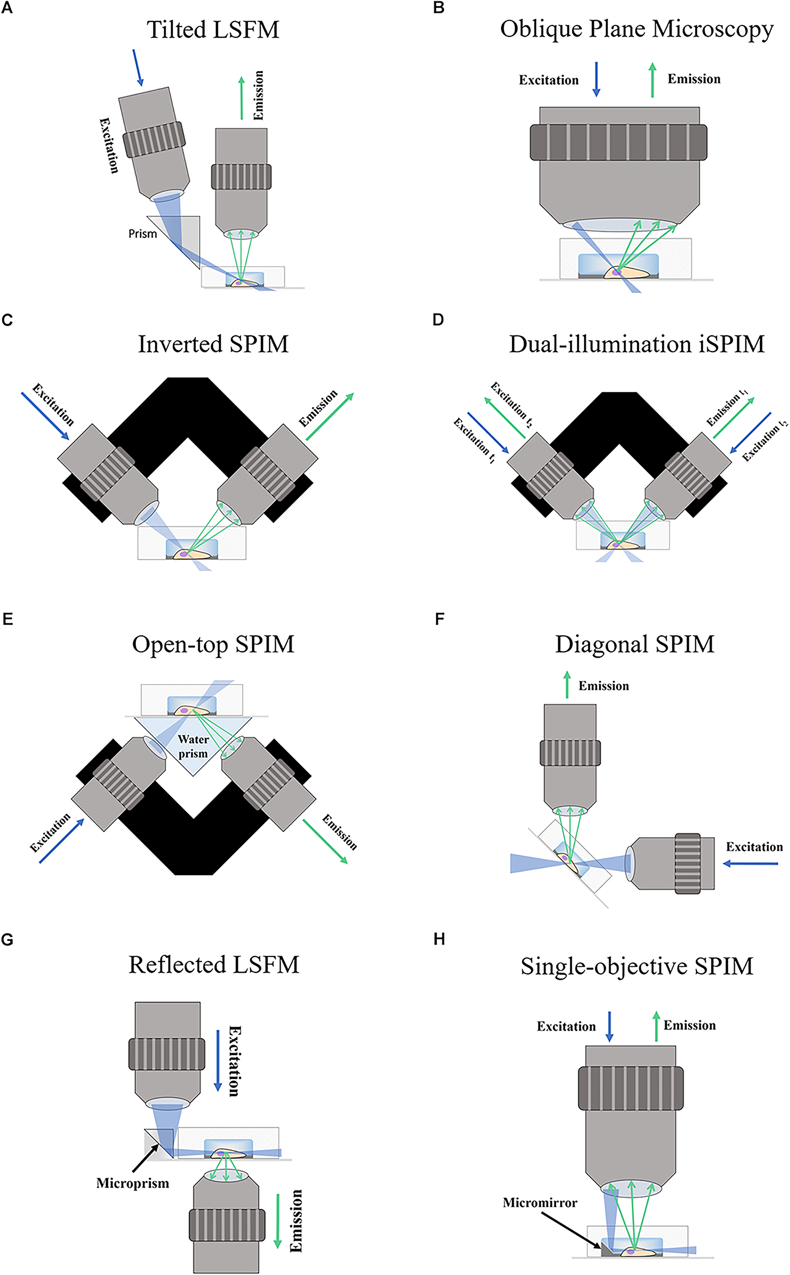
Schematic of fluorescence light-sheet microscopy techniques. The figure shows illustrations of the different LSFM techniques for imaging in microdevices.
Image detection
The traditional SPIM system in 2004 used a CCD camera for imaging. 32 With the introduction of the complementary metal–oxide–semiconductor (CMOS) camera, it is now possible to collect only that light that is confocal with the detector system and light source. 33 This is known as the rolling shutter effect, as it removes the light outside the line of excitation and enhances image quality. The detection of the image is based on the use of specific filters before fluorescence hits the camera. However, even the scattered and blocked lights too carry some information about the samples. Therefore, if the scattering is inelastic in nature, the Raman spectrum is used to study the chemical composition of 3D structures, while elastic scattering (no photon absorption) gives information about the structure of the sample and can be used for long-term monitoring.34,35 This hyperspectral detection based on the Raman signal can produce label-free images but requires a long image acquisition time, making it impractical for studies. 6
Adaptive optics is another common and widely used imaging technique to improve image quality. 36 It consists of a moving optical element (in the imaging arm of LSFM) that compensates for the aberrations produced by the optics of the sample itself. 37 These aberrations can be compensated either by directly measuring the guide star 38 or by direct modeling the aberration and inverse calculation. 39
Application and software
Imaging processing software programs, such as ImageJ and Fiji, are freely available online. The OpenSPIM configuration is included as a plugin for Fiji for 3D image reconstruction.30,40 However, one of the key challenges for LSFM imaging is a large dataset, which could be around 2 TB for a single imaging session. 41 Therefore, new ways for capturing and processing the data are needed to make the storage and processing of raw images easy and simple. 42 The variation of OpenSPIM had been introduced to address these issues, and another open-source package, namely ClearVolume, is also available for multichannel 3D visualization of the datasets.43,44
Light-Sheet Fluorescence Microscopy: A Revolution in 3D Imaging
Advantages over current imaging systems
The imaging of complex 3D structures using conventional microscopy techniques could have many challenges that restrict the quality and quantity of data. The conventional process of sectioning the 3D structures and later stitching the images together for analysis not only disrupts the sample’s morphology but also is a time-consuming procedure. Therefore, the reconstituted image could not be a real representative of complex and heterogenous 3D structures. 45 Confocal laser scanning microscopy (CLSFM) addressed these issues by virtual sectioning and stacking the optical sections to render a 3D reconstruction.46,47 However, the penetration/imaging depth is restricted to 60–80 microns.46,48 In the CLSFM technique, the excited and emitted light passes through the 3D sample, perpendicular to the image plane, thus producing light scattering. 49 The heterogeneity and complexity of cellular components within the samples add to the scattering of light and degrade the fluorescence quality. 50 Therefore, the output fluorescence quality decays exponentially with increases in the depth of the sample, making CLSFM less effective in imaging deeper tissue structures. 51 CLSFM worked well in the case of fixed samples, but image quality was insufficient for live sample imaging (Figs. 2 and 3 show the imaging depth and quality by CLSFM and LSFM for a spheroid sample). This limitation was solved using multiphoton microscopy, but it might result in photobleaching and phototoxicity of the samples in the excitation focal plane. 53 LSFM emerged to be a suitable technique for imaging heterogenous live 3D tissues.13,54 This technique supports high-speed imaging and reduces out-of-focus light, as the detection lens is placed perpendicular to the light plane, thus improving the lateral resolution. 27 LSFM can be used for optical sectioning and Z-stacking, for the 3D reconstruction of tissue models with minimal photodamage, 13 and has been widely used to study the morphology and drug penetration within spheroids.55–58
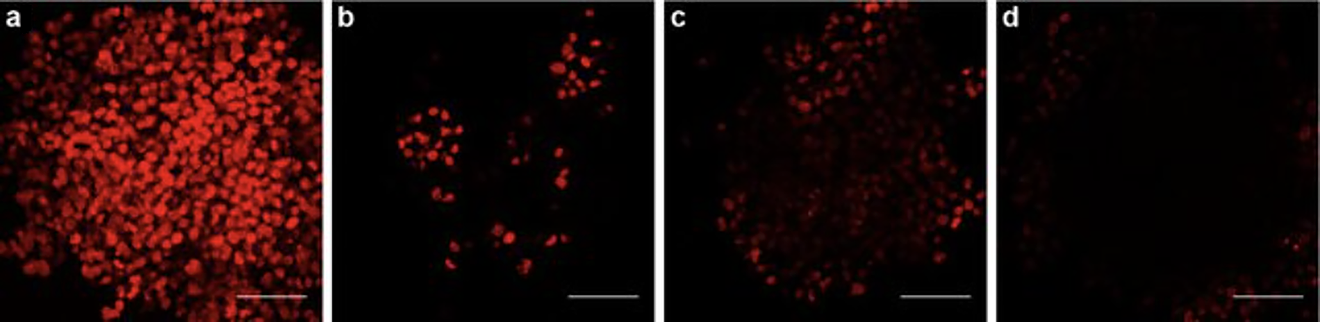
Confocal imaging of multicellular tumor spheroids (MCTS) after incubation with doxorubicin (20 μM) for 3 h at 37°C.
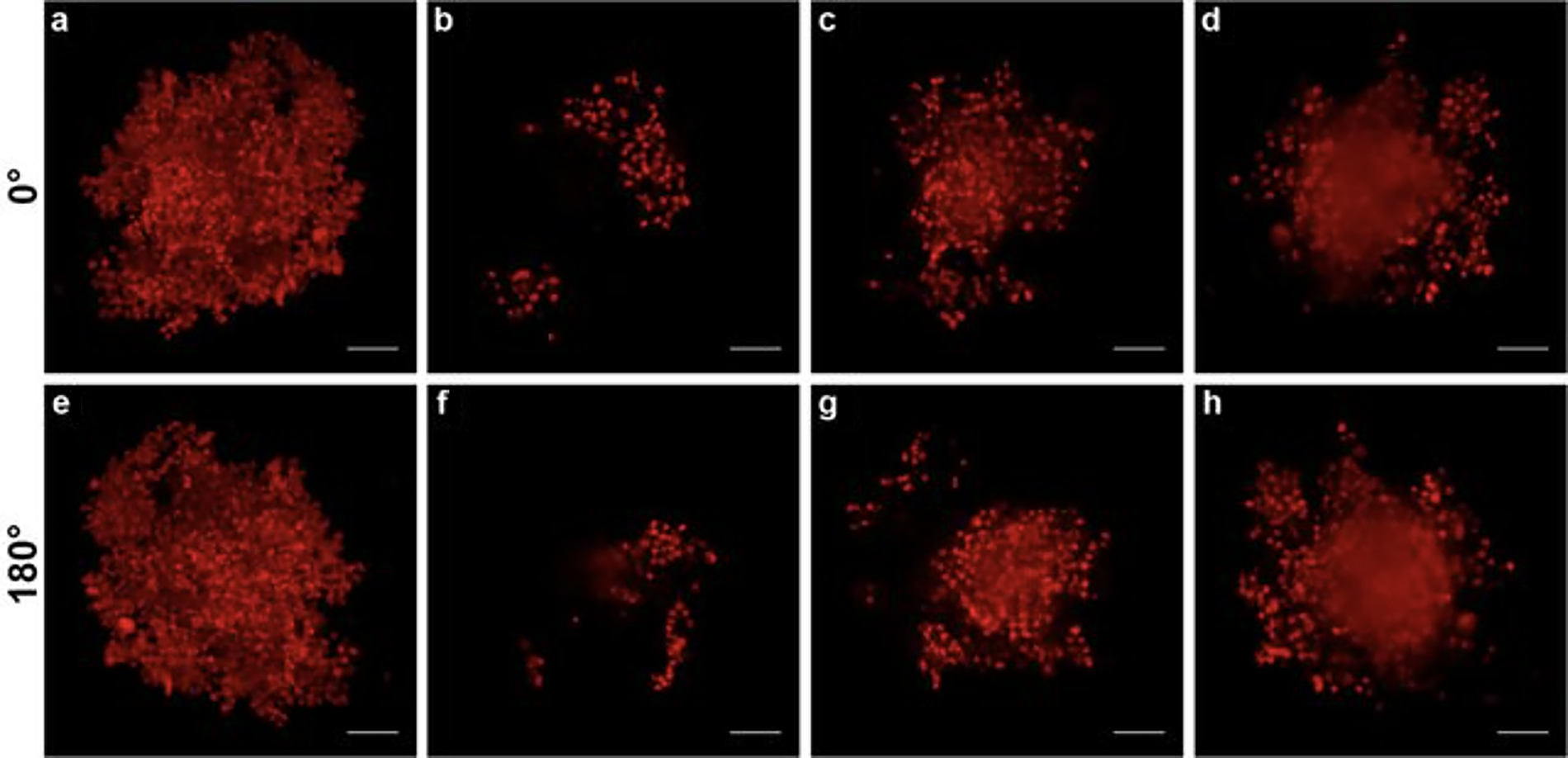
LSFM imaging of MCTS after incubation with doxorubicin (20 μM, 3 h, 37°C). Images from the Z-stack recorded at 0°:
Recent advancements in light-sheet fluorescence microscopy imaging
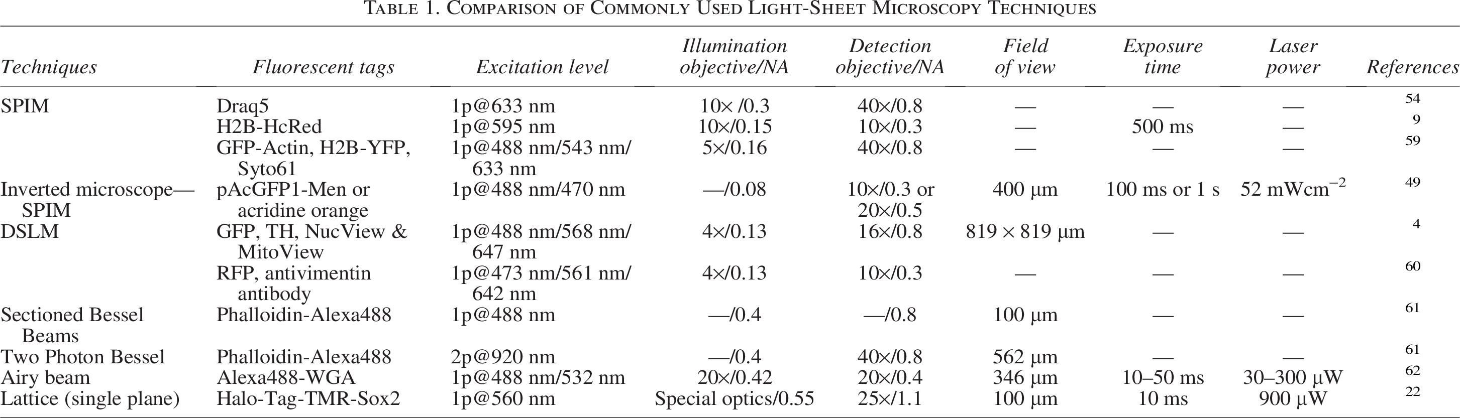
Since the inception of the LSFM technique, it underwent various customizations, depending upon the application, to push the limits of the present technology (Table 1). The cylindrical lens was commonly used to create a light sheet in the SPIM technique. 57 However, illuminating the plane from one side creates refraction and scattering of light within the tissue sample, leading to striped artifacts. The incidence of artifacts is proportional to tissue optical density. This was overcome by the introduction of multidirectional SPIM (mSPIM), thus reducing the artifacts. 58 DSLM is another variant using a Gaussian beam light sheet for scanning. 12 In opaque samples, DSLM reduces the striped artifacts more efficiently than the SPIM technique. The tussle between high resolution and illumination in LSFM led researchers to design many modifications based on their application. 29 One such type of model is a molecule localization-selective plane illumination microscope (IML-SPIM) with the lens of numerical aperture (NA)-high NAdet (1.1) limiting NAill (0.3). 13 This technique helped in achieving axial localization, but a thick light sheet limited its optical sectioning capacity.63,64 Gebhardt et al. addressed this limitation by using a pair of vertically opposed objective lenses and a 45° mirrored to redirect a thin light sheet. The lens he used was of high NA (NAdet of 1.4 in oil). 64 Another challenge in LSFM was to maintain high axial resolution across the entire field of view (FOV). The isotropic resolution could only be achieved if the light sheet is within axial confinement. 65 The Gaussian light sheet has a property to spread away from focus.
Comparison of Commonly Used Light-Sheet Microscopy Techniques
The most commonly accepted alternative to the Gaussian beam was the use of the Bessel beam. This beam consists of a narrow core surrounded by concentric rings of diminished density (Fig. 4).
67
Planchon et al. used Bessel beams in his model of LSFM with NA (0.8) in both the illumination and detection pathways to capture an isotropic resolution over a FOV spanning 40 microns.
19
The linear Bessel beam caused interference effects, and suppressing its concentric rings allowed the formation of an optical lattice. This observation leads to the conceptualization of lattice light-sheet microscopy (LLSFM), which can generate ultra-thin light sheets in a parallel manner.
13
LLSFM was paired with a high-NA detection lens (NAdet 1.1) and a fast scientific complementary metal-oxide semiconductor (sCMOS) camera, which together produced better imaging performance than traditional Bessel-beam–based techniques. In recent years, the most commonly used LSFM variant is low-NA light sheets, which have the aberrations of striped artifacts. The two-photon (2p) analogue was a substitute to this technique, as the long wavelengths are weakly scattered. This was adopted by the DSLM model later, thus providing users with many options to choose from based on their application, cost, and benefits.19,68
Gaussian and Bessel beams for light sheet generation. 
The efficacy of the current LSFM can be summarized as
69
On the basis of penetration: 1p-DSLM < 2p-DSLM < 2p-LSFM On the basis of the imaging rate: 2p-LSFM ≈ 2p-DSLM < 1p-DSLM
Application for 3D Spheroid Imaging
3D spheroid cultures and their application
The study of cell–cell interactions lay the foundation for the knowledge of cell physiology and its metabolism. 70 However, the traditional two-dimensional (2D) culture usually done on flat plastic surfaces does not mimic the three-dimensional (3D) physiological conditions. It lacks basic tissue architecture and cell-to-cell and cell–extracellular environment interactions. Therefore, to bridge the gap between laboratory culture conditions and real physiological environments, 3D cultures came to light. 71 An overview of the literature reveals an increase in the number of publications over the last decade using 3D cell cultures; either cells are embedded in a matrix or clustered to form cellular spheroids.72–75 Cellular spheroids are cell aggregates that can be obtained by techniques such as hanging drop and spinner flask method.76,77 Spheroids are easy to handle and relatively lack the complexity of the whole organism, which makes them a system of choice for biomedical and tissue engineering studies. 5 To study the specific cell-to-cell interactions and cell dynamics, cocultured spheroids came into light, which mimic in vivo conditions. 78 Currently, spheroids derived from cancer cell lines are used to test antineoplastic drugs and study cellular pathways, and their responses differ from those seen in conventional planar cultures.73,76,79–81 In the field of regenerative medicine, spheroid cultures play an important role, as they are easy to handle and inoculate compared with single cells/2D culture. Spheroids derived from mesenchymal stem cells (MSCs) of human origin show high expression of tumor necrosis factor and antineoplastic markers, such as interleukin 24, compared with 2D cultures. 82 Studies also demonstrated that MSCs cultured as spheroids enhance the osteogenic and adipogenic potential compared with 2D cell cultures.83,84
Limitations of current 3D spheroid imaging techniques
To study cell morphology and their interactions within spheroids, histological sectioning followed by fluorescence staining is commonly used. However, it has its drawbacks, such as rupture/deformation of spheroids while sectioning, poor contrast of traditional stains/dyes, and low spatial resolution. 85 Biochemical analysis is another method used to study cell viability and proliferation with spheroids. 86 However, both these techniques are inefficient for evaluating the dynamics within spheroids. The introduction of fluorescence microscopy proved to be an excellent tool for the quantitative analysis of spheroid cultures.87,88 The primary requisite for a good fluorescence image is a good penetration depth within the sample, good signal:noise ratio, fast imaging with a good spatial resolution, and low phototoxicity/photobleaching. 89 In traditional wide-field microscopy, light excites the entire sample, leading to photobleaching of the sample. 90 The advent of confocal microscopy enabled optical sectioning by point excitation and detection, while the pinhole system improved the quality of the captured image. However, the major shortcoming of confocal microscopy is the limited penetration depth of the laser within samples while using a lens of high NA. 88 These limitations restrict its use to study large spheroids in in vivo conditions. 91 Multiphoton microscopy is an alternative to image spheroids, but the low axial resolution and bleaching of the focal plane limited its usage among researchers.91–93 In recent years, many other techniques such as confocal theta fluorescence, 94 4Pi-confocal, 95 stimulated emission depletion, 96 and stochastic fluorescence 97 have been introduced, and they demonstrated excellent imaging of the subcellular structures of fixed samples. However, these techniques are not suitable to image live heterogeneous spheroid samples due to the high scattering of light within the samples. 5
Light-sheet fluorescence microscopy for 3D spheroid imaging
The LSFM technique has enabled the imaging of spheroids in their culture conditions. Handling methods have been developed to mount spheroids for live imaging. 98 These holders are based on a pipette that either holds it from above or supports it from below. The correction of the light source is taken into consideration based on the orientation of spheroids. Maizel et al. demonstrated in their study that the imaging of spheroids can be done using mDSLM along with complex manipulation required for imaging. 99 An improved imaging depth and resolution had been observed by Verveer et al. when compared with confocal microscopy. 54 Smyrek et al. advocated the use of LSFM, as they studied the distribution of protein expression and cell-to-cell interaction within a whole-mount spheroid using a digital-scanned light sheet-based fluorescence microscope (mDSLM), which ruled out the need for the traditional sectioning of spheroids. 45 High-resolution multiangle LSFM is useful in preclinical drug screening and studying the pathology of breast cancer cell spheroids. 100 LSFM, when combined with the cell membrane fluorescent protein, can be used to monitor apoptosis within multicellular spheroids. Förster resonance energy transfer can be observed within spheroids by light sheet-based fluorescence microscopy in combination with microspectral analysis and fluorescence lifetime imaging (FLIM). 101 Pampaloni et al. optimized a detailed protocol for the 3D long-term live imaging of spheroids using LSFM. This protocol laid the foundation for studies monitoring cell proliferation, aggregation, and compaction within spheroids as well as can be used to study the effect of drugs or chemicals on multicellular spheroids. 102 Similar studies were conducted by Lorenzo et al. and Deesmaison et al. using LSFM for the live 3D analysis of cell division and cell orientation within tumor cell spheroids.9,103 Therefore, based on current literature, LSFM appears to be an effective tool for studying live-cell dynamics, cellular heterogeneity, and intercellular interactions within spheroids. Following are the strategies that could be implemented to enhance the imaging quality of 3D spheroids:
Optimization of culture conditions adapted to 3D spheroids
The quality of a 3D spheroid is an important determinant for successful volumetric imaging using LSFM.5,104 Hence, optimization begins right from the upstream starting with the sample preparation. Broadly, the characterization and optimization of spheroids should be aimed at (a) generating uniform-size spheroids to reduce biological variance, (b) control over matrix formation and stiffness to reproduce optical scattering properties, (c) optimization of labeling strategies with minimum phototoxicity to ensure cellular viability, and (d) LSFM-compatible mounting schemes.
Spheroids of uniform morphology and size are crucial for high-throughput imaging. Techniques such as controlled seeding densities, microfabricated well arrays, automated spheroid sorters and microfluidic platforms allow researchers to produce spheroids with consistent diameters and cellular packing (Fig. 5).105–109 This consistency reduces the variability in light scattering, dye percolation, and ultimately improves the volumetric datasets. Similarly, fluorescent labeling also requires careful consideration. Stable genetically encoded reporters, such as H2B-GFP for nuclear labeling, provide reliable signals with minimal biological perturbation.5,110 In the case of chemical dyes, selection should ensure uniform uptake and minimal cytotoxicity, especially during long-term time-lapse imaging. Furthermore, for live spheroid imaging, the embedding medium must maintain the physiological conditions of nutrients and gas exchange without imposing stress on spheroids. Thus, the quality of live spheroid imaging is highly dependent on the embedding medium. Mismatched refractive indices between the spheroid, hydrogel, and immersion medium increase scattering and reduce imaging contrast, particularly for deep volumes. 111 Embedding spheroids in low-autofluorescence, refractive-index-matched hydrogels or mounting media enhances optical clarity while supporting stable sample placement in LSFM chambers. With recent advancements and the integration of artificial intelligence (AI) and machine learning (ML), automated algorithms can segregate spheroids based on shape and size, thereby significantly improving imaging outcomes (Fig. 6).112,113
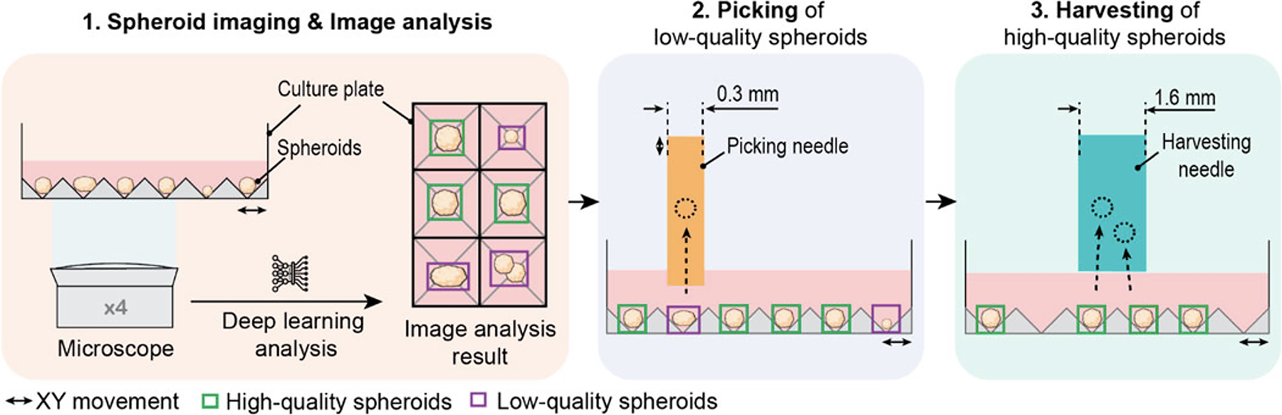
Schematic of the three-step workflow of the SpheroidSorter. The workflow of the SpheroidSorter is divided into three steps: 1. Spheroid imaging and image analysis. An inverted bright-field microscope with ×4 magnification images spheroids in the culture plate. In parallel, each spheroid is detected and analyzed by a deep learning algorithm and classified with a “low-quality” label (purple) or “high-quality” label (green). 2. Individual picking of low-quality spheroids. Spheroids labeled as low-quality are individually removed by a picking needle of 0.3-mm diameter. 3. Harvesting of high-quality spheroids. High-quality spheroids are harvested in bulk by a harvesting needle of 1.6-mm diameter. (adapted from Sampaio da Silva et al. 109 under CC BY 4.0).
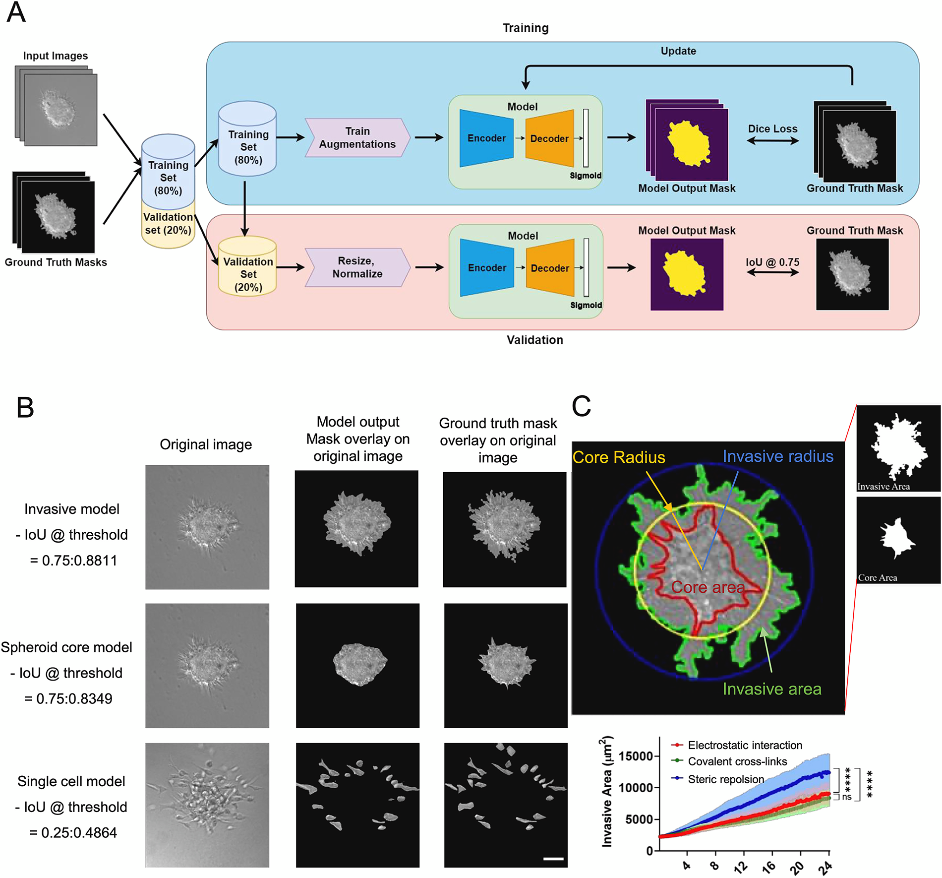
Segmentation results of the model output for invasive protrusions and core region of spheroids as well as detached single cells.
Strategies for improving imaging depth
The optical properties of spheroids include light scattering and absorption, leading to signal-to-noise ratio (SNR). Therefore, effective depth-enhancement strategies could be the combination of optical, chemical, and computational approaches. Optically, two-photon LSFM leverages nonlinear excitation at longer wavelengths to confine fluorescence to the focal plane and reduce scattering. 114 This approach enables imaging deeper into the spheroid core while maintaining the SNR. Adjustments to the light sheet, including light sheet thickness, dual-sided illumination, and adaptive positioning, further improve depth penetration. 115 On the other hand, chemical strategies include additional steps, such as the optical clearing of samples to match the refractive index, which can significantly enhance transparency, especially for fixed spheroids. 12
Complementing these physical strategies, computational tools—especially those based on AI—have become increasingly valuable. Convolutional neural networks (CNNs) and generative adversarial networks (GANs) can correct depth-dependent signal attenuation, enhance contrast, and restore image details lost in deeper layers.65,116 Some systems even incorporate real-time feedback loops that adjust illumination based on tissue heterogeneity, improving both the efficiency and quality of data. Together, these techniques allow researchers to visualize internal gradients, hypoxic regions, and necrotic zones, which are critical for understanding therapeutic responses within complex 3D systems.
Improvement of imaging resolution
Conventional LSFM offers an excellent lateral resolution but a limited axial resolution due to the thickness of the excitation sheet, NA constraints, and sample drift. 117 The integration of computational advancements, such as deep learning algorithms, perform deconvolution, denoising, and aberration correction and can even generate a “virtual” super resolution volumes from lower-resolution data.118,119 U-Net architectures are particularly effective for segmenting nuclei and cellular boundaries in 3D datasets, while GANs have demonstrated the ability to upscale image resolution and recover subcellular detail, even in dense tissue regions (Fig. 7).120,121 When combined, these tools transform LSFM from a primarily qualitative imaging approach into a robust, quantitative platform capable of mapping cellular processes in real time.
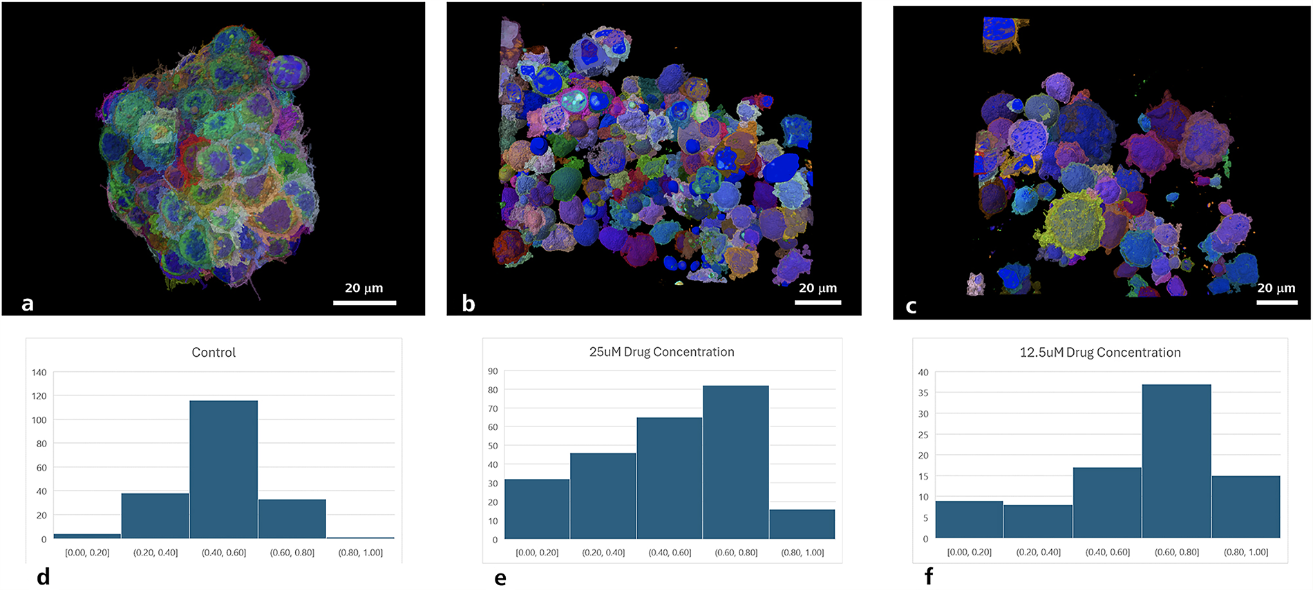
AI-based analysis of the drug response in 3D ovarian cancer cultures. Top: AI-aided segmentation of individual cells in 3D matrix cultures at different drug concentrations (cisplatin) showing cellular organization and morphology. The 3D cultures were stained for Hoechst to identify individual cells through their nuclei and phalloidin to detect the cell cytoskeleton and thus the cell boundary. Bottom: Quantitative analysis showing the effect of the drug concentration on nucleus-to-cell volume ratios. Left to right: control, 25 µM drug concentration and 12.5 µM drug concentration, demonstrating dose-dependent changes in cellular architecture (adapted from Bhattiprolu et al. 121 under CC BY 4.0). AI, artificial intelligence; 3D, three dimensional.
Optimization of in vivo imaging speed
The temporal resolution is crucial for observing dynamic biological processes within 3D spheroids. This demands for rapid volumetric imaging with minimal phototoxicity. 122 LSFM’s planar illumination and wide-field detection provide a strong foundation for high-speed imaging, but further optimization is essential for live experiments.123,124 High-speed cameras with low read noise and large fields of view enable the rapid acquisition of Z-stacks. Trade-offs between the Z-step size and the temporal resolution allow the capture of relevant dynamics while minimizing light exposure.18,125 Additionally, targeted region-of-interest scanning and adaptive acquisition strategies make data collection more efficient by focusing on the zones of biological interest. AI-driven feedback systems are increasingly being incorporated to dynamically control imaging parameters such as exposure, focus, and sheet thickness in real time. 126 These adaptive systems not only reduce photodamage but also enhance throughput and enable continuous observation over extended periods.
Integration of multimodal imaging
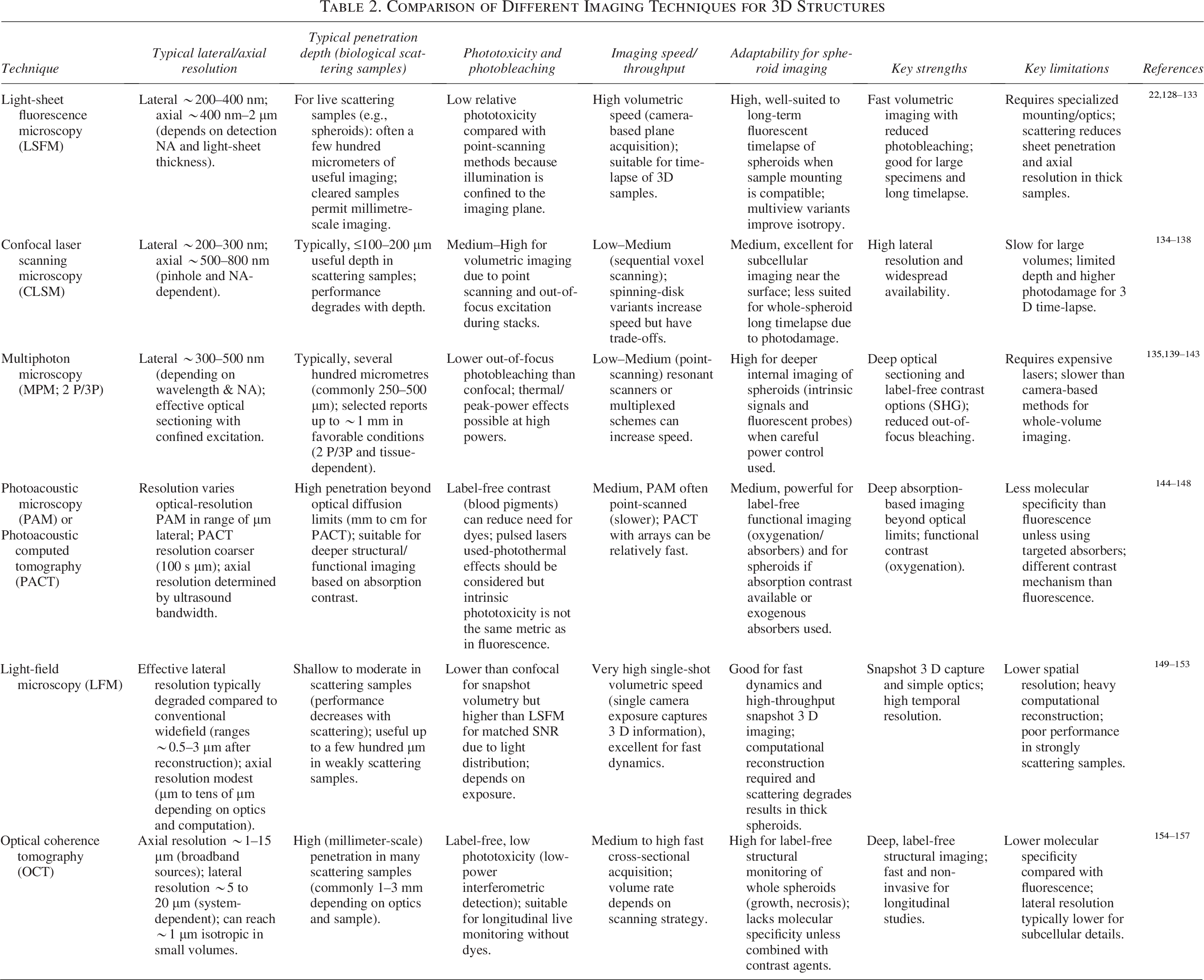
Integrating LSFM with other imaging modalities can provide complementary information that expands both structural and functional understanding. Fluorescence imaging offers molecular specificity, while label-free modalities such as optical coherence tomography (OCT) and photoacoustic imaging add structural and functional depth (Table 2). 127 OCT enables the label-free visualization of internal spheroid architecture, and photoacoustic imaging highlights absorbers such as hemoglobin, making it useful for detecting hypoxic or vascularized regions.158,159 FLIM and spectral imaging reveal insights into cellular metabolism, whereas mass spectrometry imaging provides spatially resolved molecular data.160,161 Combining these approaches with LSFM enables the coregistration of structural, functional, and chemical datasets at a high resolution. Deep learning algorithms are increasingly applied to align multimodal datasets and extract meaningful biological patterns. CNN-based frameworks can fuse OCT and fluorescent images, while AI-driven segmentation can identify phenotypic subpopulations across modalities.162,163 The advancements in deep learning algorithms have enabled the reconstruction and deconvolution of images, leading to a superior resolution (Fig. 7).121,164,165 The encoder-decoder is a powerful convolutional network that enables the capture of both high- and low-level features. 166 A popular example for this is U-Net, which is a powerful tool in image segmentation. 120 Residual Channel Attention Networks (RCAN) is an alternate model with high accuracy and has been validated for improving the reconstitution of volumetric time-lapse data. 167 RCAN was further supplemented by the incorporation of parallel connected RCAN, which enhances model efficiency and has potential to outperform CNN-based models with optimal training strategies. 168 Researchers have adapted RCAN to refine 4D super-resolution datasets, noise reduction, resolution enhancement, and acquisition of large image sequences without photobleaching. 169 However, they advocated that further refinements are needed for the neural network in terms of performance and use for imaging applications. 116 Various other models, such as the view-channel-depth neural network and Saak transform, have been developed to address the challenges with light field microscopy, such as variable resolution, artifacts, and slow reconstruction speed. These models have moderate accuracy, high speed for the reconstruction of images, and can perform efficiently with customized datasets.116,170,171 In a nutshell, AI and ML are revolutionizing LSFM by enabling a super resolution, segmentation, and speedy reconstruction of image. This will further reduce the need for heavily labeled datasets and supervised machine training. 172 Furthermore, cross-specialty collaboration in terms of innovation, software, and data sciences will further expand this field, making the advanced imaging techniques accessible to the researchers.172,173
Comparison of Different Imaging Techniques for 3D Structures
Future Prospects
Since the inception of the LSFM technique, it underwent many modifications in terms of the design, depending on the sample needs and research objectives. The integration of LSFM technology with advanced high-output culture techniques has emerged as a cutting-edge science in 3D cell culture research. 174 The combination of flow cytometry and LSFM,60,175 microfluidic devices for spheroid analysis and its 3D reconstruction, 176 and the introduction of an open-source platform for drug screening are few of the key areas of active research in 3D cultures. 177 Furthermore, the integration of 3D printing to construct a sample holder and optofluidic device for imaging spheroids paves the path for the development of compact LSFM modules, allowing the widespread use of this imaging technique in the future.176,178
Adaptative optics is a promising area to emerge and complement LSFM to reduce artifacts, aberration, and improve imaging performance. 36 The natural light-scattering properties of biological samples can be used to generate light sheets and restrict light exposure only to the targeted area within the sample.52,59,110,179–181 Researchers have come up with a spatial light modulator that can generate predefined light sheets in a different plane. This modification provides a 1.2-μm-thick light sheet at 50 μm from the fiber facet. 182 This would build scope for designing a futuristic model, which could enable in vivo endoscopic imaging at a greater depth within a biological sample.
In addition, with the inclusion of AI models and ML, future developments are expected to produced tailored options for imaging set-up designs, data acquisition, postprocessing and image reconstruction speed.128,183 Researchers, through the AutoPilot framework, have demonstrated the optimized imaging performance in living organisms. 183 Data archival, labeling, and pipeline automation are crucial, as LSFM volumes can reach terabyte scales. Cloud-based solutions and the accessibility of pertained models have solved storage issues and eliminated the bottleneck of traditional microscopy and data annotation. 121 Tools based on CNN algorithms, such as “SpheroScan,” demonstrated the power of deep learning for spheroid image analysis across multiple platforms (Fig. 8). 184 Therefore, the integration of ML/AI becomes a major axis of performance enhancement, which not only would assist image analysis post hoc but also increasingly drive acquisition, real‐time decision making, and multimodal fusion. Overall, the integration of AI gives a degree of freedom to research new configurations of LSFM with endless imaging possibilities, from the labeled to label-free region. However, it requires the thoughtful implementation of deep biological understanding, technical know-how, and rigorous validation of results by experts.
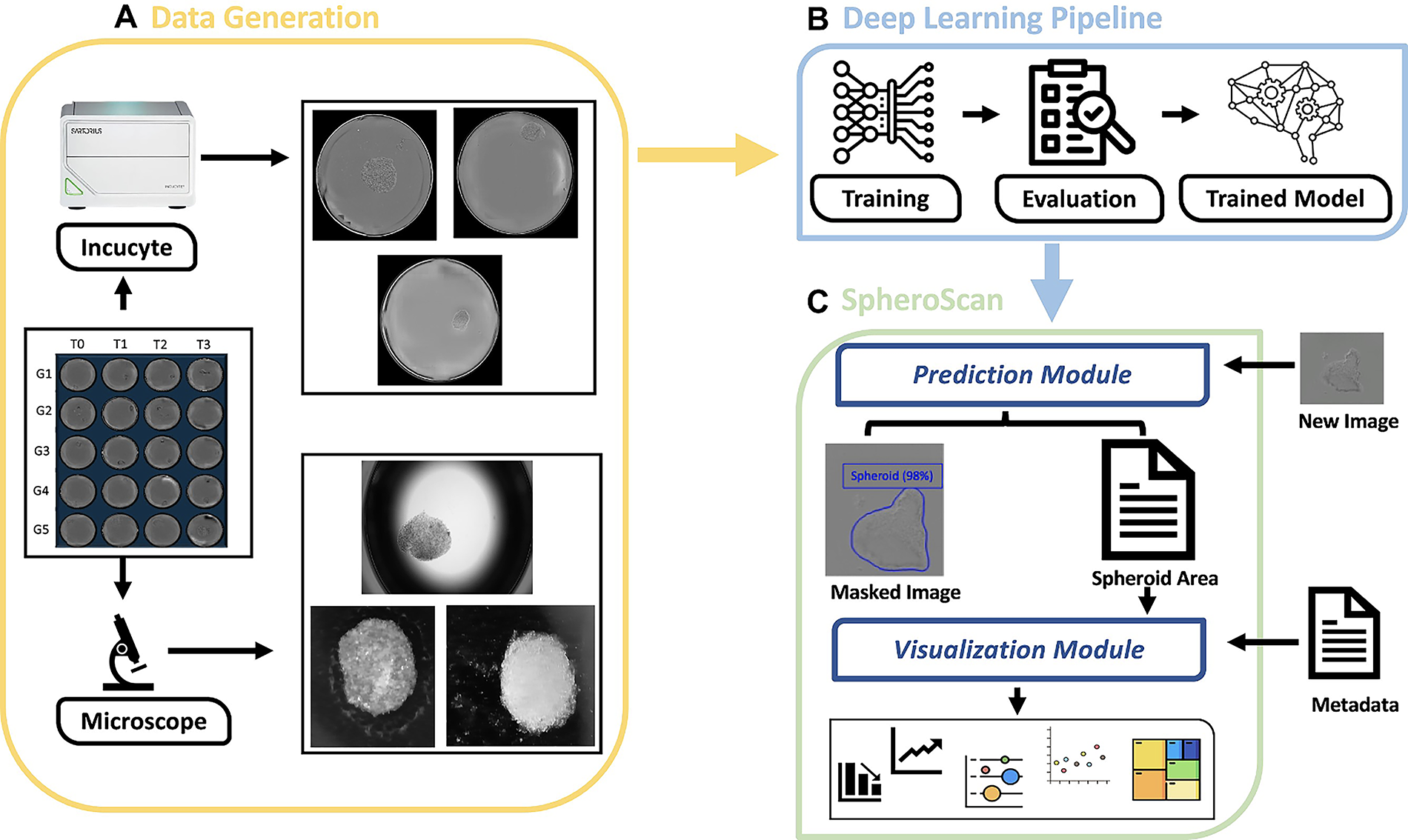
Graphical representation of the workflow for SpheroScan.
Conclusion
3D cell culture techniques and fluorescence microscopy are mainstream research areas in applied biological science. The innovations in the LSFM technique have revolutionized imaging science by its high resolution and deep penetration abilities within biological samples. The advancement in microfabrication, organ-on-chip techniques, and faster image acquisition systems aligned with modern optics will lead to the advancement in the application of LSFM in a different area of research. While significant progress has been made in resolution enhancement, adaptive control, and automated analysis, future research must address data standardization, real-time computation, and model interpretability. The development of open-access LSFM databases, cross-platform AI benchmarking, and hybrid optical–computational models will be essential for overcoming current roadblocks. As LSFM continues to evolve, its integration with deep learning and multimodal imaging will enable unprecedented insights into tissue physiology and disease modeling, paving the way for translational applications in regenerative medicine and precision oncology.
Authors’ Contributions
A.M.: Designed the study, performed the literature search, prepared the first draft of the article, and revised and edited the final version. V.A.: Designed the study and revised and edited the article. All authors agree to the final version of the article.
Footnotes
Funding Information
No funding was received for this article.
Disclosure Statement
No competing financial interests exist.