
Abstract
Biobanks serve as the cornerstone of translational research. Evolving from traditional biobanks, living biobanks—particularly organoid living biobanks—have emerged as a critical and powerful platform, characterized by their three-dimensional biomimetic architecture, long-term self-renewal capacity, and retention of key genetic and pathological phenotypes of the parental tissue. At the pivotal juncture of a paradigm shift in biomedical research, organoids, as an important component of Novel Alternative Methods, hold broad prospects for both biomedical research and clinical applications. High-quality organoids can precisely recapitulate the structure and function of native organs, ensuring the accuracy and reproducibility of research outcomes. This establishes a robust foundation for investigating disease mechanisms, drug discovery, and precision medicine. Implementing rigorous quality control is therefore pivotal for guaranteeing research reliability and clinical applicability. The present article comprehensively examines the current landscape of tumor organoid quality control, covering critical quality control checkpoints across key technical stages of the construction process, advancements in standardization, and future development trends. By synthesizing these aspects, this work aims to empower researchers and practitioners to overcome challenges in quality control, enhance organoid fidelity, and accelerate the translation of organoid technology from fundamental research to clinical implementation.
Introduction
Biobanks, serving as the cornerstone of translational research, provide the biological resources essential for understanding disease mechanisms and developing new therapies. Among these, living biobanks have emerged as a critical asset to meet the evolving demands of modern biotechnology, offering dynamic, expandable and manipulable biological systems. Tumor organoid biobanks, as a prominent form of living biobanks, are characterized by their three-dimensional (3D) biomimetic architecture, long-term self-renewal capacity, and retention of key genetic and pathological phenotypes of the parental tissue. 1 Serving not only as renewable, scalable disease modeling resources but also as precise “patient avatar” platforms, organoid biobanks play an important role in mechanistic exploration, drug development, and personalized therapy. 2 To date, organoid systems have been successfully established for multiple vital organs, including the heart, 3 liver, 4 brain, 5 lung, 6 intestine, 7 skin, 8 and stomach, 9 thereby pioneering novel pathways for disease modeling, drug development, precision medicine, and regenerative medicine. In cancer research, tumor organoids demonstrate exceptional capability to faithfully replicate in vivo tumor microarchitecture, gene expression profiles, and growth patterns. These models provide critical insights into tumor heterogeneity, metastatic mechanisms, and tumor–immune interactions, serving as powerful experimental platforms for oncology studies.10–12 In the process of drug discovery, organoids exhibit substantial advantages over conventional two-dimensional cell cultures and animal models. By more closely recapitulating human physiological and pathological states, 13 organoid systems significantly enhance the efficiency and accuracy of drug screening processes. This technological leap holds promise for shortening drug development timelines while reducing associated costs. 14
The construction process of organoids is influenced by various complex factors, such as tissue specimen acquisition, transportation and processing, cellular source characteristics, culture system composition, matrix selection, and environmental parameter regulation.10,15,16 Issues in any step may lead to significant variability in organoid morphology, functionality, and genetic fidelity. Such quality instability not only contributes to experimental discrepancies and misguides research directions, but also induces severe consequences during clinical translation and therapeutic applications. Consequently, conducting quality control (QC) research and establishing standardized and regulated QC frameworks are of paramount importance for ensuring biological consistency, enhancing methodological reproducibility, and accelerating clinical translation of organoid technologies. Starting from the critical aspects of organoid culture, this review will systematically elaborate on the strategies and technological progress of QC, while exploring key technological innovations and future development directions. The ultimate objectives are to improve the biological fidelity and experimental reproducibility of organoid models, thereby facilitating their robust translation from basic research to clinical precision medicine.
Fundamental Principles and Technical Workflow of Organoid Construction
Organoids are complex 3D cellular constructs established in vitro using embryonic stem cells/pluripotent stem cells, adult stem cells, or tumor cells from human or model animals, which recapitulate the structural and functional complexity of native tissues. 17 The construction of organoids relies on three fundamental biological principles.15,18,19 First, the self-renewal and differentiation potential of stem cells, serving as the core driving force for organoid formation; second, cell-matrix interactions, the extracellular matrix provides physical support and biochemical cues to facilitate cell polarization and 3D spatial organization; third, microenvironmental signaling regulation, through precise modulation of signaling pathways and growth factor gradients to dynamically balance stem cell proliferation, differentiation, polarity, and spatial organization, thereby inducing the formation of specific organoid types. The organoid construction process is a meticulously coordinated technique involving multiple critical steps: tissue sample collection and transportation, tissue processing, matrix embedding and 3D culture, passaging, quality assessment and validation, and cryopreservation and recovery (Fig. 1). Each step is influenced by numerous variables that directly or indirectly influence the stability and quality of resulting organoids.
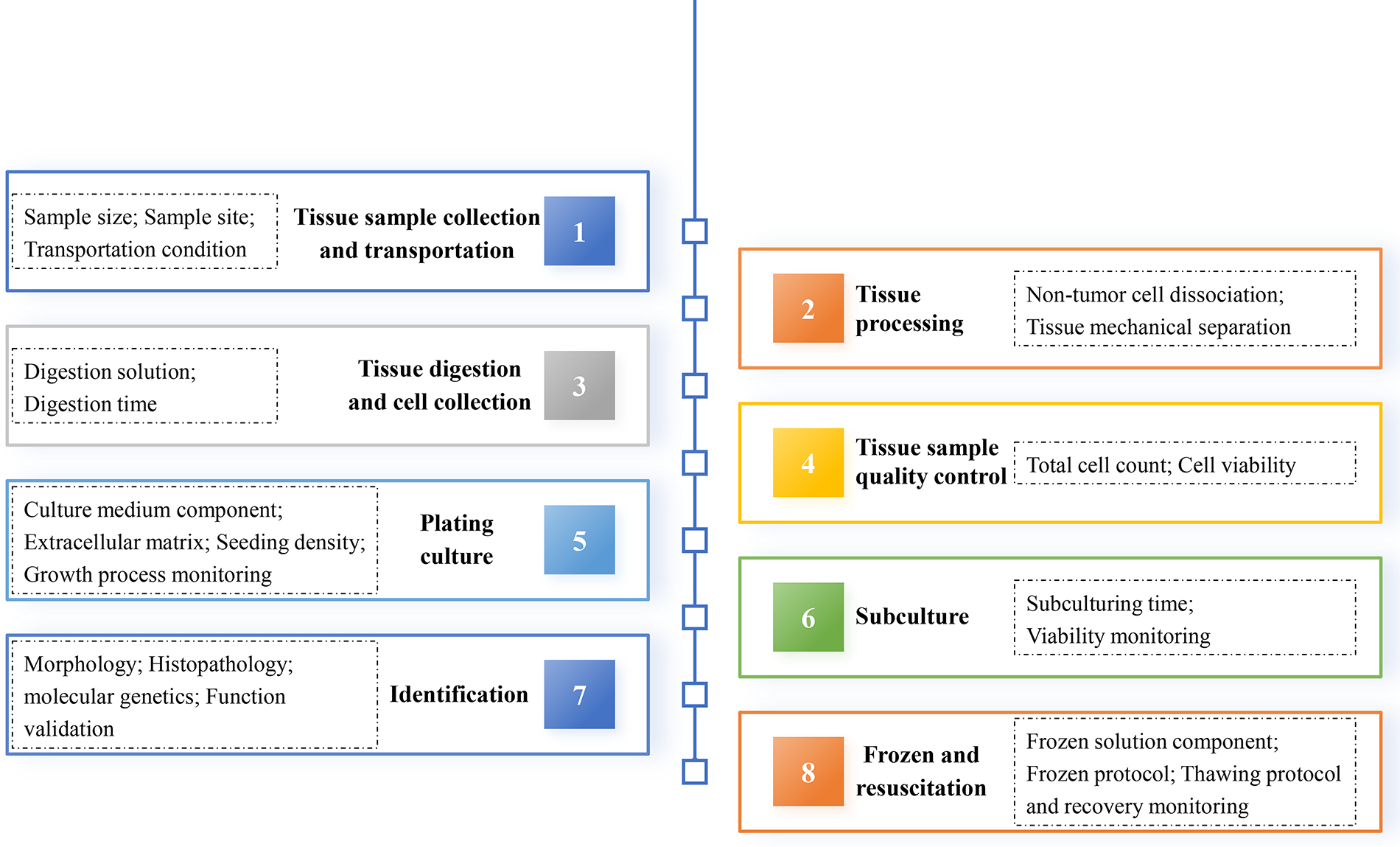
Technical processes and key quality control points in organoid construction. This schematic illustrates the sequential technical pipeline and critical quality assurance parameters for tumor organoids culture. The entire process consists of eight steps: tissue sample collection and transportation, tissue processing, tissue digestion and cell collection, tissue sample quality control, plating culture, subculture, identification, and frozen and resuscitation. Each dashed box contains the key quality control points for each step.
The Key Steps and Technical Approaches for QC of Tumor Organoids
The construction of organoids not only requires compliance and traceability of biological specimens, but also requires comprehensive lifecycle management encompassing sample collection, reception, processing, QC, cryopreservation, and resuscitation. The establishment of tumor organoid living biobanks requires a stringent QC system, 20 which is essential for guaranteeing the accuracy and reliability of research data, facilitating precision clinical decision-making, standardizing biobanking protocols, and ensuring the sustainable utilization of organoid resources.
QC in tissue sample collection, preservation, and transportation
The quality of tissue specimens directly determines the success rate of organoid culture. Therefore, standardized protocols for sample collection, preservation, and transportation are critical to ensure successful organoid establishment. Tumor tissue collection must strictly adhere to ethical guidelines, including obtaining fully informed patient consent and ensuring privacy protection.
The size and quality of the initial tumor specimen are critical determinants of successful organoid establishment. Larger specimens generally provide a higher initial cell count and viability, but they also carry a greater risk of central necrosis, which can compromise culture purity. Generally, surgical specimens should be at least the size of three soybeans, needle biopsies require a minimum of three needles, and endoscopic biopsies need at least six tissue fragments. Samples should be collected and transported on ice or at 2°C–8°C in specialized tissue preservation media to minimize ischemic stress and maintain cell viability. Strict avoidance of freeze–thaw cycles is critical for fresh tissue. While the use of fresh tissue is ideal for maximizing viability, it is noted that under specific and optimized cryopreservation protocols, the generation of viable organoids from frozen specimens has been reported in certain studies.21,22 However, the freezing process may introduce cellular differences and reduced drug response. However, whether this method is applicable to all cell types requires further validation. The choice of preservation method and solution—including commercially available options—should be optimized and validated by the laboratory and documented as part of the standard operating procedure.23,24
Rigorous aseptic technique is paramount during collection to inhibit microbial contamination. Existing research has demonstrated that the minimum amount of biopsy material plus the bacterial contaminations limited the success rate of organoid establishment to 74%. 25 Marinucci et al. 26 found that nonwashed colorectal carcinoma samples had a contamination rate of 62.5%, which decreased to 50% and 25% with phosphate-buffered saline (PBS) or penicillin/streptomycin-containing PBS, respectively. Although tumor tissues can retain viability for up to 72 hours in specialized preservation solutions under low-temperature conditions, it is strongly recommended to minimize this interval to prevent degradation of valuable samples. All transported specimens must be clearly labeled with essential identification information (e.g., tissue source, collection time) and matched with comprehensive clinical data. This information provides a crucial reference value for organoid construction and subsequent applications.
QC during the construction of tumor organoids
Obtaining high-purity tumor cells is a prerequisite for the successful construction of organoids. During tissue processing, necrotic tissue, adipose tissue, and connective tissue must be meticulously removed to ensure complete digestion of tumor tissue and prevent contamination. When digesting tumor tissue, to maintain cell viability while achieving full tissue digestion, it is necessary to select an appropriate digestion protocol for different types of tumors based on actual conditions. The number of cells obtained after digestion should be no less than 104, and a cell viability test result of over 90% can significantly improve the success rate of organoid construction. 24 The organoid culture system requires dynamic modulation of growth factors (e.g., Wnt3a, R-spondin 1, Noggin, epidermal growth factor) according to specific tumor types23,27,28 to promote stem cell proliferation and differentiation and induce the formation of specific organoids. Certain organoid types also require the addition of specific components. For example, nicotinamide is essential for prolonging the culture period of colonic crypt organoids. 29
During the organoid culture process, close monitoring of growth status is essential. Particularly, organoids are relatively unstable in the early stage of culture, so daily microscopic observation and documentation of growth status should be performed. Strict control of the timing and ratio of passaging is crucial to maintain stable organoid growth. This prevents organoid senescence or apoptosis that could affect post-passaging viability and growth status, while avoiding excessive dilution that may lead to organoid growth arrest. In addition, microbial contamination, especially mycoplasma contamination, represents a critical concern in organoid culture. Such contamination can affect organoid expansion and lead to erroneous experimental conclusions. Implementation of detection methods including polymerase chain reaction, microbial culture, and sequencing, followed by appropriate antibiotic treatment, is recommended for contamination management. 30
Sustained proliferation of organoids is a critical prerequisite for high-throughput functional testing. Quantification of adenosine triphosphate content can effectively assess organoid viability, while the application of artificial intelligence (AI)-based image analysis software enables real-time, high-precision monitoring of organoid survival status and clinically relevant morphological changes. 31 Changes in organoid volume or surface area, combined with assessments of plasma membrane integrity and esterase activity, can also reflect their viability and growth status. 32 The expression level of Ki-67 is associated with proliferation and differentiation of tumor cells. The proliferation index of organoids can be preliminarily evaluated by analyzing the percentage of Ki-67-positive cells in organoids through immunohistochemical staining. Furthermore, one of the key tasks of organoid living banks is the efficient reconstruction of cryopreserved organoids. Consequently, thawing tests should be systematically conducted on cryopreserved organoid samples to ensure viability and functionality. 33
Stable environmental conditions are non-negotiable for maintaining organoid health and ensuring experimental reproducibility. Fluctuations in incubator temperature, CO2, and humidity can induce cellular stress and phenotypic drift. Therefore, continuous monitoring and logging of these parameters are essential. As stipulated by the ISoOR-International Standards for Organoid Biobanking (6.5.4.1 Key elements of QC), “All equipment used for collection, processing, and storage (e.g., freezers, incubators, liquid nitrogen tanks) shall be regularly calibrated and maintained to ensure optimal operating conditions.”
Identification of tumor organoids
Organoids can faithfully retain characteristics of the original tissue, such as tumor-specific mutation profiles, gene expression levels, and epigenetic changes. 34 Postculture assessment and validation are essential to confirm the consistency between organoids and the original tumor tissue, as well as their ability to recapitulate the features of the native tissue. This is a crucial prerequisite for the subsequent application of organoids. The characterization of organoids can be performed from multiple dimensions, including morphology, histopathology, and molecular genetics.35,36
Morphological and histopathological characterization of tumor organoids
Morphological and histopathological analyses represent the most widely utilized, cost-effective, and rapid methods for organoid characterization. Healthy and well-growing organoids typically exhibit regular spherical, cystic, or tubular structures with clear boundaries and relatively uniform sizes. For instance, intestinal organoids often present cystic structures similar to miniaturized intestinal villi,18,29 while breast organoids generally form moderately sized and well-defined spherical aggregates. 20 Following paraffin embedding and sectioning, organoids undergo hematoxylin and eosin staining, immunohistochemical, or immunofluorescence analyses. Apart from showing consistent morphological phenotypes with the original tumor tissue, organoids should also express characteristic biomarkers consistent with the original tumor tissue. Examples include: estrogen receptor, progesterone receptor, and human epidermal growth factor receptor 2 (HER2) as biomarkers for breast cancer 20 ; Ki-67 proliferation index, caudal-type homeobox 2, and cytokeratin 20 as biomarkers for colorectal cancer 37 ; and paired box 8, P53, and Wilms tumor 1 as biomarkers for ovarian cancer. 35 Precise biomarker matching of organoids not only verifies model reliability but also guides clinical decision-making, such as predicting clinical treatment outcomes. For example, mutations in the extracellular domain of epidermal growth factor receptor in colorectal cancer organoids can predict resistance to cetuximab, allowing treatment optimization through pre-emptive organoid drug sensitivity testing. 37 Norman Sachs et al. 20 established a breast cancer organoid living biobank and demonstrated that HER2-positive organoids showed trastuzumab sensitivity significantly correlated with clinical patient responses.
Molecular genetic characterization of tumor organoids
Gene sequencing technology is the core method for verifying the genetic fidelity of organoids. Its high precision and specificity enable comprehensive analysis of organoids’ mutation profiles and epigenetic characteristics. However, their widespread adoption in organoid characterization remains limited due to challenges such as high cost, long cycle time, and complex data analysis. Hans Clevers 24 recommends performing genetic characterization of both established organoid lines and their source tissues. Technical methods such as targeted sequencing, whole-exome sequencing, whole-genome sequencing, and single-nucleotide polymorphism/short tandem repeat analysis can be flexibly selected to perform dual verification of the organoids’ biological representativeness and their matching degree with the source tissues. Consistency between organoids and source tissues can be validated by assessing canonical marker gene expression of expected cell types or cross-referencing cell populations with public tumor-type single-cell atlases. For instance, single-cell ribonucleic acid (RNA) sequencing analysis can be employed to deconvolute whether organoids contain relevant cell types of their target organ. 38 Utilizing tumor profiling panels enables not only the validation of concordance between organoids and their source tissues but also facilitates drug sensitivity screening and precision treatment planning.
Beyond genomic deoxyribonucleic acid (DNA) from the organoids themselves, the culture supernatant presents a valuable source for non-invasive QC. Exosomes and other extracellular vesicles secreted by organoids carry nucleic acids and proteins that can be profiled to monitor tumor heterogeneity and signaling pathways.39,40 For example, Alexia et al. 40 demonstrated that patient-derived tumor organoids (PDTOs)-derived cfDNA displays high similarity with patient-derived circulating DNA, indicating the potential of PDTO-derived cfDNA analysis as a non-invasive method for tumor evolution research and an important tool to support functional precision oncology. Analyzing cell-free DNA (cfDNA) released into the medium can provide a snapshot of genetic stability over time without sacrificing the culture. Rigorous genetic verification further enhances the predictive value of organoids in personalized medicine.
Functional characterization of tumor organoids
A robust and stable organoid model should not only have good scalability but also maintain the functional characteristics of the original tissue over extended culture periods. These functionalities include, but are not limited to the generation of mature cells, stress responses, and effective secretion of cytokines or hormones. 34 Functional status and maturity of organoids can be assessed through immunofluorescence-based evaluation of cell-specific marker expression, as well as normalized analyses of metabolite and drug metabolism, proteomic profiles, bioenergetics, gene expression levels, and epigenetic features.41,42 Organoids derived from chemotherapy-refractory patients should exhibit corresponding resistance to those chemotherapy agents in vitro, thereby aligning with the patient’s clinical response and confirming the model’s clinical relevance. 43 This concordance between the organoid model and the patient’s outcome serves as a powerful quality confirmation, ensuring the organoids are functionally representative tools for precision treatment. The application of biosensors enables continuous long-term monitoring of various biochemical (e.g., lactate, ions, proteins) and physical parameters (e.g., temperature, pH, oxygen levels, mechanical activity), which can be used to evaluate the functional status of organoids. 44 We summarized the specific functions required for the successful construction of some organoids, as well as the corresponding functional assessment methods in Table 1.
Specific Functional Requirements and Evaluation Methods of Organoids
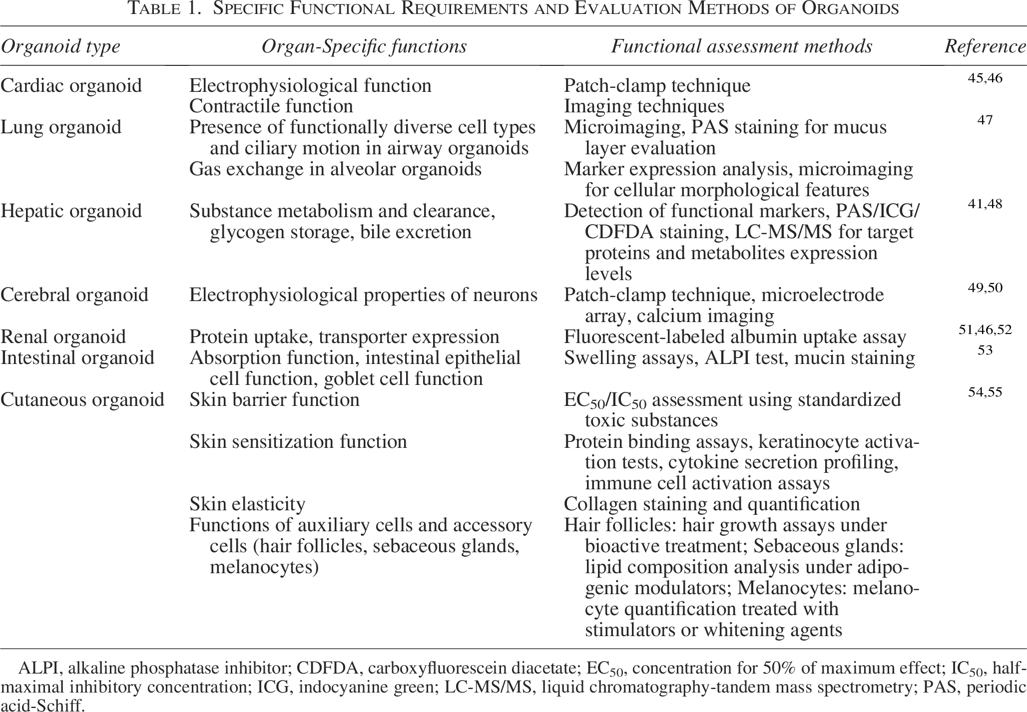
ALPI, alkaline phosphatase inhibitor; CDFDA, carboxyfluorescein diacetate; EC50, concentration for 50% of maximum effect; IC50, half-maximal inhibitory concentration; ICG, indocyanine green; LC-MS/MS, liquid chromatography-tandem mass spectrometry; PAS, periodic acid-Schiff.
Standardization and Normalization of Organoid QC
With the rapid advancement of organoid technology, its standardization and normalization process is also advancing gradually. A series of relevant standards and guidelines have been successively issued in China and abroad, providing a solid framework support for the QC of organoids. In September 2023, South Korea 56 jointly formulated the General Guidelines for Organoid Construction and Application in collaboration with relevant parties, including the academic community, regulatory authorities, and standard-setting experts. This guideline covers various aspects of organoid construction and quality assessment, and emphasizes that organoid quality assessment should involve the quality verification of source cell lines, structural maturity, cell composition, and functional maturity of organoids. It provides scientific guidance for the construction and QC of organoids, ensuring high standards in organoid construction and reliable, effective applications. In addition, guidelines covering the packaging, transportation, and storage of organoid-derived tissues have also been released. 57 Guidelines and consensuses for the construction, quality assessment, and application of organoids derived from specific tissues also have been developed—such as those for kidney, 51 heart, 45 lung, 47 skin, 54 liver, 48 brain, 49 intestine, 53 and bone 58 organoids. These documents effectively standardize the operational details of the entire process of different tissue-derived organoids, from basic processing to practical application. They provide a guiding basis for researchers to conduct scientific studies using organoids, and contribute to promoting the steady development and in-depth application of organoid technology.
Chinese researchers have also been actively advancing QC efforts for organoids. In recent years, a number of group standards and expert consensus related to organoids have been published (Fig. 2), providing guidance for the construction and QC of organoids derived from different tissues and cells. For example, the team led by Professor Wang Yunfang from Beijing Tsinghua Changgung Hospital has taken the lead in formulating multiple group standards for hepatobiliary organoids, filling the gap in standardized protocols for hepatic organoids in China. These standards specify the operating norms for the entire workflow of hepatobiliary organoids construction, QC and preservation, including the selection and identification of cell sources, precise setting of culture conditions, quality detection indicators and methods, as well as key technical points of preservation. They provide clear guidance for the standardized and normalized development of hepatobiliary organoid research and application, and effectively promote the in-depth application of organoid technology in the fields of hepatobiliary disease research and drug screening. Furthermore, the expert consensus—Chinese Expert Consensus on Quality Control Standards for Tumor Organoid Diagnosis and Treatment Platform (2022 Edition)—has also been published. This consensus document standardizes the QC of organoid diagnosis and treatment platforms and guides the clinical application of organoid drug sensitivity testing in selecting precision treatment strategies for cancer patients in China.
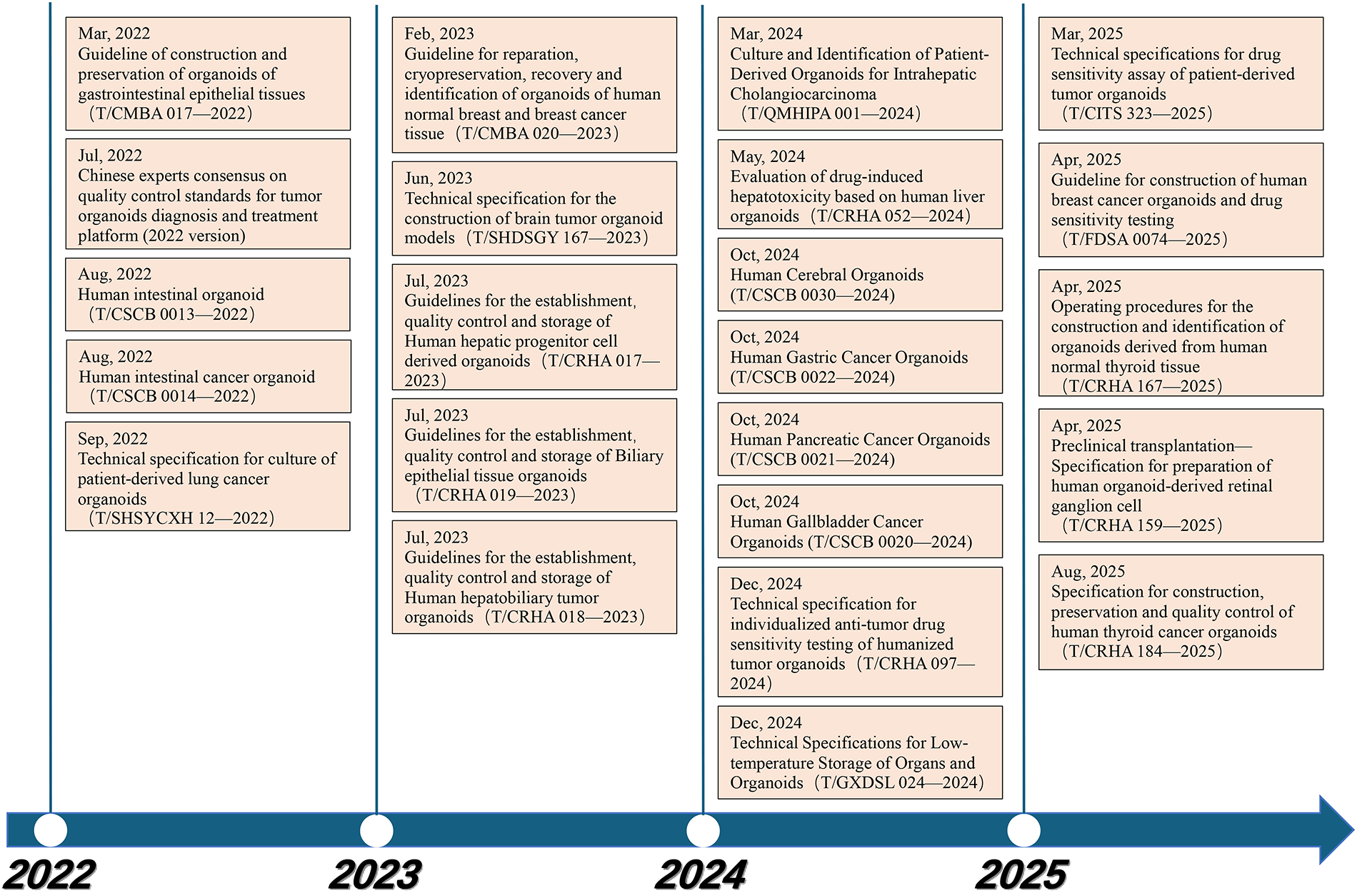
Domestic group standards and expert consensus related to organoids. This timeline summarizes the progressive establishment of standardized frameworks for organoid technology, as reflected by the sequential publication of technical specifications and consensus documents. Each entry represents an official standard or expert consensus addressing specific aspects of organoid construction, preservation, quality control, identification, or drug sensitivity testing. Each entry contains the release date, name and code of the group standard or expert consensus. The abbreviations in the standard code (such as China Medicinal Biotechnology Association [CMBA], Chinese Research Hospital Association [CRHA], Chinese Society for Cell Biology [CSCB]) are group codes, the unique identifier of the publishing group.
Despite these advancements, several hurdles remain in the full standardization of organoid technology. Proprietary issues concerning specific culture media formulations and protocols can restrict their widespread adoption. Furthermore, significant variability in resources and technical expertise between institutions can lead to inconsistencies in organoid quality and data interpretation, hindering uniform implementation. 59 To overcome these barriers, it is crucial to emphasize the importance of sharing standardized protocols and best practices within the scientific community.
Future Development Directions of Organoid QC
Global collaboration in standardization systems
The translation of the vast potential of organoid technology into clinical applications relies fundamentally on quality reliability, which depends on rigorous and standardized QC systems. Currently, there remains a lack of unified standards for organoid culture and detection. Variations in culture protocols across different laboratories lead to significant heterogeneity in organoid quality and characteristics. Additionally, the methods and metrics for organoid characterization and assessment differ among laboratories, resulting in a lack of objectivity and accuracy in quality evaluation. 60 To address the key issue of poor reproducibility and comparability in current organoid research and applications, the core approach lies in establishing internationally recognized standards, standardized operating procedures, and rigorously validated reference materials. Examples include widely accepted, thoroughly validated standard culture media or matrices, as well as the development of globally recognized standardized protocols and minimum standards for organoid culture and QC. These efforts will provide a consistent foundation for the entire field, ensuring the comparability and reliability of data generated from organoids of different origins and batches.
Automated and intelligent organoid culture and QC
To meet the needs of large-scale applications such as high-throughput drug screening and to enhance the objectivity and efficiency of QC, automation, AI, and microfluidic organ-on-a-chip technologies will serve as key driving forces. By using automated equipment and technologies, the entire process—from organoid culture and passaging to functional detection—can be automated. This will minimize human-induced variability while improving throughput and consistency. With the support of AI, big data analytics, organ-on-a-chip, and microfluidic technologies, automated analysis of organoid microscopic images can be achieved. These technologies can also monitor parameters (e.g., metabolite levels, cytokine secretion) and identify abnormal states in real-time.
Simultaneously, by integrating multi-omics data and functional data, the consolidation of multi-omics and functional data will facilitate the identification of key QC biomarkers and the development of predictive models. Such integration will support automated assessment of organoid functional status and drug responses.
Establishment of deep-layered and multidimensional evaluation system
Although organoids can simulate the characteristics of native organs to some extent, they still fall short of replicating the full complexity of real organs. The fidelity and functional complexity of organoid models need to be further improved, and determining the extent to which organoids represent organs remains challenging. 61 In the future, QC for organoids will extend beyond superficial morphological observations to deeply evaluate molecular composition, cellular heterogeneity, spatial organization, and core functions. This includes employing multi-omics technologies to precisely compare and evaluate whether the diversity of cell types, differentiation status, gene expression profiles, and epigenetic status of organoids match those of source tissue. It is also necessary to deepen the functional verification of organoids, including their physiological functions, disease-specific characteristics, and drug response prediction. Such comprehensive evaluations will more scientifically and holistically reflect the similarity, stability, and application reliability of organoids compared with real organs. The establishment and widespread adoption of publicly available reference control organoid lines might be a promising step too. These well-characterized organoid lines, analogous to reference cell lines used in other fields, would allow laboratories worldwide to benchmark their culture and QC pipelines, thereby improving inter-laboratory reproducibility and the reliability of data generated from organoid biobanks.
Breakthroughs in clinical translation and personalized medicine
The core value of organoid technology lies in constructing “patient avatars,” and providing a revolutionary tool for precision medicine. However, the current application of organoids is still in the basic or preclinical research stage. 62 In the future, the advancements in organoid QC systems will directly promote their translation in precision medicine and regenerative medicine. By enhancing the modeling fidelity of organoids, constructing more complex and dynamic multi-organoid interaction systems, establishing clinical-grade drug sensitivity testing and drug screening procedures, and integrating patients’ genomic data, clinical information, as well as organoid drug sensitivity and functional profiles, this technology will truly guide personalized treatment decision-making.
Summary
As a groundbreaking advancement in the biomedical field, organoid technology stands at a critical juncture of transition from laboratory investigation to clinical application. Tumor organoids, capable of closely mimicking key characteristics and functions of source tumors, demonstrate significant potential in disease modeling, drug screening, and personalized medicine. This potential aligns with the global push toward Novel Alternative Methods to complement or reduce reliance on animal models as discussed in recent policy initiatives by the FDA and NIH. However, challenges such as functional incompleteness, lack of standardization, and industrialization bottlenecks still restrict their development. The large-scale application of tumor organoid living banks relies on the establishment of unified standards and rigorous full-chain QC systems, which deserve widespread attention from researchers. In the future, through interdisciplinary innovation, combined with the construction of international standard systems and breakthroughs in automated production, organoid technology will be promoted to achieve a leap from morphological simulation to functional recapitulation. Ultimately, a new era of patient-centered personalized medicine will be realized. Achieving this goal not only requires the deep integration of biotechnology and engineering technology but also relies on the simultaneous advancement of interdisciplinary collaboration, global standardization cooperation, and policy supervision, so as to unlock the clinical transformation code of organoid technology.
Authors’ Contributions
R.F.: Writing—original draft. S.Z.: Visualization and writing—review and editing. P.A.: Writing—review and editing. X.Y.: Visualization and resources. M.Y.: Resources. Y.W.: Resources. C.W.: Conceptualization, supervision, project administration, and funding acquisition.
Footnotes
Author Disclosure Statement
The authors declare no competing interests.
Funding Information
This study was supported by National Key Research and Development Program of China (2024YFC3607500), the Basic Strengthening Plan Technology Field Fund Project(2019-JCJQ-JJ-066) and Naval Medical University “San Hang” Military Medical Talent Project (2019-YH-09).