
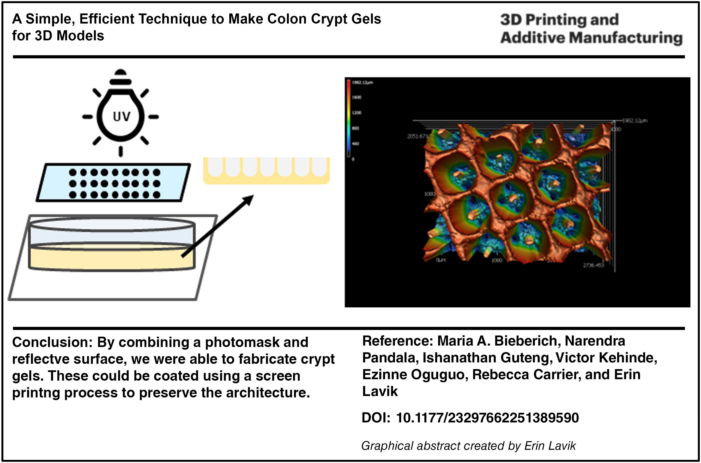
Abstract
An in vitro colon model, particularly one suited to high-throughput screening, has the potential to enhance understanding of cellular mechanisms and functions important in intestinal health and can be used for drug testing and drug permeation studies. While extensively studied, traditional monolayered cultures using immortalized colon cancer cell lines on transwell plates fail to accurately replicate the native intestinal epithelium’s complex architecture. To address this limitation, we have developed a novel, facile photopolymerization technique to fabricate scaffolds that closely resemble colon crypts. We have further developed a method using screen printing to be able to coat these scaffolds while preserving the crypt architecture in order to vary the surface chemistry of these systems. This article focuses on the development of 3D crypt models that can be made with simple equipment and with chemical precursors that are commercially available to make building tissue models more accessible to the broader research community.
Introduction
The Centers for Disease Control and Prevention estimates that at least 3.1 million people have inflammatory bowel disease (IBD)1,2 and over 1 million people have ulcerative colitis (UC), a type of IBD, in the United States. 1 UC can cause debilitating symptoms and increases the risk of colorectal cancer. 3 While there are therapies available to treat UC, many are expensive and only work in a subset of patients.4,5 Patients who do not respond to traditional therapies including steroids and immune modulators 6 must go through a trial and error process to identify effective treatments that is expensive and exhausting.7–9
Ideally, one would have an efficient platform for modeling diseases of the colon, such as UC, and screening potential therapies. The intestinal epithelium is essential to the development of UC. The epithelial tissue directly confronts the microbial flora, pathogenic bacteria, and environmental factors such as food antigens, which transit through the intestine. The intestinal epithelium largely consists of four cell types: enterocytes, goblet cells, enteroendocrine cells and Paneth cells. 10 Enterocytes are the most abundant cell type in the epithelium and regulate nutrient absorption. 11 Goblet cells secrete mucin, helping to maintain the protective mucus layer and prevent bacterial infection. 12 The GI tract is the largest mucosal surface in the body, serving as the main interface through which nutrients are absorbed,13,14 and this mucosal component is essential to its function. Enteroendocrine cells produce and export peptide hormones that aid in metabolism as well as appetite regulation. 11 Lastly, Paneth cells are primarily located at the base of the crypt structure in the small intestine and secrete digestive enzymes, growth factors and antimicrobial peptides such as defensins. 12 These peptides and enzymes secreted by Paneth cells help to regulate the autochthonous flora of the intestine and protect against pathogenic invasion. 12 Due to the number of antigens and microorganisms exposed to the gastrointestinal (GI) tract, the GI tract also accommodates a large population of immune cells. 15 The gut immune system includes both inductive immune sites such as Peyer’s patches, where primary innate immune responses take place and effector sites such as lamina propria, where large numbers of lymphocytes are present and adaptive immune responses occur. 16
While the colon is complex, relatively simple in vitro models of the colon have been extremely helpful in enhancing understanding of disease and screening potential therapies. 17 These models enable in-depth study of cellular mechanisms in both healthy and diseased states, providing crucial insights into the underlying pathology of gastrointestinal disorders and aiding in therapeutic development. 18 Controlled or slow-release drugs that spend extensive time in the large intestine19–21 also motivate the need for colon models. Many colon models to date have relied on Caco-2 and HT29 cells in 2D culture systems. While these are human cell lines, they recapitulate a subset of colonic cell behaviors.22–24 Caco-2 and HT29 cells are human colon cancer cell lines that have been extensively used to model both the large and small intestines.25,26 Caco-2 cells are known to form tight junctions and express enzymes, characteristic features of enterocytes, and the HT29 cells secrete mucus, making them a good model for mucus-secreting goblet cells. 27 Due to these features, cocultures of Caco-2 and HT29 cells have been used to model the intestinal epithelium in vitro in 2D.14,28,29 However, the 3D structures of the colon are essential to their function.
The outermost surface (i.e., closest to the lumen) of the large intestine is a single layer of epithelium arranged in a crypt-like pattern.11,30 These crypts are invaginations that serve as the functional unit of the colon.31,32 The base of the crypt structure contains the proliferation compartment where colonic stem cells reside. 33 After differentiating, cells move along the basal-luminal axis guided by a gradient of various growth factors such as Wnt, Bone morphogenic protein, and Notch.33–36 Thus, it is not surprising that there has been elegant work looking at recapitulating the 3D structures of the colon.
Organoids have played a critical role in understanding and modeling the colon. Organoid cultures exhibit responses to drugs such as metformin in modulating tight junctions not seen in 2D cultures 37 and have been used to study metabolism and drug permeability.38,39 Organoids can be grown from stem cells in patient tissues or induced pluripotent stem (iPS) cells. Organoids include some of the architecture of a tissue and at least some of the cell types present in the tissue. 40 Historically, one of the challenges with organoid work was the need for feeder cells but, in recent years, methods have been developed to mitigate the need for feeder layers for both adult stem cells 41 and iPS cell-derived organoids, 42 which make organoid culture more accessible. Culture media and signaling molecules play critical roles in what cells are present in different organoid cultures. 40 At a minimum, epithelial cells are included, 40 but most models include mucosal cells and many include immune cells and, potentially, microvascular components. 43 While organoids are powerful tools, they can be challenging to interrogate, in part because the apical surface of the epithelial layer typically faces the enclosed organoid lumen. Thus, researchers have worked to develop 3D systems that recapitulate the architecture of the colon and are more amenable to high-throughput screening methods.
The architecture is important to cell behavior in the colon. Stem cells differentiate and migrate along the crypt-villus axis in vivo, guided by a gradient of growth factors, cytokines, oxygen, and pH. 18 Because these gradients are typically altered early in conditions or ailments of the intestine, the ability to alter these gradients in vitro could facilitate the development and testing of novel treatment approaches.18,44 Building crypt structures has the potential to replicate the gradients that are important in colon stem cell migration and the tissue response to diseases.
Three-dimensional printing, or bioprinting, allows the printing of cells and hydrogels, which has emerged as a promising technique for recreating complex architectures.45,46 Traditionally, hydrogels are preferred materials used to recreate the extracellular matrix (ECM) in vitro, as they possess similar mechanical properties and allow for the diffusion of molecules.18,44 Three-dimensional printing of colon structures has been pursued to reduce the time to make models compared with some organoid approaches. These printed structures, typically incorporating hydrogels, can promote formation of villi 47 as well as the combination of crypts and villi. 48 These 3D printed colon structures are a major step forward in modeling the colon.
Bioprinting, in which materials and cells are printed together, enables one to develop patterns mimicking those seen in vivo, but it requires that the materials and cells be extruded through relatively fine openings as well as specialized equipment. While the cost of bioprinters has come down, the extrusion process requires bioinks that are compatible with the shearing associated with the approach as well as materials that protect cells during this process. 49
To reduce cost and complexity and decrease the time to fabricate structures, we sought to determine if we could build crypt structures from common, commercially available hydrogel materials using simple, inexpensive equipment in a manner that might make 3D culture systems accessible to a broader group of researchers. In our previous work, we utilized screen printing to construct 3D structures with high cell survival. Building on this foundation, we have now developed a novel approach to rapidly create a preform that can be further modified with screen-printed layers as needed, instead of building 3D structures 10–20 μm at a time.
Our method involves fabricating a crypt structure based on a poly(ethylene glycol) diacrylate (PEGDA) hydrogel system using a simple mold, a UV lamp, and photomasks with arrays of dots. To determine the components of the approach that lead to a reproducible colon-crypt gel structure, we investigated the architecture, the ability to alter the surfaces with simple screen printing approaches, and cell survival. This work provides the foundation for efficiently building 3D models of the colon that are flexible, adaptable, and cost-effective.
Experimental Section
Materials
Polyethylene glycol (PEG) diacrylate (Mn∼7500 g/f) was purchased from JenKem Technologies. Solvents, media components (MEM alpha, minimum essential medium and Dulbecco's Modified Eagle Medium), fetal bovine serum, nonessential amino acids, calcein-AM/ethidium homodimer (live/dead) stain, Triton X-100, 4% paraformaldehyde, DAPI, bovine serum albumin, and dialysis tubing were purchased from ThermoFisher Scientific, USA. Normal goat serum was purchased from Vector Labs, USA. Poly-L-lysine (PLL) (70–150 kDa), and HT29-MTX-E12 cells were purchased from Sigma Aldrich. Caco-2 cells were obtained from American Type Culture Collection, USA. All other chemicals were purchased from Sigma Aldrich.
Synthesis of PEGDA hydrogels and crypt scaffolds
A 5 mg/mL solution of Irgacure in deionized water was prepared. A 10% w/vol solution of PEG diacrylate in Irgacure supplemented with 2% v/v collagen solution (5 mg/mL) was prepared and then set under UV light (365 nm) for 10 min. To obtain the crypt structure, the same PEG solution was kept in a polytetrafluoroethylene (PTFE) cylindrical mold covered with a photomask and exposed to UV for 10 min. The photomask was made by printing a grid of dots of 600 µm and 1000 µm diameter onto a transparent sheet using an inkjet printer in saturated black. The mold was made of white PTFE with dimensions of 1.5 mm thick and a diameter of 20 mm. After the gels were set, they were washed with Phosphate buffered saline (PBS) or culture media (3 times, 5 mins each) before use. An identical procedure was used with a glass photomask as with the inkjet transparency mask.
The PLL–PEG coating was created by making 5% w/v solutions of each polymer (polylysine, PLL, 110,000 mw from Sigma) and 10 kDa SG-PEG-SG (4-arm from JenKem) in PBS and mixing the solutions together in a 1:5 ratio of PEG to PLL before printing gels that gelled within minutes following our previous work. 50 When the PLL/PEG was screen printed, the two components were mixed immediately before pushing them through a 200 mm mesh nylon screen following our previous work. 50
The collagen–PEG coating was made by making 5% w/v solutions of collagen (Type I, gift from J. Madri from cow skin preparation) and SG-PEG-SG (3400 Da, JemKem) in PBS. The solutions were mixed in a 4:1 ratio of PEG to collagen immediately before screen printing onto a PEGDA/collagen crypt gel. About 100 µL of solution was used to coat each 2 cm crypt gel.
Imaging hydrogels
The lateral and top views of the crypt scaffold sections were taken by using a laser scanning confocal microscope VK-X1000 (Keyence, Japan).
Cell experiments
The information on cell experiments is in the Supplementary Data.
Results
Formation of crypt structures
To create the crypt-shaped scaffolds, the precursor solution containing PEGDA and the photocrosslinker was pipetted into a white PTFE mold, and the photomask was placed covering the precursor solution. The entire setup was exposed to ultraviolet light at 365 nm for 10 min (Fig. 1A). The light went through the transparency but was blocked by the black dots on the photomask (Fig. 1B). The use of the white mold scattered light near the surface of the mold resulting in crypt-like structures in the gel (Fig. 1C–E) rather than pores that traversed the entire gels. The use of a black mold, by contrast, led to pores through the gels rather than the crypt structure.
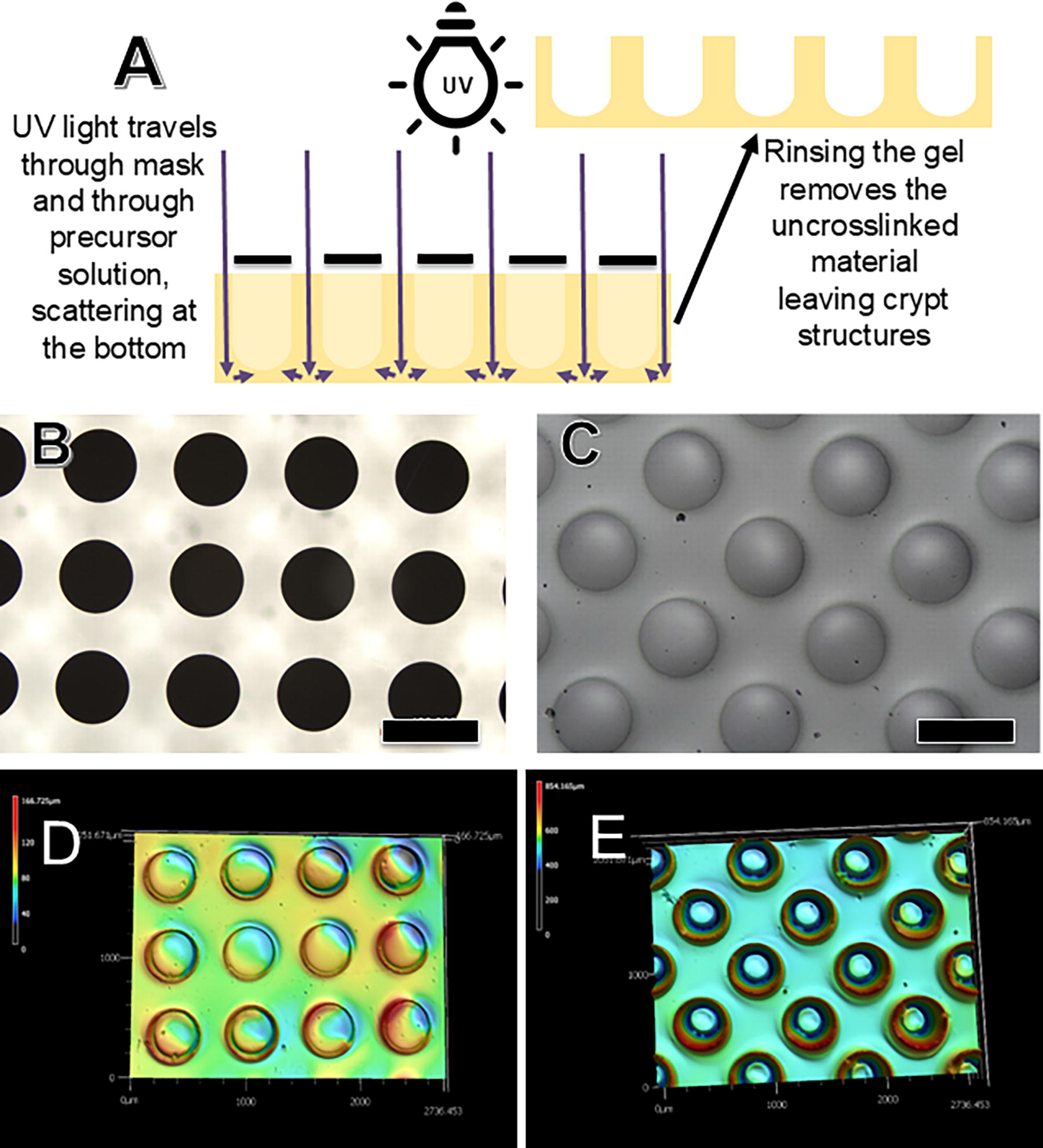
By filling the molds completely, the gel precursor solution wetted the glass photomask leading to very sharp columnar structures with a thin gel bottom. Heatmaps were made using the laser scanning feature on the Keyence microscope. However, it was very difficult to get reproducible heatmaps from the crypt gels. The heatmaps suggested that the crypts were only ∼100–300 μm deep, and the scans were highly variable even with the same crypt gel (Fig. 1D). Once the gels were stained with eosin, the heatmaps were reproducible with crypt depths of approximately 800 μm. The eosin stain produced contrast that made it easier to see the crypts and to scan them with the laser scanning microscope. We scanned six gels made by two people and saw that the structures appeared similar and the height of the gels by heatmap was 800–1000 μm across the six gels.
Coating crypt gels
With our simple molds and acrylated PEG, we could make six gels at a time within 10 min. The speed at which one can produce gels is one of the attractions of the approach. However, surface chemistry is essential to cell behavior. While one could easily adapt a number of different photopolymerizable gels to this approach, there may be a great deal gained from being able to quickly alter the surface chemistry on the crypt gels or vary it across gels in a simple fashion.
Coating the gels would be the simplest way to alter the surface chemistry, but applying a coating directly to the gels led to nonuniform coatings that filled many of the crypt structures. We have experience screen printing gels and cells into patterns,50,51 and we had previously observed that screen printing led to very uniform, thin layers of gels. We hypothesize that the very thin coatings created by the screen printing process50,51 allow the crypt structures to be coated without filling in the crypts. A schematic of the screen printing process can be seen in Figure 2.
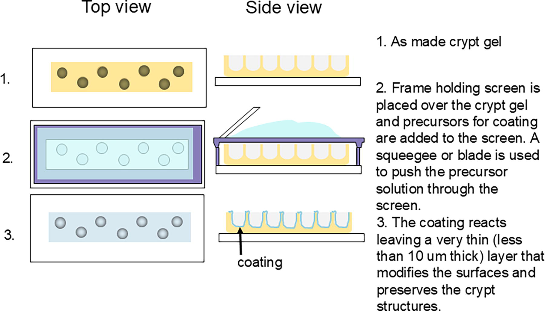
Schematic showing the screen printing process. The crypt gel is formed and placed in a mold. The stretched screen with a mesh size of 200 μm is across the top. The gel is added and pushed along the screen with a squeegee, which allows a small, uniform amount of the gel coating to move through the screen and attach to the crypt gel below.
Chemically reactive precursors are mixed and pipetted onto a screen. In this case, the screen has a pattern that is the same dimension as the crypt gel. The gels are 1 cm in diameter, and the screen pattern is also 1 cm in diameter. By having the crypt gels in the molds, the screen is able to just touch the gels without deforming or damaging them when the precursor solution is spread across the screen. The precursor solution is designed to gel within 4–6 min, which is enough time for the screen printing process to be completed.
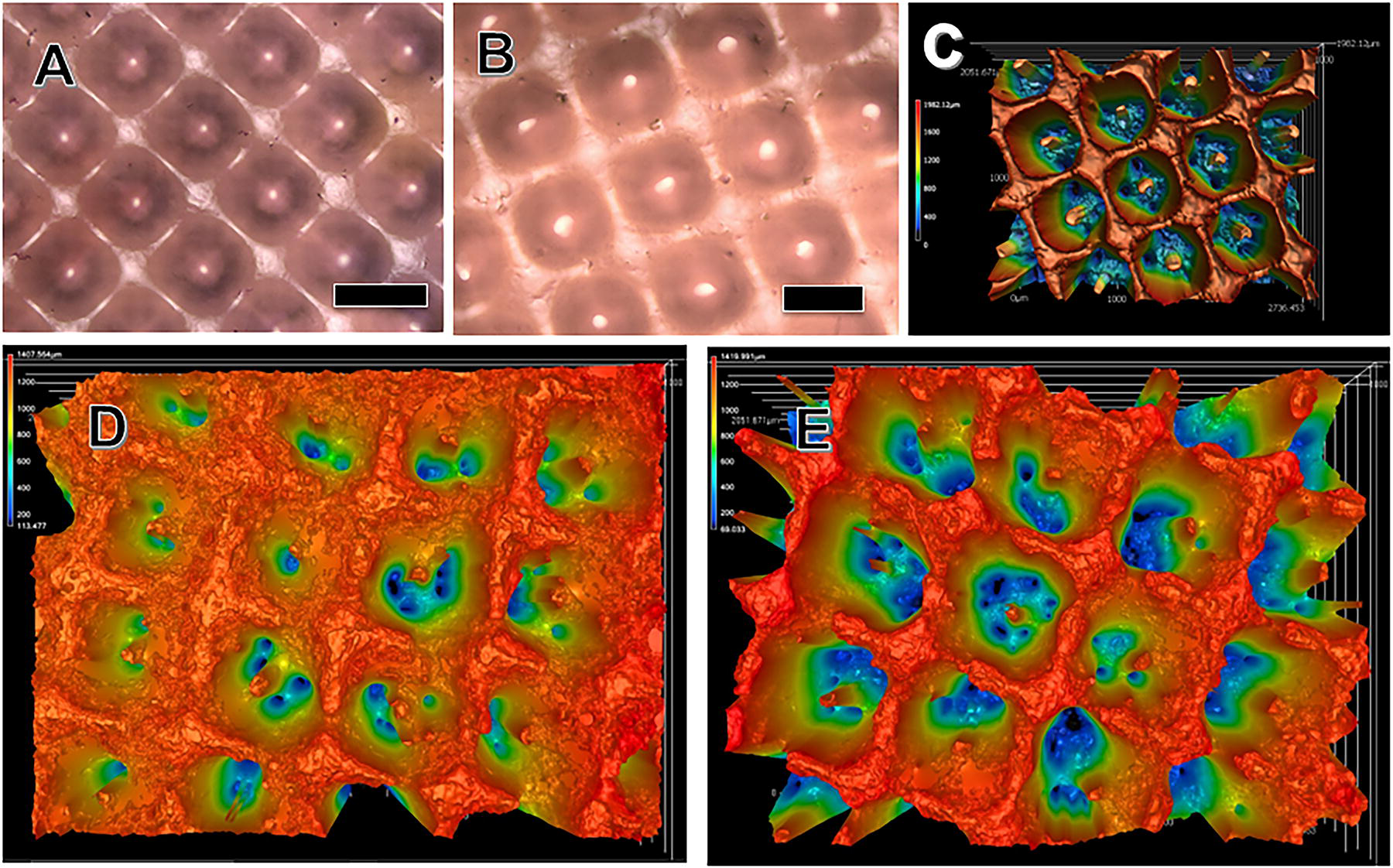
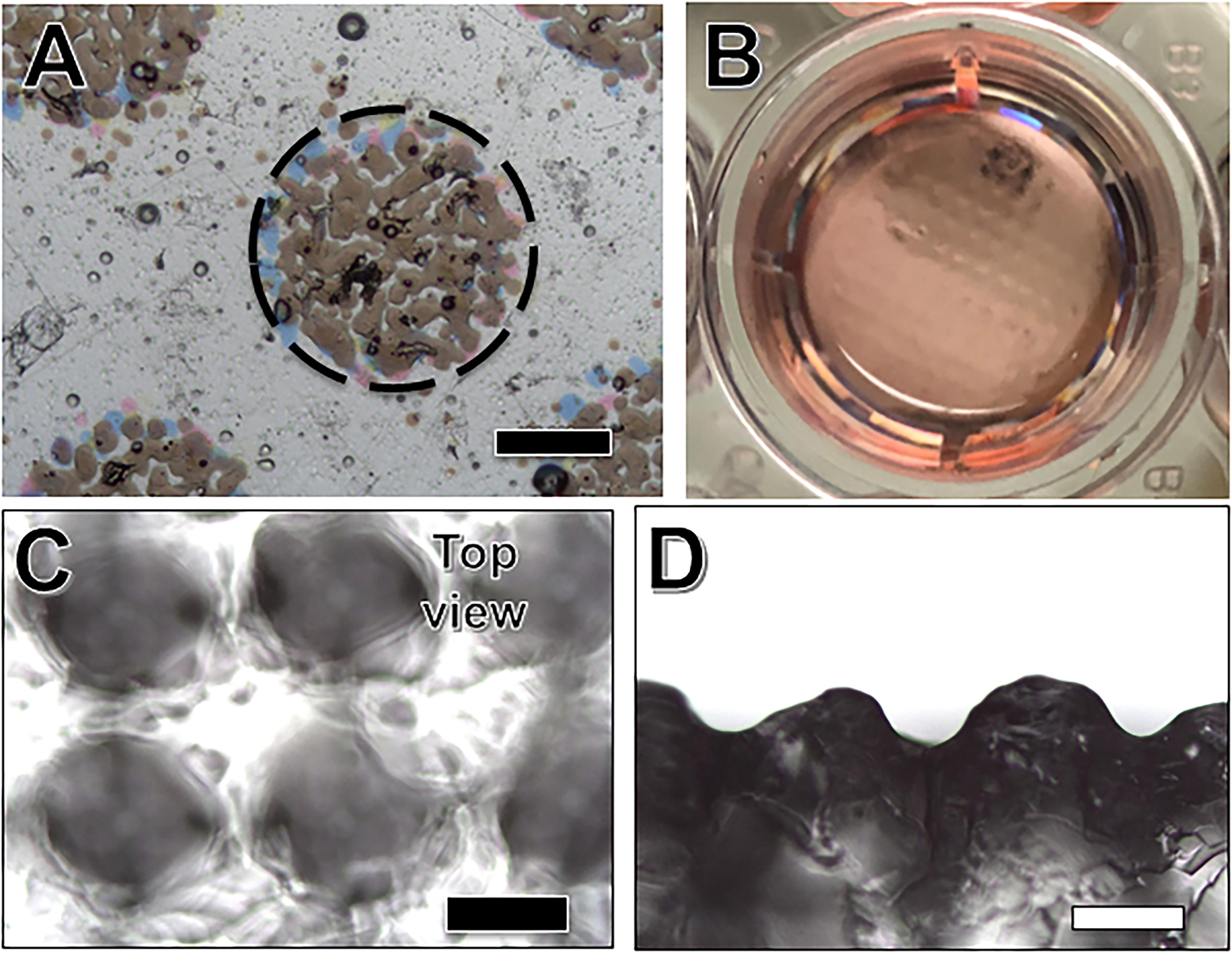
We found it challenging to coat the high-feature structures of the initial crypt gels in Figure 1. By reducing the volume of the precursor solution added to the molds for the photopolymerization of the crypt gels from 400 to 380 µL, the gel solution no longer touched the photomask. Since the gel solution did not touch and therefore wet the surface of the mask, the fabricated gels exhibited structures (Fig. 1) that flared at the top, which we believed would facilitate introducing coatings within the crypt structures (Fig. 3A). The gel in Figure 3A was made from diacrylated PEG blended with 2% collagen. We term these photopolymerized gels PEGDA/collagen gels. The collagen was incorporated for cell culture studies in subsequent work since PEG gels are nonadherent.
Photopolymerized PEGDA/collagen gels using the glass photomask.
Screen printing allows one to add a controlled amount of gel coating that preserved the crypt architecture. Figure 3 shows examples of uncoated crypt gels (Fig. 3A, D) and coated crypt gels (Fig. 3B, C, E). The heatmaps in Figure 3D and E show that the geometry of the crypts was well preserved. While the differences in the heatmap suggest that the coating is present, we also used crypt gels without dye and added dye to the screen-printed coating. Images of crypt gels coated with PLL–PEG screen-printed coating containing blue food coloring can be found in Supplementary Figure S1. Unfortunately, there was enough noise in the measurements that we could not determine the exact coating thickness in this work. However, from measurements of coating thickness in our previous work, we expect that the thickness of the coating was approximately 10 μm.50,51
We used both a PLL–PEG coating and a collagen–PEG coating in this work. Both precursor solutions could be screen printed easily and preserved the crypt architecture. One of the benefits of screen printing the coating was that excess material was left on the screen. The minimum material that we found to work was 100 μm per 2 cm gel. When we used more solution, we saw no apparent differences in the preservation of the crypt architecture, but there was more material left on the screen. With less than 100 μL of solution, we often saw areas that appeared uncoated. We chose the PLL–PEG gel and the collagen–PEG gel formulations for coating based on our previous work showing that PLL improved cell adhesion,50,52 and that collagen is a critical component of the ECM of the colon. 53
Making crypt gels with low-cost inkjet photomasks
While crypt gels can be made with a photomask, a glass photomask is a significant investment that may not be easily accessible to all researchers. As an alternative, we investigated making the crypt gels with inkjet-printed transparencies. A used Epson WF-3640 inkjet printer from 2016 was used.
This color inkjet printer makes a black color by overlaying all of the colors on the transparency. Viewing the transparency with a white background in the Keyence microscope demonstrated the heterogeneity of the black dots (Fig. 4A). Thus, it is not surprising that the crypt structures do not have crisp edges of the glass photomask system (Fig. 4C). A side view of the crypt gels demonstrates the crypt-like pattern (Fig. 4D). While the crypts are less uniform and sharp compared with the glass photomask system, this low cost approach still leads to gels with crypt structures to facilitate 3D crypt models. Inkjet printers that print more consistent black dots would approach the quality seen in the glass photomask case, but it is worth noting that even these poor-quality masks are enough to lead to reproducible crypt gels. We fabricated five gels with 600 μm dots and five gels with 1000 μm dots, and we measured the crypt diameters. They were within 50 μm of the dots across all of the gels synthesized.
Attachment and survival of cells
Since PEG gels are nonadherent, we incorporated a 2% collagen solution into the PEGDA gel precursor solution. To test for the potential cytotoxicity of the PEGDA/collagen gels, we used gels without any crypt structure and compared them to ones with crypts of different dimensions. Live-dead staining 3 days following cell seeding showed no significant cell death in ratios of Caco-2 and HT29 seeded on the hydrogel as compared with controls (Supplementary Fig. S2). Likewise, there were no significant differences observed between the crypt gels.
Screen printing the PEG/collagen gels with PEG–PLL led to better cell attachment but impacted cell survival (Supplementary Fig. S3). This has been seen in other cell types and is often a function of the density of amines.50,52,54 Being able to vary coating easily would allow one to explore the optimal chemistry for both cell attachment and survival. Coating with collagen/PEG provided a less toxic alternative; however, one of the shortcomings of this work was that we did not quantify cell survival. While it appeared close to complete cell survival on the gels, we should have quantified this for completeness.
In the absence of a coating, while there were areas of confluent monolayers of cells (Supplementary Fig. S5), there were also spheroids that were seen (Supplementary Fig. S6). The collagen/PEG coating promoted more consistent monolayers of cells. However, immunostaining suggested that there were no tight junctions, as marked by Zonula Occludens-1 (ZO-1) staining and no signs of ezrin staining in the Caco-2 and HT29 cell cultures. Future work will focus on optimizing the cell choice and mechanics of the crypt gels to produce colon gel models based on these crypt structures. Notably, these crypt gels can be handled easily, which allows seeding of macrophages on the noncrypt side of the gels, which opens the possibility of not only investigating the epithelial layer but also adding immune components to the crypt gels.
Discussion
In this work, we have developed a simple method for creating crypt structures in hydrogels and coating them with molecules of interest while preserving the underlying crypt architecture. This approach has broad applications for 3D printing and fabrication of tissue models. For example, one could develop a preform that exhibits the key architectural features of a tissue of interest and then add the appropriate matrix with controlled chemical and mechanical properties while preserving the architecture. One can also use the screen printing process to pattern cells in the structure,50,51 which opens possibilities for replicating other aspects of the colon.
The basis for building in vitro models hinges on two critical factors: recreating physiologically relevant culture conditions and structures and the selection of appropriate cell types. In this work, we focused on applying the preform/coating approach to building crypt-like structures as the basis for modeling the colon. By using a scaffold that could be fabricated easily without complex tools or chemistries and modified simply as the basis for a 3D model of the colon, we have the basis for building in vitro models even in laboratories with limited resources. Making technologies available to the broader research community is essential. In vitro colon models hold significant potential for a wide range of applications, including drug testing for colon-related ailments, permeation studies for nutrients and drugs absorbed through the large intestine, and potentially elucidating complex colon–microbiome interactions.19–21 Creating a flexible, easy-to-use platform makes it possible to investigate the complexities of the colon in novel ways.
The 3D architecture of the crypts is critical to the behavior of cells in the colon. Given the right conditions, cells derived from organoids can recapitulate the complex architecture of the intestine, 55 but this process typically takes substantial time and requires highly controlled conditions. This can make it challenging to scale systems efficiently for screening applications. 3D printing offers a method to have extraordinary control over the structures and to replicate the colon architecture, but 3D printing of photopolymerizable hydrogels generally involves a 3D scannable laser system to direct the gelation at specific locations.56–58 It creates exquisite structures, but it can be a significant investment for researchers. Having a platform technology that guides the cells has the potential to reduce the complexity, cost, and time involved in building colon models. We have extensive experience with PEG-based photopolymerizable gels, and we used materials we were deeply familiar with in the development of this platform. We had previously optimized the curing time and other parameters with these gels, independent of the mask.52,54,59 One of the promising aspects of this work is this introduction of the glass or transparency mask did not impact the curing time of the gels. It appears to only have introduced the crypt structures. The ease with which the crypt platform can be coated offers the potential to coat the resulting scaffolds and incorporate them into gut-on-a-chip systems, allowing one to introduce flow into these systems.
As important as the structure is, the cells are equally important. In this work, we examined cell lines, Caco-2 and HT29, that have been used extensively to model aspects of the colon.60–62 While important data and screening can be obtained with human cell lines, modeling involving colonic stem cells have been shown to more accurately replicate the human colon. 63 Colon stem cells were challenging to obtain, but the development of human iPS cells in 200664,65 opened exciting doors for regenerative medicine. Human iPS cells have been shown to model the physiology of the colon better than other cell types42,66,67 and have been shown to better model the pathology of diseases such as IBD. 68 Protocols that allow researchers to access large numbers of progenitors quickly hold promise for developing systems for high-throughput screening. 69 In future work, it will be important to test these 3D structures with colon stem cells.
One of the significant shortcomings of this work is that we have tested it only with immortalized human cell lines. It is entirely possible as this system is tested with other cell types, the chemistry or mechanics of the gel may need to be altered. The mechanics of the colon are complex, as it is a tissue that must be extremely elastic and strong. 70 Soft gels (2 kPa) and stiffer materials (1–2 GPa) seem to maintain the epithelial structure while intermediates promote migratory phenotypes.71,72 The stiffness of the gel is known to impact the behavior of cells of the colon. 71 This gel formulation has a stiffness of approximately 20 kPa, which may not be optimal for these cells.54,73 Lack of ZO-1 expression with Caco2 and HT29 cells has been associated with the stiffness of the matrix on which cocultures were cultured.74,75 While matrix stiffness is an extremely important feature, we did not investigate the impact of matrix stiffness in this work and focused instead on the architecture of the system. However, it is simple to alter the mechanics of PEG-based photopolymerizable gels.52,54 Being able to screen print coatings on gels opens up the possibility of not only altering the chemistry of the gels in simple manner, but it also allows one to consider patterning the surfaces with different materials. 50 In future work, it may be interesting to determine the stability of coatings over time.
One of the exciting aspects of the facility of screen printing onto these gels is that one can consider not only printing gels and cells but also electronic structures, which could allow integration of electronics to provide real-time analysis of tissue models. For example, one could include electrodes for transepithelial electrical resistance (TEER) measurements. TEER plays a critical role in examining the permeability in colon tissue models.76,77 In many of the tissue models, immune cells play a critical role, and it is exciting that immune cells can be incorporated directly into this colon structure by simply adding them on the apical side of the crypt model.
The presence of flow, a critical component in vivo, can play a role in the tissue development and behavior in vitro. Flow, which can be introduced using gut-on-a-chip models, has been shown to promote the formation of villi in monolayers of Caco-2 cells. 78 Pulsatile flow could mimic flow in vivo and thus has the potential to introduce critical mechanical cues that can impact cell behavior as well as the behavior of microbiome cultures. 79 Being able to combine the 3D architecture of the colon, particularly the crypt structure, with flow has the potential to better mimic the complexity of fluid movement in the colon and more accurately represent the in vivo environment. 80 A critical step in achieving this complexity is to be able to efficiently build these 3D crypt structures.
Conclusions
This work provides a biofabrication process without complicated equipment. All the molds and the photomasks used for this process are obtained from off-the-shelf parts. Given the facile nature of the synthesis and the low toxicity of the gels, these gels can be used to work with not only tumor-derived cells but also translated with stem cells and other primary cells. Since these PEG-based hydrogels are nondegradable this system can be integrated into a microfluidic device without losing the structural features. The addition of screen printing allows one to modify the chemistry of these crypt gels as well as to consider introducing electronics into the system.
Authors’ Contributions
M.A.B. and N.P. carried out experiments, analyzed results, and wrote sections of the article. I.G. synthesized crypt gels, coated them, and performed imaging and characterization of them, as well as helped image immunocytochemistry. V.K. and E.O. carried out cell-seeded experiments and performed immunocytochemistry. R.C. helped design the experiments and edited the article. E.L. helped design experiments, imaged immunocytochemistry, made gels, analyzed data, and edited the article.
Footnotes
Acknowledgments
The authors thank Dr. Joseph Madri (Yale, School of Medicine, USA) for kindly providing the type-1 collagen obtained from bovine fetal skin. The authors also thank William Lavik-Rice for providing help with making hydrogels when his mom’s arm was broken.
Author Disclosure Statement
The authors do not have any conflicts of interest to report.
Funding Information
This work was supported by the National Science Foundation (Award No. 1804743) and the NIH Award R01EB021908 (Carrier).
Data Availability Statement
The data supporting this article have been included as part of the Supplementary Information.
Supplemental Material
References
Supplementary Material
Please find the following supplemental material available below.
For Open Access articles published under a Creative Commons License, all supplemental material carries the same license as the article it is associated with.
For non-Open Access articles published, all supplemental material carries a non-exclusive license, and permission requests for re-use of supplemental material or any part of supplemental material shall be sent directly to the copyright owner as specified in the copyright notice associated with the article.