
Abstract
Vision impairment caused by cornea-related diseases seriously affects patients’ quality of life, and the shortage of corneal donors prompts the search for more substitutes. The three-dimensional bioprinting technology has rapidly developed in recent years and provides new hope for corneal transplantation and regeneration. This review discusses the crosslinking methods of different bioinks, such as ion crosslinking, physical crosslinking, and photocrosslinking for bioprinting. We then summarized the characteristics of biomacromolecule-based bioinks, including decellularized extracellular matrix and natural polymer-based bioinks, such as alginate, gelatin, gelatin methacrylate, hyaluronic acid, and collagen, highlighting the respective disadvantages of single-component hydrogel inks and improvements (mechanical strength, biocompatibility, and light transmittance) for cornea bioprinting. We also focused on the assistant bioinks, including support baths and sacrificial inks, and explored their potential in high-fidelity bioprinting and the preparation of porous hydrogels. In addition, bioinks for corneal structure bionics, regenerative functions, and clinical applications are discussed. Finally, we discuss the evaluation of bioinks for creating functional corneal substitutes and look forward to the combination of bioprinting and other biofabrication methods. In summary, this review presents the latest advancements of corneal bioinks, discusses the potential preparation strategy, and challenges in the development and evaluation of new generation bioinks in the field of corneal regenerative medicine.
Introduction
Eye diseases greatly affect the quality of vision and life (Fig. 1). The cornea is the outermost transparent layer of the anterior segment of the eyeball,
1
and it possesses a high degree of structural integrity and clarity. These physiological characters make it more vulnerable to external damage as a barrier against contaminants and pathogens, resulting in reduced or even loss of vision. At present, the number of corneas donated is far from meeting patient demand in many developing regions, and artificial corneas are only suitable for end-stage corneal transplantation.
Different eye disease perspective.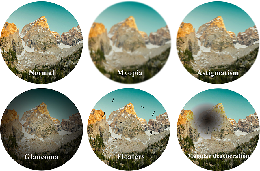
The development of three-dimensional (3D) bioprinting and biofabrication technologies has brought hope for the mass preparation of corneal substitutes. 2 3D bioprinting is to prepare a scaffold with spatial depth by layer-by-layer stacking, which can improve the interaction between cells compared with traditional fabrication techniques. 3 The printing materials that contain cells or biomolecules are called bioinks, 4 and the formulation of bioinks is an important part of the implementation of successful 3D printing. Cell-loaded bioinks have been used in a variety of printing methods, particularly extrusion printing. The inks used in extrusion-based bioprinting should have shear thinning properties, also known as pseudoplastic, because excessive shear forces and extrusion pressures could affect the activity and function of loaded cells. 5 Although the methods of inkjet printing and laser-assisted bioprinting (LaBP) are more suitable for low-viscosity materials, and the printing accuracy of these methods is relatively high; the printing speed is slow. As another 3D printing technology, stereolithography (SLA) requires a large number of cells to fill the entire bath volume with ink. This means there is a high demand for cell culture.
Due to its relatively low thickness and lack of vascularization, natural corneal tissue is suitable for preparation by 3D bioprinting. In contrast, as the precise geometric structure and high transparency, the bioprinting of cornea poses some challenges, so the preparation strategy for corneal substitutes needs to be well designed. In addition, the basic requirements of bioinks for printed corneal substitutes are transparency, printability, fidelity, cellular compatibility, and proper mechanical strength. Among them, printability is the most basic requirement. Printability is used to describe the properties of a material that can be extruded into continuous filaments to maintain the 3D structure with good shape fidelity and integrity. The performance requirement for the ideal printability of ink is the ability to produce a suitable well-defined jet, droplets, or continuous filaments.
6
Extrusion and inkjet printing systems have certain requirements for the rheological properties of materials, especially viscosity. Hydrogels or their precursors are hydrophilic polymers with 3D networks that can capture large amounts of water or biological fluids for cytocompatibility requirements and emulate the extracellular matrix (ECM) of cornea,7–9
so they are mostly used as bioinks in regenerative medicine. Crosslinking is a common way to cure bioinks into hydrogels after/during printing. The polymer backbone of hydrogels is connected with different hydrophilic functional groups, including -OH, -CONH, -COOH, -SO3, and -CONH2, which can be crosslinked physically or chemically.
10
The following are the common crosslinking mechanisms in bioprinting:
Ion crosslinking: Sodium alginate is a typical representative of ion crosslinking. The most commonly used method is “external gelation.” However, the crosslinking speed of external gelation is too fast for the preparation of fine structures. In contrast, the internal gelation method improves the fidelity and increases printability of alginate hydrogels. For example, precrosslinked hydrogels were prepared by mixing sodium alginate, CaCO3, and D-glucono-δ-lactone in a ratio of 2:1:1. A loosely precrosslinked alginate network enabled it to maintain a certain shape during the extrusion process and further crosslinked with calcium chloride after printing. The results showed that it could enhance the printability without affecting the viability of the embedded 3T3 cells.
11
Physical crosslinking: The crosslinking of gelatin is a representative example; by regulating the temperature of the printing sleeve, the temperature of the air in the environment, and the temperature of the receiving plate in the printing process separately, hydrogels can be formed through hydrogen bonds between molecules.
12
Photocrosslinking/radical polymerization: According to the timing of the printing process, it can be classified as postcrosslink, precrosslink, or in situ crosslink. Postcrosslinking is commonly used at the last moment of each layer or during overall printing. Appropriate photoinitiators are required to crosslink hydrogels into solids in minutes or even seconds to minimize cytotoxicity. The mechanical properties, degradability, and biological properties of hydrogels can be adjusted to a certain extent by adjusting the ratio of methacrylamide or photopolymerization time.13,14 In addition, precrosslinking irradiates ultraviolet light before extrusion, but it leads to high and inconsistent extrusion forces, making it difficult to form a uniform structure, with a cell survival rate of only 47%. In situ crosslinking by replacing the printing needle with a transparent material such as glass, rapid crosslinking can be performed before the material is deposited, and the precrosslinking strategy can enhance the viscosity of the ink, thereby improving its printability.
15
In addition, there is a trade-off between printability and biological function, in which weak materials are usually more suitable for 3D cell culture but exhibit poor shape fidelity when printed in air. Briefly, functionalized bioinks require the incorporation of bioactive substances or signals to induce cell behavior, including adhesion, migration, and differentiation, and ultimately achieve autologous corneal regeneration. The different types of biomacromolecule-based bioinks for corneal construction are introduced below.
Co-dECM-Based Bioinks
The corneal ECM is composed of collagen, glycoproteins, sulfated glycosaminoglycans (sGAGs), and a variety of proteins. Different components play different roles in the corneal structure, for example, proteoglycans can regulate the orientation and structure of collagen in the ECM. 16 The corneal decellularized ECM (corneal dECM/Co-dECM) is the component closest to the natural tissue.
Corneal-specific bioink prepared from the components of the acellular cornea was evaluated. Co-dECM gels showed similar transparency (over 75%) with human cornea in the visible spectrum of light due to the thin collagen fibrils. In the printability test, the Co-dECM bioink also showed concentration dependent property, the gelation time was in 5 minutes at 37°C–,8.7 times shorter than 0.5% Co-dECM, and the viscosity value of high concentration Co-dECM ( 2.0% ) could reach 64.99 Pa s at shear rate of 1 s−1. 17
The acellular matrix-derived bioink was used to print the film and implanted into the rabbit corneal stroma layer for 4 weeks. Optical coherence tomography was used for noninvasive monitoring in vivo, and the results showed excellent biocompatibility. 18 Other researchers have tried to use acellular matrix and tissue engineering methods to simulate the layered 3D structure of the natural cornea.19,20
To date, numerous methods have been reported to remove cells from tissue, including nonionic, ionic, and zwitterionic detergents, as well as enzymatic and physical methods. 20 For xenotransplantation, in order to meet the safety requirements of transplantation, the dsDNA residue of 1 mg dry tissue should be <50 ng, the length of the DNA fragment should be <200 bp, and the nucleus should not be stained with DAPI (4',6-diamidino-2-phenylindole) or hematoxylin and eosin. 21 However, existing decellularization methods cannot effectively remove all cells and DNA fragments after processing. 16 In addition, different decellularization methods still cause damage to natural components to various degrees. According to the literature, after sodium dodecyl sulfate (SDS) treatment, the loss of sGAGs in the corneal stroma is as high as 47%. 22
Although other studies have shown that the content of collagen and glycosaminoglycan after treatment is not much different from that of the natural cornea, decellularization treatment also reduces the content of other cytokines, including bone morphogenetic protein, FGF, IGF, TGF, and VEGF (Table 1). The absence of these cytokines may have an impact on the biological activity of the bioinks.
Cytokine and Growth Factors in Natural Bovine Cornea Before and After Decellularization (pg/mg) 17

BMP, bone morphogenetic protein; FGF, fibroblast growth factor; IGF, insulin-like growth factor; TGF, transforming growth factor; VEGF, vascular endothelial growth factor.
In addition, the low viscosity of the dECM bioink would also affect the mechanical properties and fidelity of the printing scaffold, 23 so further improvement is needed. Alternatively, acellular matrix can not only be used as a scaffold structure, but also has the effect of promoting corneal regeneration. 24 For example, dECM can be converted into additives of particle components (corneal tissue-derived ECM microparticles) and added to fibrin hydrogels for promoting corneal epithelial and matrix repair. 25
Natural Polymer-Based Bioinks
Some natural water-soluble polymer materials from biomacromolecules (sodium alginate, hyaluronic acid [HA], starch, collagen, gelatin, polylysine, etc.) can be crosslinked under certain conditions (heating/chemical/physical crosslinking) to form hydrogels with good biocompatibility similar to the ECM structure. The thixotropic shear thinning behavior of hydrogels is suitable for the extrusion process and can be processed into various shapes. Hydrogels formed through crosslinking also have a 3D network structure that can absorb liquids with a dry weight of 1000 times. These characteristics make them suitable for use as bioinks and cell carriers.26,27
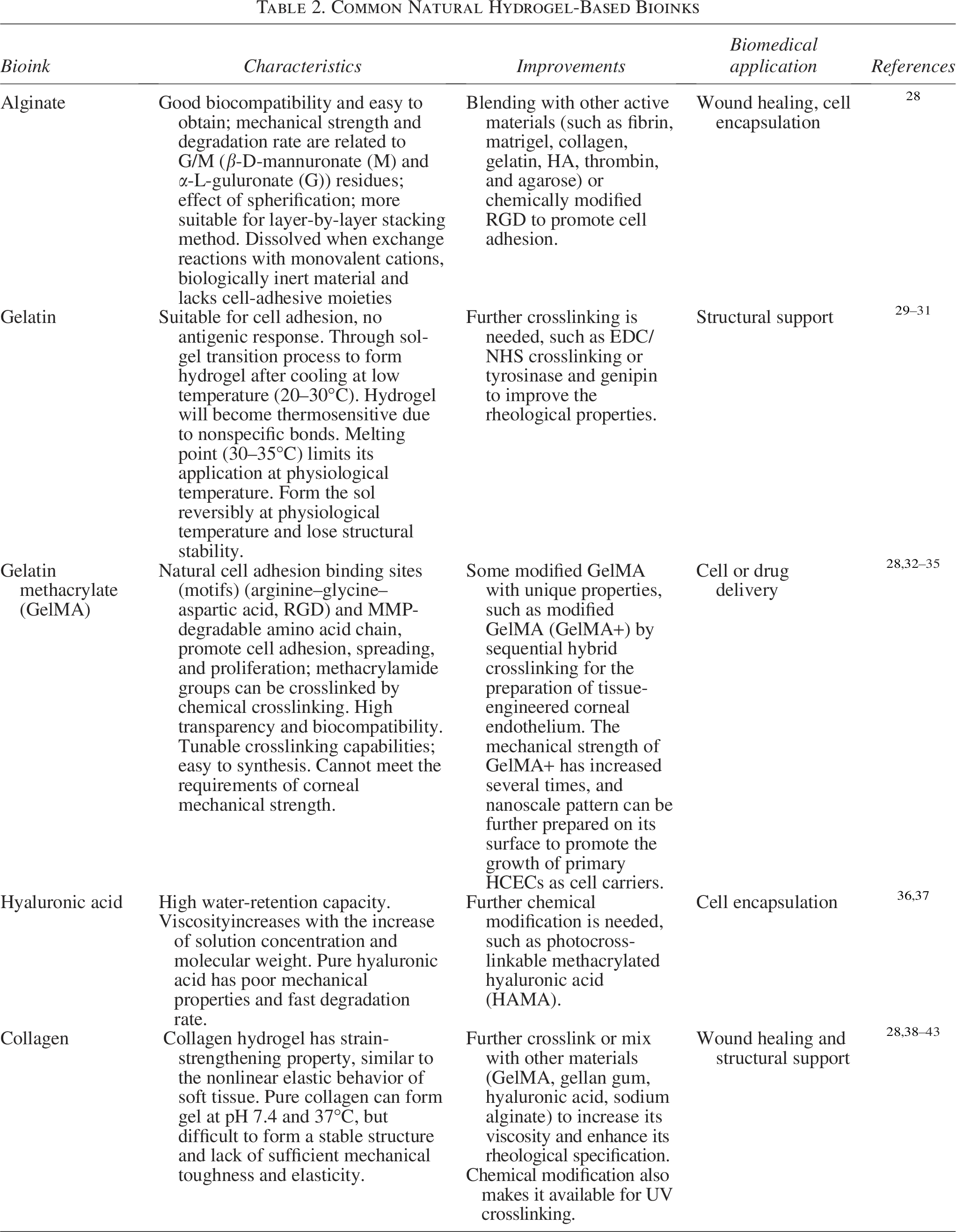
Natural hydrogels can simulate a 3D tissue environment, making them good candidates for corneal bioprinting inks. However, the high water content, limited structural stability, and viscoelasticity of natural hydrogels pose challenges for printing scaffolds with high shape fidelity and porous structures. Furthermore, natural hydrogels have biocompatibility advantages, but their mechanical properties are generally weak. So far, single-component hydrogel inks have been unable to meet people’s printing needs before further blending or modification to form a composite hydrogel (multicomponent hydrogel). Table 2 describes the characteristics and improvements of natural hydrogel-based inks.
Common Natural Hydrogel-Based Bioinks
By preparing a composite hydrogel material, the following advantages can be obtained.
Improvement in mechanical strength
In a study, a natural silk fibroin nanofiber structure (nanofibrils) was mixed with gelatin methacrylate (GelMA) and crosslinked by UV to obtain a tissue-engineered cornea with high transparency (light transmittance >85%) and good mechanical properties (tensile strength [3.8 ± 1 MPa]) and E-modulus (36.2 ± 7 MPa). 44 In the digital light processing (DLP) printing process, GelMA–GelMA homogeneous double-network hydrogel was prepared by adding GelMA with a low modification degree to GelMA with high concentration and high modification degree and photocrosslinking twice, which proved a better fatigue resistance and printability. 45 In another study, poly (ethylene glycol) diacrylate (PEGDA) and GelMA were mixed to obtain a two-component ink, as the copolymerization of long-chain PEGDA with GelMA, its compressive modulus close to the native cornea (100.7 kPa vs 115.3 kPa). 46 In order to improve the mechanical and biomimetic properties of the hybrid hydrogel, an electrospun collagen nanofiber membrane was applied, then the load of the break of hydrogel was increased by 40% and the markers of the encapsulated corneal stromal cells can be upregulated over time. 47 By dually crosslinked strategy of physical and moderately chemical, the mechanical properties of gelatin-carbohydrazide alginate hydrogel could be remarkably increased (tensile strength 1.068 MPa) compared with 0.129 MPa of gelatin-alginate hydrogel. 48
Improvement in biocompatibility and light transmittance
Since a single material is often unable to simultaneously meet both biocompatibility and light transmittance properties in cornea bioprinting, the combination of different materials could make up for their respective defects. 49 For example, the formula of acellular matrix–GelMA could greatly improve the light transmittance of the acellular matrix treated by SDS. 16 In addition, synthetic and semisynthetic materials have not received sufficient attention and application, as the high risk of transplant rejection. Compared with other polymers, aliphatic polyesters are biodegradable and biocompatible. For example, PLGA electrospun membrane can promote corneal epithelium regeneration in vivo, 50 but the degradation rate should match the requirements of medical applications. In another study, poly-ε-caprolactone (PCL) scaffolds would lead to upregulation of scar-related genes in stromal cells. 51 The transparency of Poly(lactic-co-glycolic acid) (PLGA); or PCL is relatively poor, which is also a problem that needs to be overcome in the future to meet the requirements of corneal transmittance.
“Removable Inks”: Assistant Bioinks
As a precision optical structure, bioprinting of the cornea puts forward higher requirements for high-fidelity bioinks. At present, a corneal prototype with curvature can be perfectly fabricated using computer software. 52 Corneal scaffolds with a certain curvature can be better spread and integrated seamlessly with autologous corneal tissue and restore vision effectively. A study also showed that the printed convex corneas could inhibit the transformation of cornea stromal cells into fibroblasts compared with flat corneas. 53 However, as an overhang structure, corneal bioprinting still faces challenges. To reduce the damage to cells, low-viscosity inks are often used, and these inks cannot self-support before one or even multiple crosslinks, 54 because the self-gravity of the hydrogel often leads to the collapse of the printed structure. 55
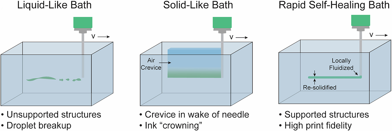
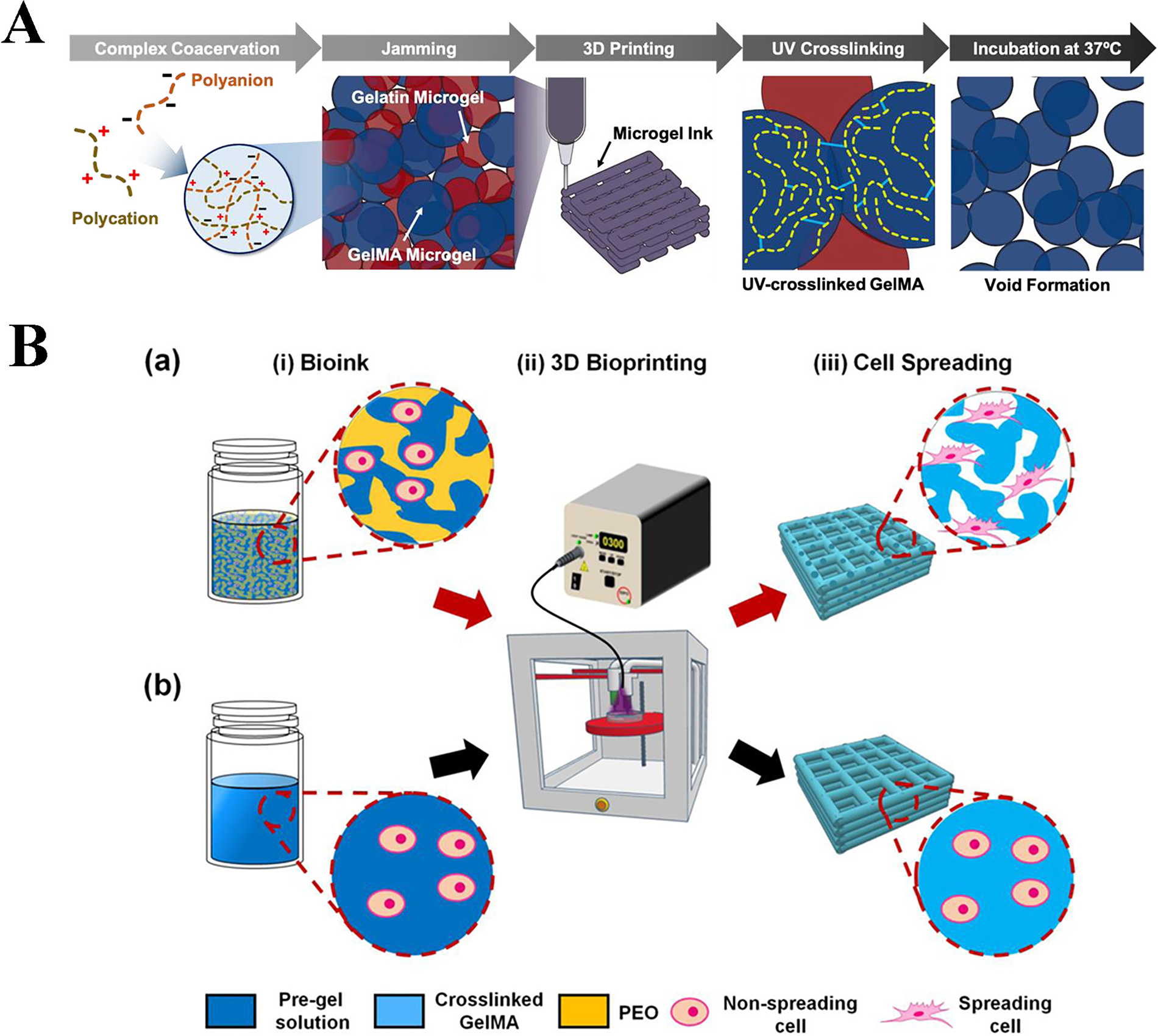
A new type of 3D bioprinting material and form could provide assistance for bioinks before they are removed from the printed structure. According to the “extent of assistance” (material usage), assistant bioinks can be divided into two types: support bath and sacrificial ink. With the “temporary assistance” of the materials, the support of bath relaxes the strict material requirements for the printability of bioinks by preventing structural collapse and improving print fidelity, so that otherwise unsuitable bioinks may be used to print into larger and more complex constructions with high fidelity (Fig. 2).

Corneal 3D bioprinting of
Support baths (suspension bath/3D embedded bioprinting)
In contrast to traditional extrusion printing that is exposed to air, 3D embedded bioprinting limits the enclosed deposited ink by the physical constraints of the supporting bath and reduces the interfacial tension between the ink and the surrounding environment, thus avoiding the limitations of printing in the air to improve the resolution and shape fidelity. This can prevent the structure from collapsing and make it print more complex features. This method eliminates the self-supporting requirements of inks, the use of a support bath medium allows the weak materials with poor structural integrity to be used as bioinks, including some lower viscosity materials—ECM-based bioinks with only a 2.80 Pa·s viscosity, 56 which can also construct complex 3D structures, and the printed objects are closer to the target computer-aided design (CAD) model.
Since the first use of synthetic polymers, Pluronic F-127 was used as a support bath to print interconnected microvascular network structures (microvascular networks) in 2011, 57 this kind of 3D embedded printing has developed rapidly. When the nozzle tip of the printer is supported by the bath liquid material, the bath liquid can heal itself, resolidify around the printed ink, and fix it in an appropriate position. Most of the support baths are granular gel support baths, as support tanks composed of jammed microparticles have recently become popular. In this system, hydrogel particles are usually mixed with aqueous, cell-compatible buffers and then centrifuged to compress the particles into a blocked state, usually called a slurry. When the printer nozzle passes, the filled particles are temporarily fluidized and no longer blocked. Once the shear stress of the nozzle was removed, it was restored to a blocked state (Fig. 3). In addition to the volume rheological behavior that affects print quality, factors such as particle size and shape also affect print resolution and fidelity. 58
Assistive materials for 3D bioprinting. Reprinted with permission from Brunel et al. 58
Greater particle size and greater polydispersity have been shown to result in highly variable printed filament morphologies, as the ink can flow into the gap between adjacent support groove particles. 59 Reducing the particle size and increasing its uniformity can reduce the printing resolution to 20 μm and reduce the surface roughness of the printing filament, resulting in the printing structure closer to the expected CAD model. 58 The time required for the bath material to be scratched by the nozzle and return to the initial state (resolidification) is called the recovery time (self-healing time or thixotropic time). Materials with a shorter mechanical recovery time after removing the applied shear stress (e.g., nozzle movement) are more suitable as support baths because they can quickly return to the initial solid state after a sudden change in shear stress. This ensured that the bioink material remained supported after printing. This support bath makes non-Newtonian fluids a candidate for bioinks. At present, support bath materials often yield stress materials, and the range is extended to include alginate, agarose, gellan, laponite nanoclay, acrylamide, nanocellulose, xanthan gum, and even slurries of cell spheroids.55,58
In addition, when printing layer-by-layer in air, discontinuities often occur because the interface between layers that are deposited sequentially is not seamless. For example, the strip pattern formed by extrusion printing and the weak connection between the rows affect the transparency of the corneal scaffold. Even if the printing resolution is as low as 10 μm, it appears as a stair or terrace for the curvature of the cornea. However, in a water-containing supporting bath, a small amount of ink diffusion can occur before curing, this is conducive to the improvement of the staircase effect and make it easier for a single filament to crosslink into a sticky structure.
In another study, 60 the printing fidelity of GelMA bioink was assessed in multiple parameters, including the diameter and the uniformity of printed strands. The results showed that as the concentration of Carbopol bath increased, the deformation of printed strands reduced significantly compared with conventional air printing. A high granular particle number in support bath is also attributed to the formation of an uniform line (Fig. 4). Some new support bath and technologies are also emerging; “Freeform reversible embedding of suspended hydrogels” (FRESH) technology is a suspension printing technology supported by gelatin particles, which can print 20 um thick collagen filaments and print human hearts using suspension hydrogel FRESH and collagen biological ink. 59 HA is modified with adamantane or beta-cyclodextrin (β-CD), so that when the two components are combined, they form a supramolecular, self-healing hydrogel support bath. 61 The supporting bath can be washed away by competitive binding with soluble β-CD.
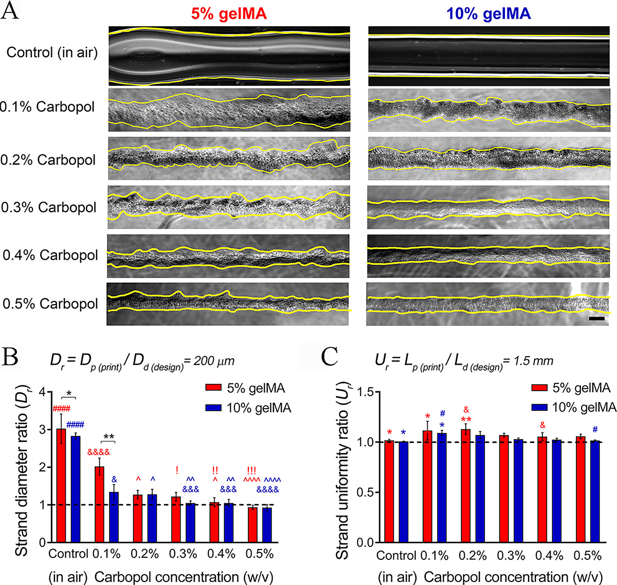
Characterization of printing fidelity of GelMA bioink in Carbopol support bath. (
Although most bioprinting strategies require removal of the printed structure from the support bath, some studies have incorporated the bath into the final fabricated structure. This alternative strategy can be used to prepare bioinks with a higher cell density or even bioinks with only cells, and the support bath will continue to provide structural support throughout the culture period.
Sacrificial inks
Sacrificial inks, also known as fugitive inks, are mainly printed separately from bioinks in 3D extrusion bioprinting and create hollow internal structures such as vascular-like networks during printing. 58 Sacrificial inks are mainly used to create void spaces and patternable perfusion networks, but they can also be mixed directly with other bioinks to temporarily increase viscosity and slow down cell sedimentation to change their mechanical properties to improve printability or increase porosity to improve the diffusion, migration, and proliferation of encapsulated cells. Similar to the supporting bath, sacrificial inks can be used as physical supports for overhangs and hollow structures until the bioink is crosslinked to self-support. 62 It prevents the collapse of the printed bioink geometry, and the removal mechanism is similar to that of a support bath. At present, a series of sacrificial ink materials have been used to make other printable materials, including alginate, agarose, and gelatin, to the bioink formula.58,63 Because sacrificial inks usually do not contain cells, some nonhydrogel materials with more stringent manufacturing conditions are also used as sacrificial inks, particularly for sacrificial molds.
Porosity is an important matrix property that affects many aspects of cell behavior and can be adjusted by including sacrificial ink components. Compared with nanoporous hydrogels, microporous hydrogels have several advantages in 3D cell culture, including improved nutrient exchange and increased cell growth, diffusion, and migration. Notably, sacrificing microgels 62 or movable, immiscible aqueous phases in bioinks64,65 makes it possible to print structures with interconnected pores (Fig. 5).
Sacrificial inks were used to prepare porous structures. (
When the printing is completed and after the surrounding nonsacrificial material is solidified into its expected geometry, the sacrificial inks are removed from the structure via a removal mechanism. For sacrificial ink and a support bath, the removal mechanism is critical to the final printing quality of structure and biological function of embedded cells. Removal mechanism includes physical extraction, chelating agents, dissolution in aqueous solution, washing with water, elevated temperatures (37°C), lowered temperatures (4°C), and dissolution in aqueous solution. First, the acidity and alkalinity of sacrificial ink or support bath need to be unified with the bioinks. Second, if the assistive materials are removed by temperature changes, the crosslinking mechanism of the bioinks should not depend on temperature. Otherwise, the structural integrity of the print cannot be maintained during the removal process. For instance, Pluronic F-127 can be removed without destroying collagen fibers due to thermoreversibility and extrudability, when it is combined with type I collagen to form a bioink. 66 In another study, the microgels are composed of crosslinked polyacrylic acid copolymer (Carbopol). 67 After printing, the printing structure is cleaned by stirring in water to remove the carbon wave support bath, but the methods that rely on stirring or physical removal may not be suitable for printing with fine or detailed features. Any microgels can be further removed by adding multivalent cations and reacting with the residual Carbopol. 58 Similarly, methods that induce large changes in pH, temperature, or ion concentration may reduce cell viability and affect cell behavior. 58 So, these methods may not be suitable for cell types that are particularly sensitive to such changes in the environment. Another challenge is the relative difficulty of visualizing when bioinks are extruded. If the support groove is not optically clear, it is difficult to identify problems, such as needle blockage, bubbles, or defects during the printing process.
In the future, assistive printing materials can also be designed to provide biochemical and biophysical clues to surrounding or encapsulated cells to help them mature to the desired phenotype. The interface of the bioink bath and the assistive printing materials can also undergo crosslinking reaction through modification of the reactive groups. For example, carbohydrazide-modified gelatin ink (Gel-CDH) can rapidly cross-react with oxidized alginate (OAlg) in a supporting bath. 68
Evaluation and Development of New Generation Bioinks
Corneal structure bionics
For corneal stroma reconstruction, the most challenging aspect of bioprinting technology is arranging collagen fibers to construct bionic scaffolds with high transparency. Transparency is an important biological characteristic of the cornea. When exposed to light, the transmittance of the human cornea is approximately 90% at 700 nm. 69 The unique ultrastructural features of the cornea, that is, the highly oriented arrangement of collagen fibers, are crucial to the transparency of the cornea. In addition, it guarantees the biomechanical properties and affects the shape of the cornea.
The cornea is formed by the orthogonal arrangement of collagen fibers, namely, the lattice pattern. Approximately 250 collagen lamellae of 1–2 μm thick form the central matrix.70,71 Although the individual difference in human corneal elastic modulus is great, the measurement results showed that there is a biomechanical gradient in the stroma lamellae at different positions. 72 This is caused by the difference in the arrangement of collagen fibers in the anterior and posterior parts of the corneal stroma. The packing density of the anterior lamella is higher. It is undulating and interwoven, which makes it more mechanically stable. 73 In contrast, the lamellar structure of the posterior stroma is looser, making it easier to expand. The orthogonal arrangement of corneal stromal fibers results in the lower light scatter. Therefore, imitating an orthogonal arrangement is helpful for improving the transparency. Researchers have attempted to use 3D bioprinting to accurately deposit layers and affect the arrangement of fibers through shear-induced and prepare collagen fibers with oriented structures. Nevertheless, a relatively small diameter needle would increase the shear force and lead to fibroblastic characteristics of stromal cells. 74
Composite materials have been used to better simulate the highly heterogeneous composition of natural corneas. HA bioinks containing human adipose tissue-derived stem cells (hASCs) and cell-free inks have been used to construct composite scaffolds with heterogeneous microstructures. 75 In addition, a porous structure would contribute to implant integration into host tissues seamlessly, and appropriate porosity and pore size are needed to support the diffusion of oxygen, carbon dioxide, and nutrients. But if the pore size is too small, it will only adhere to the corneal epithelium and endothelium and will also affect the integration of host tissue. 76
Promoting corneal regeneration
For corneal regeneration, cells loaded in bioinks play an important role,77,78 especially considering that the proliferation ability of human corneal endothelial cells in vivo is limited. Using gelatin as the printing medium, corneal endothelial cells overexpressing ribonuclease 5 (angiogenin) were deposited on freeze-dried amniotic membranes. The transplantation experiment of rabbit corneal endothelial decompensation model showed that corneal thickness and edema were significantly improved. 79 In the other two experiments, another corneal cell, stromal keratocytes were also added into collagen-based and GelMA bioink.80,81 GelMA has good compatibility with corneal stromal cells and reported the potential to synthesize biglycan and decorin motives. 82
High-throughput printing of gelatin-collagen ink was achieved using a combination of various preparation techniques (SLA and extrusion). Corneal stromal cells maintained high activity (>95%) within 2 weeks, with high transparency, smoothness, and a certain curvature. 83 In addition, loaded cells also facilitate the integration of the printed corneal stroma and host tissue. After cornea-mimicking tissues were prepared by the 3D LaBP method, two kinds of human stem cells were loaded into the ink, and after 7 days of porcine organ culture in vitro, immunohistochemical staining showed that the printed matrix successfully adhered to the host tissue, and hASCs tended to migrate to the host tissue. However, the composition of the ink was complex and required polymer sheets such as PET as a stable matrix for printing tissues, indicating that it was only suitable for static culture conditions in this study. Anni et al. showed that the adhesion performance of ink was improved by covalent grafting of dopamine to the polymer backbone on the main chain, and human pluripotent stem cell-derived neuronal cells (neurons) were loaded on the periphery. The results of in vitro pig corneal transplantation showed that after 21 days of culture, it had good attachment and adhesion to the host cornea. Human adipose stem cell-derived stromal cells in the scaffold contribute to axonal growth and innervation development. 84
There is close intercellular communication between different corneal cells, and the factors secreted by endothelial cells can promote the complete differentiation of epithelial cells and the formation of the epithelial basement membrane. Keratinocytes in the anterior corneal stroma secrete basement membrane proteins that promote epithelial repair. The coculture experiment of corneal epithelial cells and stromal fibroblasts showed that there was obvious secretion of provisional matrix components and extracellular vesicles after 4 weeks. 85 So, multitype cell printing is contributed to provide a more suitable microenvironment for cell communication. By mixing corneal epithelium, stroma, endothelium, and limbal stem cells (LSCs) with ink, 3D bioprinting enables the construction of an accurate 3D model of the cornea for in vitro drug screening and toxicological studies of corneal tissue engineering and corneal regeneration. 86
Although the cost of derivation, differentiation, cloning derivation, clone characterization, and differentiation process of hiPSCs are still high, for iPSCs that are sensitive to physical forces, the shear force generated during the printing process may change their gene expression profiles and adversely affect the cells, 87 inducing hiPSCs to obtain specific corneal cell phenotypes is still promising in large-scale cell cultures. 88 In addition to stem cells, exosomes secreted by corneal stromal stem cells could also promote the regeneration of transparent stromal tissue and block the infiltration of neutrophils into damaged corneas and scaffolds. 89 Compared with only stroma or mesenchymal stem cells (MSCs), MSC exosomes contain cytokines/miRNA that also contribute to corneal stromal cell activity and stroma regeneration.90,91 Other studies have also demonstrated that mixing with biodegradable polymeric materials, such as methylcellulose, could promote cell adhesion and proliferation. 92
Oriented to clinical needs
At present, early intervention is becoming surgical trend to reduce the risk of postoperative complications, and the development from penetrating keratoplasty to lamellar keratoplasty reduces the immunogenicity of xenotransplantation. 93 The change in surgical protocol is required to maximize the retention and mobilization of the regenerative capacity of the natural cornea. A more targeted and personalized treatment is needed. Personalized preparation strategy is an advantage of 3D printing. By using custom design through computer tomography scan, and developing personalized corneal substitutes, including drug delivery strategies, 94 geometry control, and even refractive ability design based on corneal injury or lesion type, it would contribute to shortening postsurgery management and rapid recovery of vision (Fig. 6). The external location of the cornea makes it susceptible to several pathologies. Understanding the clinical treatment and demand of corneal common diseases is helpful for better designing and preparing bioinks.
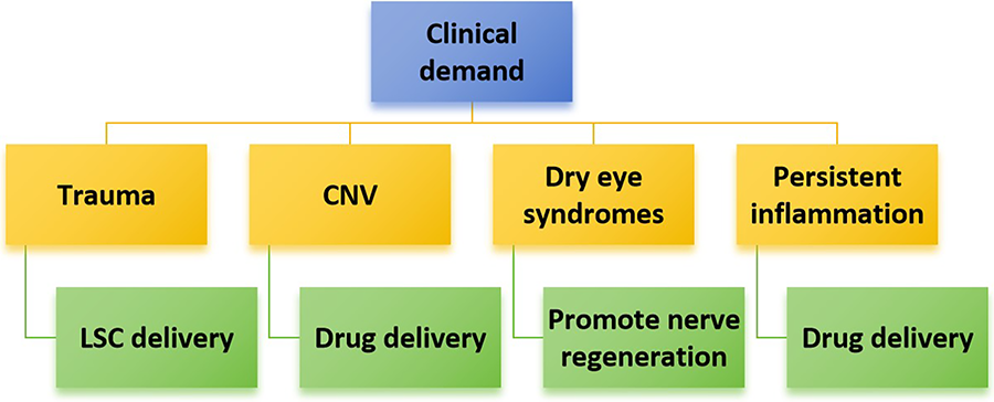
Clinical requirements of 3D-printed corneas.
Trauma
After the destruction of the LSC niche, inducing LSC deficiency, the corneal epithelial cells cannot be updated, and the conjunctiva and blood vessels overgrow, accompanied by severe pain and photophobia, 95 finally causing traumatic corneal blindness.
Regeneration of the corneal epithelium is very important because it can stabilize the tear film and reduce the risk of infection. At the same time, the activation/quiet state regulation of embedded LSCs is required. 96
Corneal neovascularization
The cornea needs to remain avascular to prevent pathological blood vessels from growing. The triblock copolymer PLGA–PEG–PLGA was used as a carrier of metformin and levofloxacin hydrochloride (MET/LFH @ Thermogel) to form a thermosensitive hydrogel at physiological temperature, which could be continuously released in vitro for 1 month. Subconjunctival injection of the mouse corneal alkali burn model inhibited angiogenesis and significantly inhibited the formation of corneal neovascularization. However, its transparency is poor. 97
Dry eye syndromes
Cornea is the most abundant and sensitive tissue of nerve fibers.98,99 Innervation is distributed in the corneal epithelium and stroma, and the stromal nerve trunks have a density of 33–71/mm2. 100 The corneal stroma and epithelial nerve plexus are cut off in a large range during corneal transplantation. Long-term dry eye after surgery may be related to incomplete repair of damaged corneal nerves. For example, neurotrophic factors secreted by innervated tissues and glial cells (trophic factors such as neurotransmitters and neuropeptides) contribute to the regeneration of the subepithelial nervous system of the graft after surgery. It can also increase the number of goblet cells and tear secretion and reduce the apoptosis of corneal epithelial cells.101,102 The addition of nerve growth factor to the scaffold helps guide neuronal extensions. In another study, a corneal tissue model that can be innervated by nerves was constructed using a silk fibroin scaffold. 103 Besides, electroactive hydrogels, containing conductive polymers, could promote corneal nerve regeneration. 104
Postoperative persistent inflammation
Due to surgical injury and infections, there is often a persistent inflammatory response on the ocular surface. Patients often require glucocorticoids to inhibit T cell activation and reduce the expression of inflammatory factors. In severe infectious keratitis, loading protein inhibitors could reduce scar formation in the healing stage, 105 and loading strategy could maintain the sustained release of MET and LFH in thermoresponsive hydrogels. 97
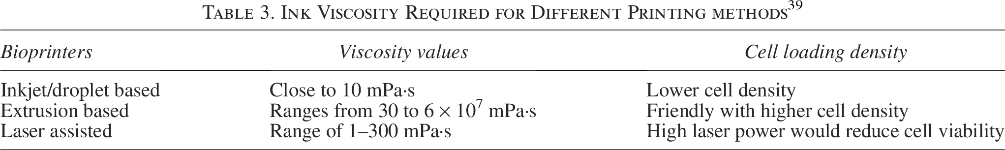
At present, the number of bioinks suitable for cornea is still limited, and more types of bioinks still need to be developed. Most of the evaluation methods for printed materials come from the methods established and widely used in tissue engineering and regenerative medicine, such as gelation time, biocompatibility, mechanical strength, swelling characteristics, and degradation profile. 54 Many experiments simply evaluate the printability and light transmittance of the inks. The improvement of the evaluation method also facilitates the development of ink specificity, including new materials within the printable range (as shown in Table 3); in addition to evaluating the fiber formation ability and rheological evaluation (viscosity can be controlled by adjusting the molecular weight/polymer concentration/additive/temperature and precrosslinking), 106 the permeation of small molecular nutrients, such as glucose/albumin, and clinical complications, such as stromal melt and long-term calcification, should also be considered.
Ink Viscosity Required for Different Printing methods 39
In addition to the characteristics of the hydrogel ink, the crosslinking method and crosslinking agent used are also important factors affecting the printing results. Studies have shown that the residual initiator after crosslinking will have an adverse effect on cell proliferation. 107 It needs to be evaluated to avoid cytotoxicity to the embedded cells during and after crosslinking. Most of the experiments have a short investigation time and focus on the cell compatibility of the material, and evaluation of the long-term performance of the ink is needed before clinical application. Owing to the lack of blood supply in the natural cornea, whether the transplanted cells can differentiate and proliferate for a long time remains to be further investigated.
Although the necessity of corneal scaffold degradation remains controversial, some researchers have explored the regulation of material degradation rate. Type-I collagen and gelatin were used to prepare bioengineered corneas, and the water content and enzymatic degradation can be adjusted by controlling the ratio. The results showed that the composite hydrogel formed by EDC/NHS crosslinking had better transparency and an obvious porous structure than the pure gelatin hydrogel, which was beneficial to the adhesion of human bone marrow MSCs. 108 In addition, the degradation of the hydrogel can be regulated by adding sodium citrate to the collagen–gelatin–sodium alginate hydrogel. 109 In a bandage contact lenses research, 110 the incorporation of PEGDA (Mn700) improved the GelMA lenses’ resistance to handling and prolonged the release of loaded drug, and higher concentrations (10% and 15%) of PEGDA could extend the degradation profile of lenses up to more than 14 days. The results also provide an inspiration for the preparation of controllable degradability and drug release corneal substitutes. Degradability matching has a positive effect on corneal regeneration. With the increase in the number and type of loaded cells, degradability has also become an important factor to be considered. Controlled degradation allows cells to gradually replace the printed structure through the ECM secreted in the body.
Future Perspective and Conclusion
With the continuous expansion of bioprinting technology, it will inevitably intersect with other preparation technologies. A new integrated 3D cornea printing system, such as DLP in combination with extrusion, has been used to print 3D corneal substitutes. 111 By combining LaBP and inkjet printing techniques, accurate localization of single limbal cells and reconstruction of niches can be achieved. In addition, some nonbioprinting technologies also help improve the final performance, such as solvent casting, 112 direct writing, 113 and electrostatic spinning. 114 Aerosol jet printing, commonly used in electronic printing, can further improve the printing resolution of pure collagen ink. 115 Microelectric scanning and microelectric writing can accurately control the fiber orientation at the micro/nanoscale, which is an ideal method for reconstructing corneal ultrastructure. 86
It is necessary to further subdivision of hydrogel materials to investigate the performance differences between subclasses, such as the selection of type III collagen with finer fiber diameter and more resistance to digestion to prepare more tightly packed hydrogels. 116 Supramolecular hydrogels that can self-heal immediately after printing, with higher print fidelity, are emerging candidates. Furthermore, to meet the requirements of suturing, clipping, and other transplantation operations, nanoparticles have been used to improve the biomechanical properties and transparency of the artificial cornea. 117 In situ printing also has unique advantages in this field. 118
The low metabolic demand and avascular structure of the cornea are advantages over other tissue-printing methods. With the establishment of a series of proof of concepts for 3D bioprinting of human corneal structures,119–121 systematic evaluation of bioink formulation, rheology, and harmonization with all the parameters, and standardized use of bioinks will lead to better printability and fidelity without affecting corneal transparency and homeostasis.
Finally, the functional modification of biomacromolecule-based bioinks will also promote the application of advanced next-generation inks in personalized corneal preparation and regeneration.
Authors’ Contributions
L.J.: Literature review, data curation, and writing. J.Z.: Data curation, investigation, and writing. W.Z.: Methodology and supervision. X.L.: Project administration and conceptualization. All authors have reviewed and edited the article and approved the final version for publication.
Footnotes
Funding Information
This work was supported by the National Nature Science Foundation of China (32101101), Science and Technology Project of Tianjin Health Committee (TJWJ2021QN073), and Tianjin Key Medical Discipline Construction (No.TJYXZDXK-3-004A-3).
Disclosure Statement
The authors declare no conflicts of interest.
Ethical Approval
This study did not require ethical approval as it did not involve human or animal subjects.