



Abstract
Holins and spanins are bacteriophage-encoded enzymes that control the onset and efficiency of bacterial cell lysis in the late stage of the bacteriophage life cycle. Their ability to compromise bacterial membranes has attracted attention in various domains. With rise in antimicrobial resistance surpassing the development of new antimicrobials, phage-encoded lysis proteins, especially holins and spanin, have now become prospective for implementation of next-generation therapeutics because of their specific activity and low impact on the host. Furthermore, understanding their complex regulation, structural diversity, and controlled lytic activity has led to the expansion of their potential in fields such as vaccine development, drug delivery, bioprocessing, and other medical or industrial applications. This review highlights recent insights into holin and spanin biology, focusing on how these lethal, membrane-disrupting proteins function, and discusses their potential use in a wide range of applications.
Introduction
Bacteriophages (phages) represent a highly diverse class of viruses that specifically target and infect bacterial hosts. Frederick Twort and Felix d’Herelle independently reported agents capable of bacterial lysis. D’Herelle named the agent a bacteriophage, meaning “bacteria eater,” and began using it to treat bacterial infections. With the identification and large-scale development of antibiotic compounds, research on bacteriophages continued in some countries such as Georgia, but they were disregarded as significant therapeutic agents in the West, mainly because antibiotics were easier to administer. 1 The emergence of bacteria resistant to multiple drugs has renewed attention toward nontraditional antimicrobial approaches. One of the potential replacements for antibiotics is the use of bacteriophages. The advantages of phages—including specificity, nontoxicity, and self-dosage—are considered an effective strategy against infections caused by antibiotic-resistant strains. 2 However, despite their potential, phage therapy faces certain limitations, including narrow host range, delivery challenges, and the risk of bacteria evolving resistance to phages. 3
To overcome these limitations, recent research has turned to the bacteriolytic and bacteriostatic proteins encoded by phages, which comprise a variety of phage-derived proteins, such as endolysins, holins, virion-associated enzymes targeting peptidoglycan, and depolymerases that act on bacterial polysaccharides. Phage-derived enzymes have gained attention as potential clinical agents due to their selective bacterial targeting, wide range of antimicrobial activity, minimal likelihood of resistance development, and fast-acting bactericidal effects, and can be engineered for enhanced efficacy and specificity. 4 Endolysin and holin, two key lytic proteins, play crucial roles in their host lysis, and are the most commonly used lytic enzymes. Endolysins can degrade bacterial peptidoglycan during the final stage of phage propagation, enabling the release of newly formed viral particles. 5 Besides, holins and spanins are membrane-spanning proteins that build up within the bacterial membrane to compromise its integrity, regulate host cell lysis, and work together with endolysin to break down the bacterial cell wall. Although holin and spanin show advantage potential, endolysin is the most frequently used protein. A number of endolysins have already been employed as antibacterial agents, including Staphefekt SA. 100 against Staphylococcus aureus skin infections, CHAPK against S. aureus colonization, P128 against S. aureus nasal isolates, and the artilysin LoGT-008 against multidrug-resistant Pseudomonas aeruginosa and Acinetobacter baumannii.
Self-lysing bacterial strains for vaccine delivery, therapeutic protein secretion, and metabolic product recovery have been engineered by combining holins and endolysins in programmable genetic circuits that respond to environmental stimuli such as light, metabolites, or metal ions. These systems allow precise control of the duration and intensity of lysis, thereby enhancing safety and efficacy in clinical, pharmaceutical, and bioengineering applications. The modular design of these lysis tools makes them attractive for integration with future microbial platforms. As a result, there has been growing interest in endolysins, holins, and spanins in recent decades.
This review summarizes recent advances in our understanding of holin and spanin proteins, focusing on their structure, diversity, mechanisms of action, and potential applications not only in antibacterial therapy but also in vaccine development, food safety, and industrial biotechnology.
Function of Holin
The term “holin” was first introduced to describe a class of small membrane proteins thought to control when host cells undergo lysis. This concept was mainly developed through molecular and genetic investigations using bacteriophage λ in Escherichia coli. 5 In the classical lysis model, holins gradually accumulate in the bacterial inner membrane and, at a defined time, form large pores, 6 that allow endolysins to pass into the periplasm and degrade the peptidoglycan, leading to cell lysis. 7 In Gram-negative bacteria, this process also requires spanins to disrupt the outer membrane (OM) as the final step. In contrast, an alternative system known as the pinholin–signal-anchor-release (SAR) endolysin pathway exists, where SAR endolysins initially accumulate in the periplasm as inactive, membrane-anchored proteins. When pinholins trigger membrane depolarization, the proton motive force (PMF) collapses, releasing and activating SAR endolysins, which then hydrolyze the MurNac–GlcNac bonds of peptidoglycan. 8 Thus, in such phages, holins only need to disrupt the membrane potential to control lysis timing. 9 Overall, while both systems depend on holins (or pinholins) for precise timing and on endolysins for cell wall degradation, their molecular mechanisms differ fundamentally.
Classes of Holins and Their Functional Roles
Holins and spanins are phage-encoded membrane proteins required for host cell lysis. In contrast to endolysins, the biochemical characteristics and structures of which have been well characterized, the diversity, topology, and action mechanisms of holins and spanins are much less known. It is important to understand their specific structures to take full advantage of their potential applications. The known holins vary in length from 60 to 145 amino acids. 10 Despite high sequence variability among different bacteriophages, holins typically contain one to three hydrophobic transmembrane domains (TMDs). 11 According to the number of TMDs, holins are classified into three categories: class I, class II, and class III (Fig. 1). 12
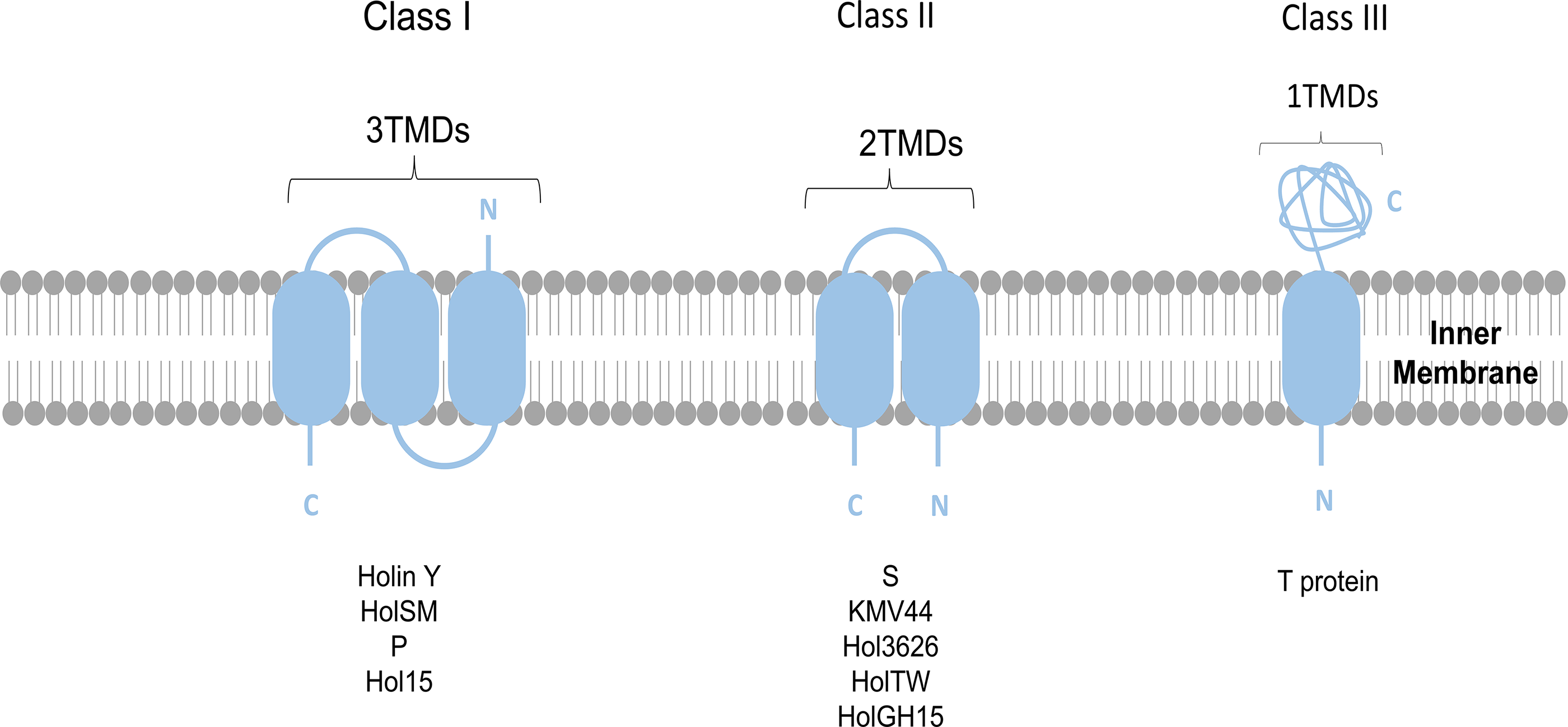
The topology of various holin classes. Class I holins possess three transmembrane domains (TMDs), with their N-terminus oriented externally to the membrane and the C-terminus internally (N-out/C-in). Class II holins, which possess two TMDs, have both ends—the N- and C-termini—positioned on the cytoplasmic side (N-in/C-in). Class III holins are characterized by a single TMD and have an N-in, C-out arrangement.
Class I holins
Holins grouped in class I are between 90 and 130 amino acids, with three hydrophobic TMDs. The N-terminus of these holins is situated in the periplasm, while the C-terminal domain, which is short, charged, and hydrophilic, is found in the cytoplasm. The λ phage holin S105 is a well-characterized representative of this class. 13 Additional examples of this category include holins such as HolSMP from the Streptococcus suis phage SMP, 14 Y from coliphage P2, 15 and Hol15 from S. aureus phage P68. 16
Class II holins
Class II holins are shorter, between 65 and 95 amino acids, and have two TMDs arranged in an N-in/C-in configuration.14,17 This group includes pinholins, which may work in a way that is quite different from canonical holins. 18 Pinholins have two transmembrane regions, TMD1 and TMD2, and are distinguished by their N-in/C-in membrane topology. 19 A well-studied example of a pinholin is the S2168 protein, which is produced by the S21 gene found in the lambdoid bacteriophage 21.17,20–25
KMV44, derived from P. aeruginosa φKMV, is predicted to have two TMDs. With a compact structure of ∼66 amino acids and a highly positively charged region at the C-terminus, it suggests that it is a typical pinholin and SAR endolysin combination. 26 Hol362, from Clostridium perfringens phage φ3626, is a protein consisting of 125 amino acids that features two TMDs and is found outside the cell membrane, classified as a group II holin. 27
HolTW, encoded by S. aureus phage Twort, is a holin protein with a length of 185 amino acids, making it the largest known class II holin. It has two possible hydrophobic, antiparallel transmembrane regions, along with a highly negative, charged C-terminus. 10 The protein encoded by HolGH15 in the GH15 phage of S. aureus is a 167-amino-acid protein that potentially contains two hydrophobic transmembrane helices. 28
Class III holins
Class III holins were initially identified, based on structural analyses of the T protein from bacteriophage T4, which is very different from classes I and II. 29 These holins are typically N-in/C-out oriented and have a single TMD. A 34-amino-acid cytoplasmic domain, a 20-amino-acid TMD, and a 163-amino-acid periplasmic domain make up the atypically large (218 amino acid) T holin. A characteristic of class III holins is the regulation of lysis timing through interaction between the periplasmic domain and an antiholin. 12 Antiholin RI directly binds to the periplasmic domain of holin T and inhibits holin oligomerization and pore formation. This inhibition blocks lysis until it is relieved by membrane depolarization. Holins from the KP15 and KP27 phages that infect Gram-negative bacteria are more examples. These proteins, which are ∼215 amino acids (∼24 kDa), have one TMD and a large 168-residue periplasmic region, aligning with the characteristics of the T-holin superfamily. 30
Holin Pathways in Phage Lysis: Canonical Versus Pinholin Mechanisms
Holin-mediated membrane disruption has been explained by two different paths: the canonical (Fig. 2) and the pinholin (Fig. 3). These pathways are linked to different hole creation and lysis time patterns. 31
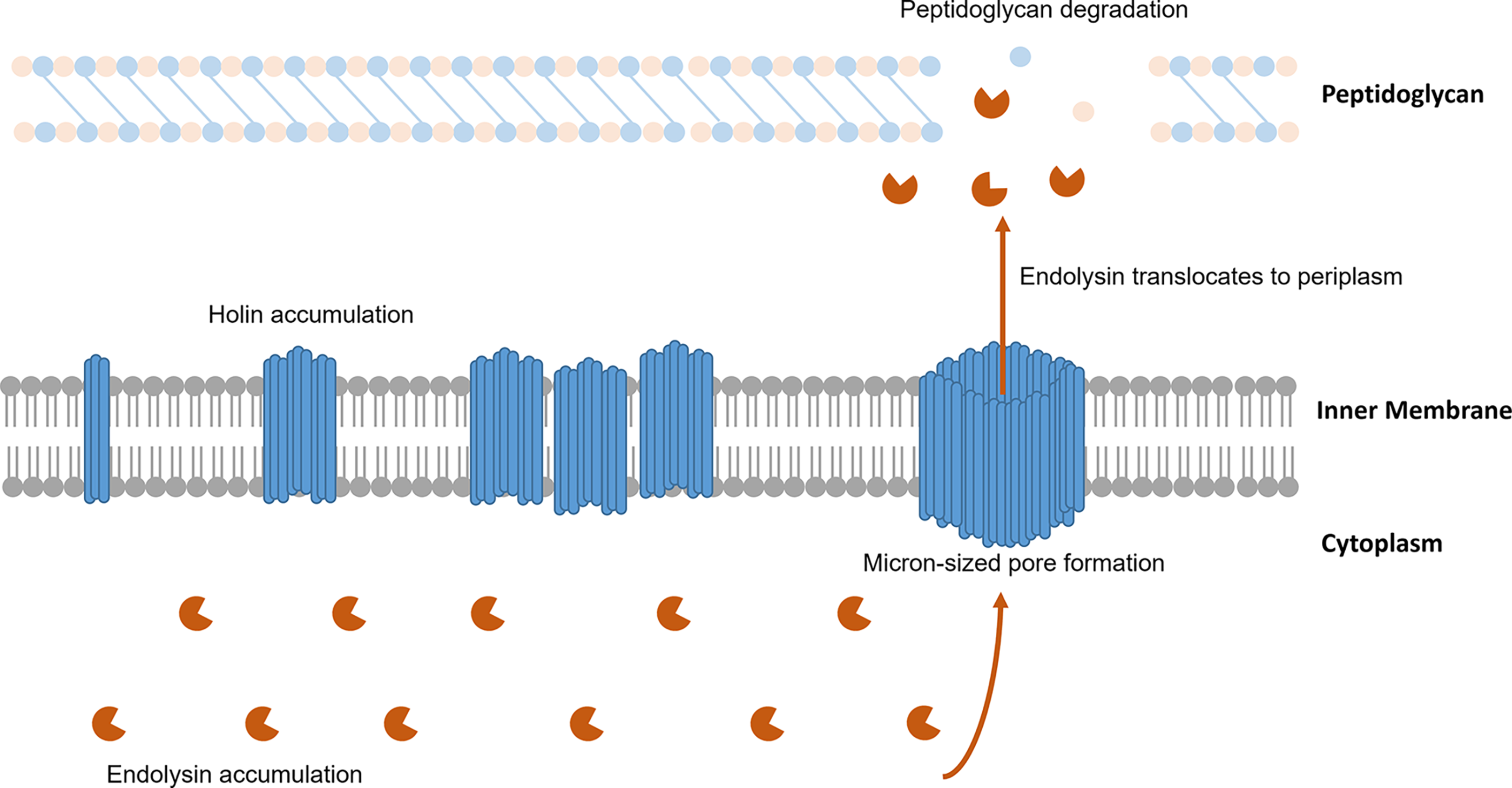
A schematic representation of the canonical lysis pathway shows that endolysins build up in the cytoplasm while canonical holins gradually accumulate in the inner membrane. At a certain point, the holins create large holes in the inner membrane that enable the endolysins to move into the periplasmic space and break down the bacterial peptidoglycan layer.
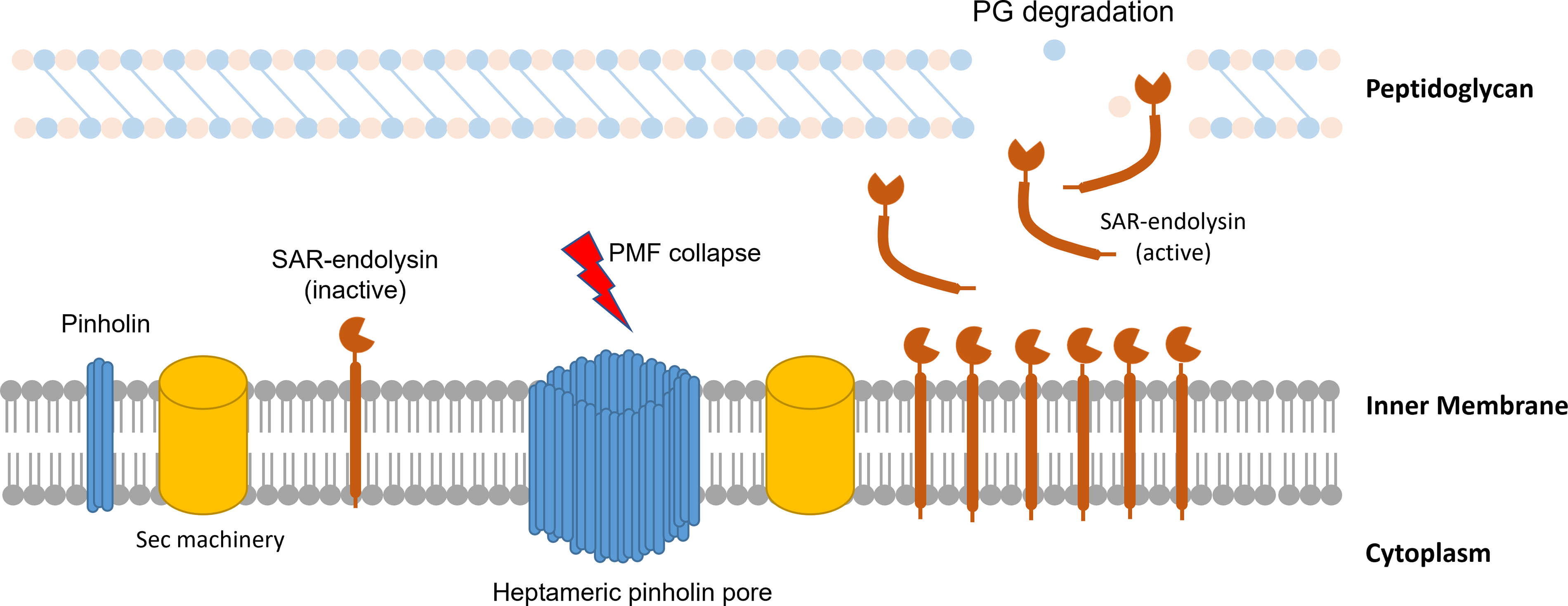
Schematic representation of the pinholin-mediated lysis pathway: pinholins and inactive SAR endolysins accumulate in the inner membrane. The proton motive pump is disrupted at a specific period in the lytic cycle when pinholins form heptameric channels ∼2 nm. Due to their sensitivity to the proton motive force, SAR endolysins are activated by this disturbance. Once activated, these enzymes degrade the peptidoglycan layer.
Canonical holins
Lambda bacteriophage holin, S105, represents the prototype of canonical holins. In order to allow prefolded, enzymatically active endolysin to get into the periplasm, where it breaks down the peptidoglycan layer (PG) and starts host cell lysis, it creates big, nonspecific holes in the inner membrane. 31 The oligomerization of S105 is essential for its lytic function. 9 During the late phase of gene expression in the phage lytic cycle, S105 builds up in the inner membrane as homodimers and later organizes into two-dimensional microdomains known as death rafts. 32 These rafts, composed of ∼1000 to 3000 holin molecules, exhibit local lipid depletion due to the dense packing of proteins, which compromises membrane integrity. This reorganization results in ion and proton leakage, causing localized depolarization and a decrease in the PMF. 31
As depolarization progresses, the death rafts reorganize, resulting in the rapid development of a micron-sized opening in the membrane. 33 In bacteriophages such as λ and T4, these pores are sufficiently large to permit the translocation of large proteins, including a 480 kDa R-lacZ fusion protein. 32 Imaging by cryo-electron microscopy (cryo-EM) of lesions induced by S105 confirmed their extraordinary size, average diameters being 340 nm, with certain lesions surpassing 1 μm. 34 Thus, holins serve as a “molecular clock” for the bacteriophage life cycle, precisely regulating the timing of host cell lysis at a genetically predetermined moment. 35
Pinholins
The holin S21 from phage 21 creates small pores in the membrane that disrupt the membrane potential, and is categorized as a pinholin, unlike large-hole-forming holins such as S105. 10 These structures serve primarily to depolarize the membrane without physically rupturing it. Structural studies using negative-stain transmission electron microscopy demonstrated that the active form of S21 consists of symmetric heptameric assemblies with a pore diameter of ∼2 nm, which are too small to permit the passage of large proteins such as λ-endolysin or GFP-fused periplasmic markers. 36 In a study by Pang et al., pinholin was tagged with a fluorescent marker to demonstrate that during viral replication, the protein accumulates uniformly and without harm in the bacterial membrane until it abruptly clusters into small foci (rafts), triggering membrane depolarization and initiating cell lysis. 37
In contrast to the classical holin–endolysin model observed in λ and T4 phages, which relies on large pore formation for endolysin translocation, some phages—such as P1 and 21—employ a SAR endolysin mechanism. The host Sec system exports these SAR endolysins to the periplasm, while they remaining membrane-bound and inactive via their N-terminal SAR domain. 38 Activation is triggered not by large pore formation but by holin-mediated depolarization, which induces the translocation of endolysin by the SAR domain into the periplasm, where it becomes active and soluble. 39 Consequently, the holins from phage 21 and P1 enable lysis exclusively when combined with SAR endolysins. Thus, it is unnecessary to generate large membrane disruptions, such as those caused by Sλ triggering. 32
Function of spanins
In Gram-negative bacteria, it is necessary to disrupt the OM. This disruption usually happens through a spanin complex. One suggested method for how spanins disrupt the OM is by helping the inner and OMs fuse. Without spanin activity, lysis does not occur, which leads to progeny virions being trapped inside dead cells. 40
Phages utilize either two-component spanins or unimolecular spanins during the last phase of lysis in Gram-negative hosts. Examples of two-component spanins include Rz-Rz1 from phage lambda, 40 while gp11 from phage T1 serves as an example of unimolecular spanins (Figs. 4 and 5). The key feature distinguishing two-component spanins is their arrangement as separate, interacting subunits. Unlike the two-component system, unimolecular spanins contain a lipoprotein signal sequence at the N-terminus that targets the OM, and a transmembrane segment at the C-terminus. Because of this arrangement, they are able to extend across the periplasm as a single, covalently bound polypeptide chain. 41 The NCBI reference dataset was found to comprise 528 two-component spanins and 58 unimolecular spanins, according to a recent survey. 42 The spanins that are most well characterized are Rz and Rz1 from the λ phage, which were the first ones discovered and include an integral membrane protein in the cytoplasm (Rz) and a lipoprotein in the OM (Rz1). These two proteins create a structure that extends across the periplasm and are known as subunits of the spanin complex, with Rz serving as the model i-spanin (inner membrane subunit) and Rz1 as the model o-spanin (OM subunit) (Fig. 5a).
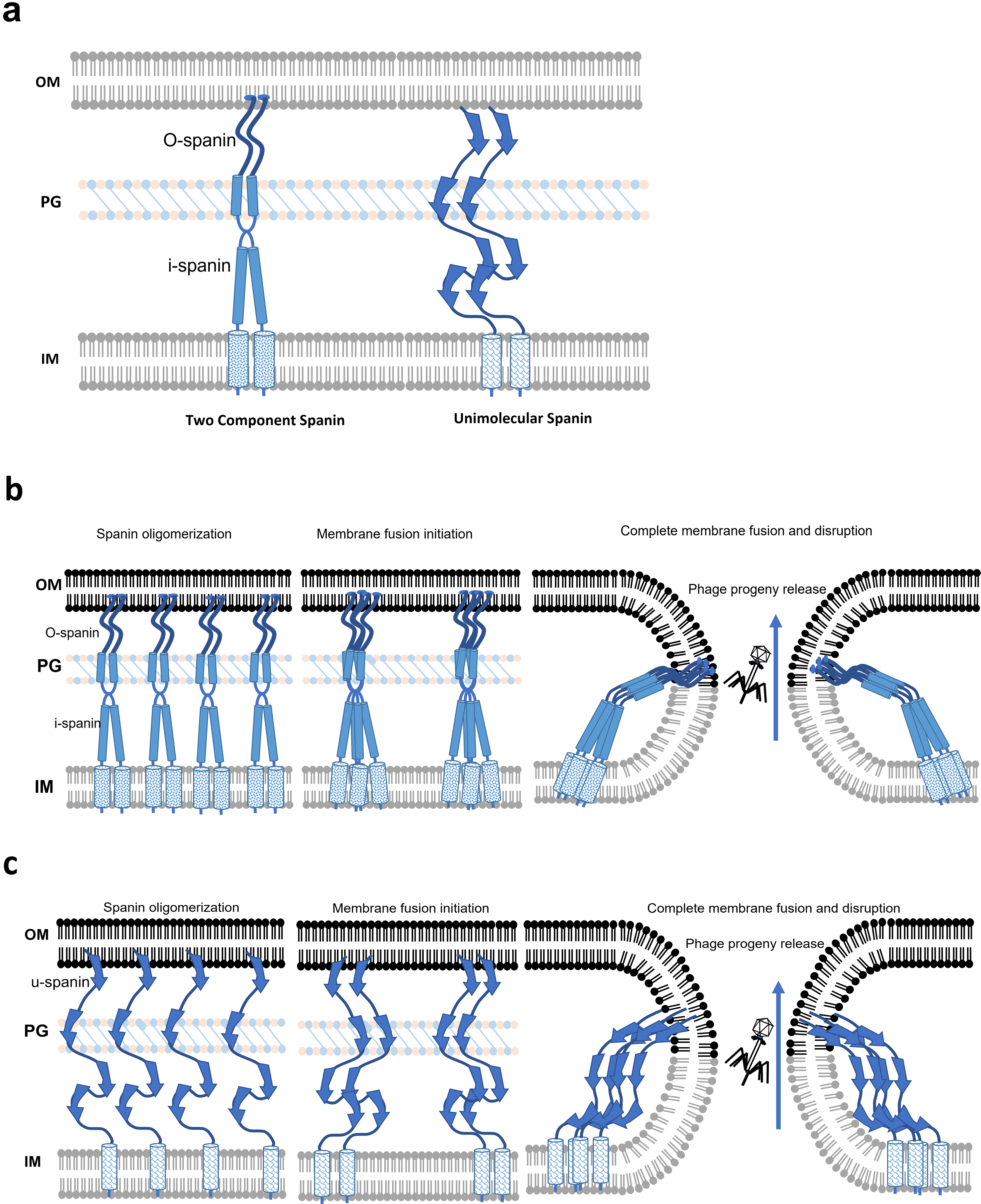
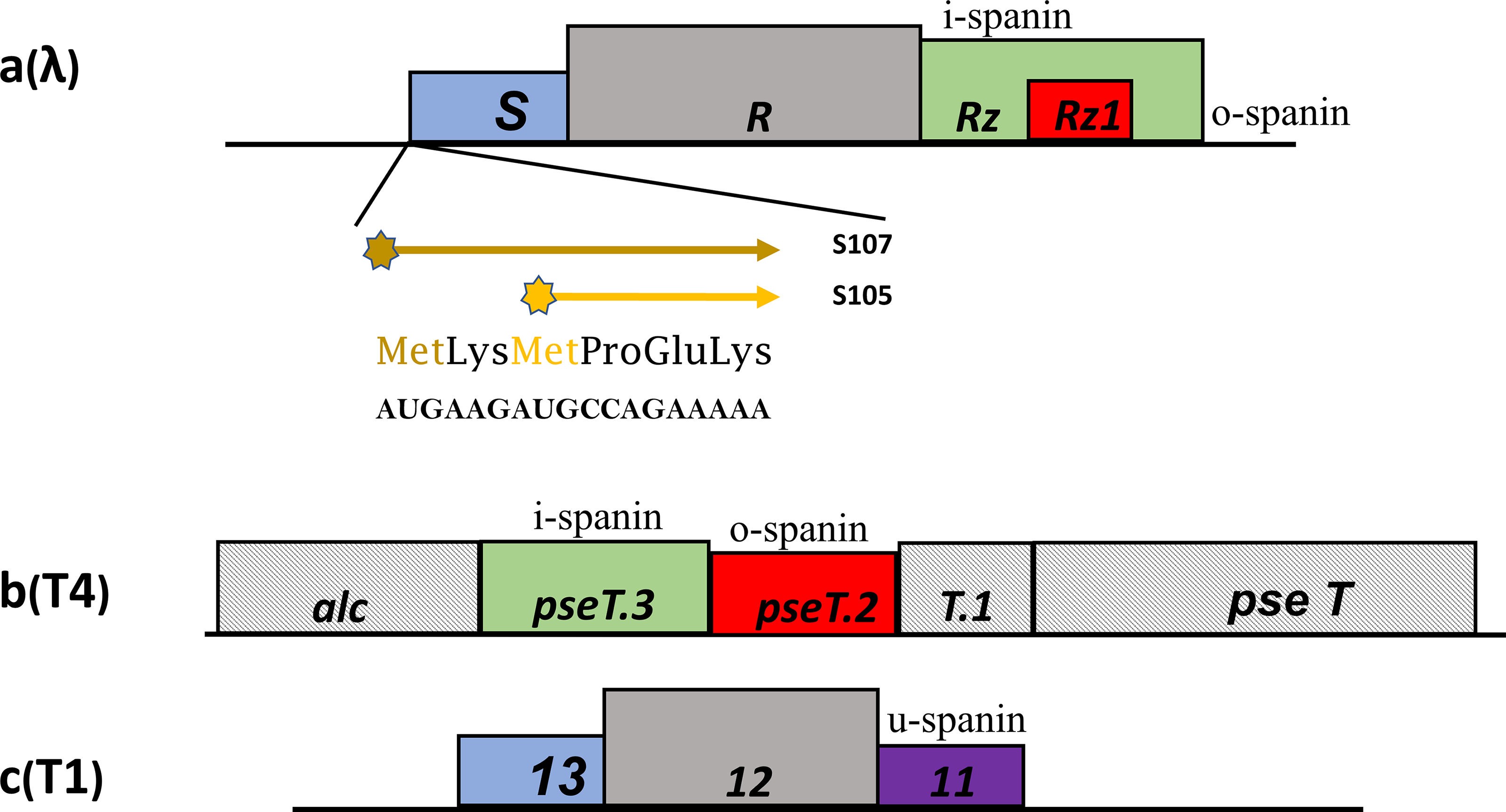
Comparative depiction of phage lysis cassettes:
The Rz-Rz1 genes in phage lambda and ϕ21 encode proteins that have been shown to be necessary for complete lysis in both known lysis pathways. 43 Disruption of either gene resulted in a complete lysis failure. The roles of holin and endolysin were long believed to be both essential and adequate for cell lysis, as the PG provides the cell with its shape and mechanical stability. 32 However, the data show that the spanin complex then functions to disrupt the OM after the endolysin destroys the PG. 44 Research has demonstrated that Rz and Rz1 are capable of facilitating fusion between two membrane bilayers. It has been suggested that following the degradation of PG, spanin complexes experience a conformational shift that draws the opposing membrane bilayers closer together, allowing for fusion (Fig. 4b, c). 45 The infection cycle ends with a spherical cell morphology without the spanin function, where the inner membrane has been fatally disrupted by holin, the peptidoglycan has been degraded by the endolysin, yet the OM remains intact. 44
Regulation of lysis
Phage lysis is a tightly regulated process that begins when holins form lethal holes in the bacterial membrane to allow endolysin to reach the peptidoglycan. Holins are like molecular clocks that decide when a bacteriophage will burst its bacterial host, and that timing can be regulated by antiholins and by changes in the environment. In bacteriophage λ, the S gene uses dual-start mechanism to generate both S105, a holin, and S107, an antiholin, coordinating the precise timing of lysis. Phage T4 employs a distinct approach. Lysis inhibition (LIN) is activated if an infected cell experiences superinfection. Lysis is postponed until the host-to-phage ratio becomes optimal for the release of new viral particles. These strategies illustrate how phages balance timely lysis with maximized progeny yield (Fig. 6).
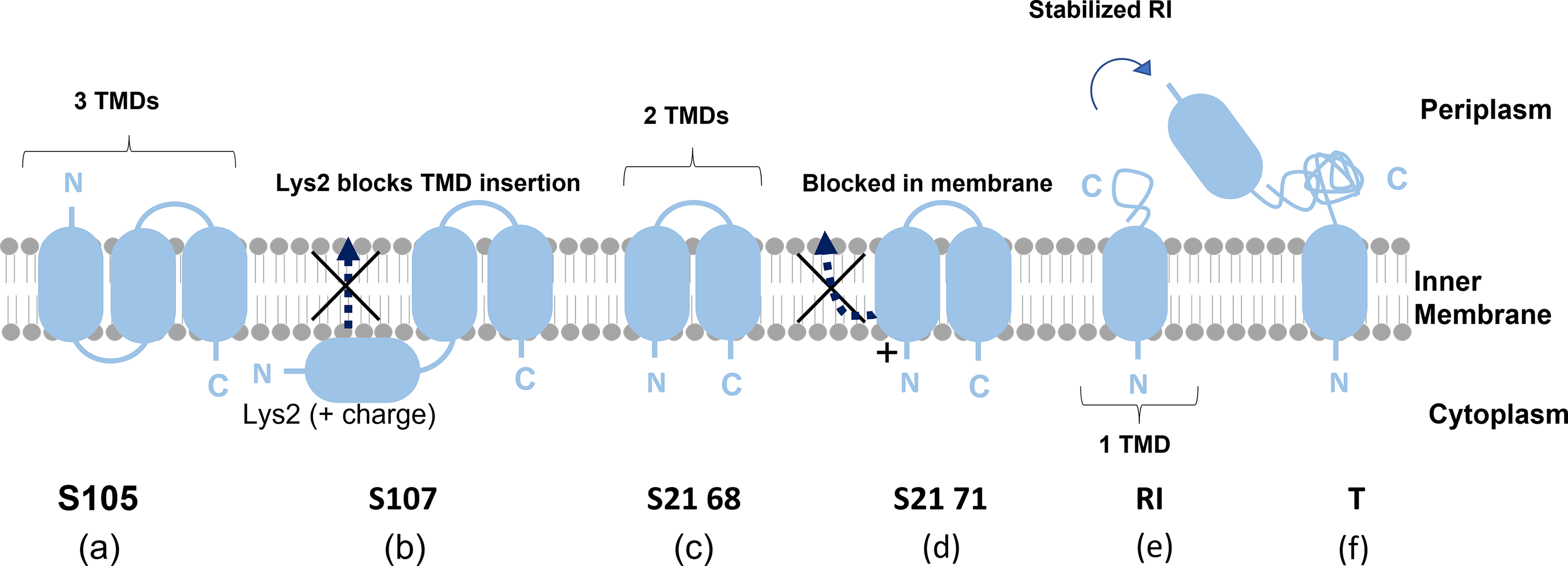
Topological features of holins and their corresponding antiholins. This figure illustrates the membrane topologies of three holin–antiholin pairs: S105/S107, S2168/S2171, and T/RI.
Dual-Start Regulation
Lambda phage (λ) and phage 21
Dual-start regulation is a mechanism utilized by many phages to yield both holins and antiholins from the same gene through alternative translational initiation. This mechanism allows precise timing of lysis of host cells without releasing the phage prematurely or without being able to reach maximal phage yield.
The lambda phage S gene encodes two membrane-associated proteins, S105 and S107, by alternative translational initiation in the same open reading frame (Fig. 5a). S105 is the classical holin, spanning the membrane via three TMDs and features an N-out, C-in topology, which is necessary for creating membrane lesions and inducing lysis. In contrast, S107, functioning as the antiholin, contains only two TMDs due to an N-terminal extension that disrupts the stable embedding of TMD1 within the membrane. This modification in membrane topology, largely influenced by the Lys-2 residue, slows N-terminal translocation and blocks both signal sequence cleavage and functional activation.46,47 This blocks pore formation and thus retards lysis. The S105:S107 expression ratio, generally at ∼2:1, is crucial for the timing of lysis to be precise, early enough to effectively lyse the host but not so early that it results in low phage yield. S107 at higher concentration suppresses lysis and additionally enhances phage production.9,23,48
A similar dual-start mechanism occurs in the pinholin S21 gene. Initiation of translation at the Met-1 of gene S yields a 71-amino-acid peptide (S2171), having a Met-Lys extension at the amino terminus and having the ability to inhibit lysis. In contrast, Met-4 initiates to produce a 68-amino-acid peptide (S2168), which acts as a holin, inducing membrane permeabilization and lysis at a specific time.19,49
Thus, λ and phage 21 both deploy a conserved regulatory principle in which dual translational start sites create holin–antiholin pairs to fine-tune the timing of lysis. This regulation allows phage replication to be maximized by achieving an optimal balance between burst size and the correct release time.
Conditional lysis inhibition
Phage T4
In phage T4, which infects E. coli, lysis is controlled by two key proteins: holin (T) and antiholin (RI). Under normal conditions, holin T accumulates harmlessly within the host’s membrane until the most suitable moment for lysis. At that time, it forms holes in the bacterial membrane, allowing phage-encoded enzymes that degrade peptidoglycan to escape into the periplasm and reach the cell wall. 29 The absence of the cell wall triggers the activation of the spanin complex (pseT.2 and pseT.3), leading to the disruption of the OM and final release of the progeny. 40 The antiholin RI modulates the function of the T4 holin by binding to its soluble domains. 50 RI contains a SAR domain at its N-terminus, is secreted to the periplasm in a form tethered to the membrane, and is subsequently released from the bilayer. 51
However, when the same host is infected again shortly after the first infection (early superinfection, around 3–5 min after), the phage delays lysis through a system called LIN—a situation occurs when there are more phages than hosts in the environment, making it less advantageous to release new phages. It has been suggested that a signal from the superinfecting phage is recognized, resulting in the inhibition of lysis by stabilizing the RI antiholin protein from the original phage. 12 RI protein binds to holin T and blocks its activity.
Once released from the membrane, RI becomes unstable. The periplasmic protease DegP recognizes its exposed SAR domain, leading to the rapid degradation of RI. 51 In conclusion, the stabilization of RI following a superinfection results in an increase of free periplasmic RI, which can bind to the periplasmic domain of T, inhibiting its ability to form pores. This gives the phage more time to replicate before killing the host, helping it adapt to situations where host cells are limited or multiple infections occur.12,51
A general view of holin control systems, including dual-start motifs, antiholin, and host signal regulation, not only improves our view of bacteriophage lysis control but also provides the conceptual foundation for their applied application. By connecting the detailed molecular processes involved in hole formation to potential treatments, these insights show how we might use holin regulation for developing new antimicrobials or innovative biotech tools. Building on that foundation, the next part explores how these biological principles can translate into real-world applications, focusing on the antimicrobial potential of holins and how they could be used in therapy or industry.
Antimicrobial activity
In addition to their essential involvement in phage-mediated lysis, holins have been reported as having a direct antibacterial effect. 28 Expression or application of these proteins often leads to growth inhibition and, in multiple instances, robust bactericidal activity. Observed responses are a fast turbidity decrease, a 2- to 4-log decrease in bacterial viability, ultrastructural damage and cytoplasmic leakage, and biofilm disruption in various bacteria. For instance, HolB, HolP30, and Holμ1/6 caused a significant reduction in E. coli survival, but holins such as Hol-4086 and HolSSE1 exhibited wider lysis against not only S. aureus but also Enterococcus faecalis and some Gram-negative pathogens. Furthermore, coexpression or combination of holins and endolysins has been demonstrated to enhance and accelerate bacterial lysis as well as to extend the host range. Table 1 summarizes the main in vitro findings related to the antimicrobial activity of representative holins.27,35,52,54–59,61–67,69,70
Studies on Holin and Spanin Proteins

Potential applications
An understanding of the biology of holin and spanin indicates a complex mechanism for membrane lysis that could provide temporal regulation and implementation of bacterial cell lysis in bacteriophage infections. Their antimicrobial activity against multidrug-resistant pathogens and membrane targeting has triggered various applications. Here, we review these developing applications and discuss how these distinctive biological properties of holin and spanin proteins are being exploited to develop beneficial tools. An overview of the current development stages of the potential applications, along with the main challenges limiting their clinical translation or commercialization, is summarized in Table 2.
Progress and Challenges of Potential Applications of Phage-Derived Holin and Spanin

Medical applications
Vaccine development
Bacterial ghosts are genetically inactivated vaccines generated through the regulated activation of the E lysis gene, cloned from PhiX174 bacteriophages in Gram-negative bacteria. 71 The lysis gene E encodes the E protein, which forms transmembrane pores that allow the release of cytoplasmic contents. The resulting empty bacterial shells retain their original shape and immunogenic components. 72 Due to their biosafety and capacity to trigger immune responses, these bacterial ghosts have been developed as candidates for nonliving human vaccines. 73 The lysis gene E is not consistently effective in eliminating all target bacteria and transforming them into bacterial ghosts. To improve the lysis efficiency, the holin–endolysin-mediated lytic mechanism is used to develop a novel acellular vaccine candidate. A novel vaccine was developed using Salmonella Enteritidis ghosts engineered to display the globular domain of the HA1 subunit derived from the H1N1 influenza virus. The codon-optimized open reading frame for HA1 was inserted into the ghost plasmid pJHL420, which contains the holin, endolysin, and the gene E derived from phage PhiX174. 74 A plasmid for lysis—named pJHL464—containing an R lysing cassette with the S, R, Rz, and Rz1 genes was inserted into a vaccine strain of S. typhi Ty21a, resulting in a new apoptotic vaccine candidate for typhoid fever. 75 A new ghost plasmid was created that encodes both protein E and holin–endolysins in a consecutive manner. 76
Compared with using gene E alone, the combined sequential expression of these lysis genes significantly accelerated and enhanced the efficiency of cell lysis.75,76 In the Salmonella Enteritidis–HA1 construct, enhanced immunogenicity was observed due to efficient antigen display. 76 In the S. typhi Ty21a system, the pJHL464 plasmid produced effective apoptotic vaccine candidates. 74 In comparative studies, ghosts generated with combined E + holin–endolysin expression provided superior immune protection: 70% of immunized mice survived a lethal challenge, while 100% mortality occurred in mice immunized with E-only ghosts. 75
These results demonstrate that combining bacteriophage-derived lysis systems can transform bacterial ghost platforms into highly efficient acellular vaccine candidates. Such strategies enable targeted antigen display and improved immunogenicity, highlighting their potential in developing next-generation vaccines against both bacterial and viral pathogens.
Cancer treatment
Gene therapy approaches for cancer may employ cytotoxic proteins that compromise cell survival, and the bacteriophage lambda-holin protein, which is known to disrupt bacterial membranes, has been evaluated for its impact on eukaryotic cells. In Tet-inducible human cell lines, lambda-holin expression triggered over 98% cell death in vitro and dramatically inhibited tumor growth in vivo, highlighting its strong cytotoxic potential for cancer gene therapy—pending further safety assessment and optimized delivery methods. 77
Drug delivery
Minicell purification
Bacterial minicells are attractive drug and gene delivery vehicles due to efficient targeting and low cytotoxicity. However, minCDE-derived minicells are hard to purify due to parental cell carryover, which resists multistep centrifugation. By expressing the iEPS5 holin/lysin system to kill only active parent cells, contamination dropped >400-fold, and simple centrifugation yielded over 2 × 10¹⁰ high-purity minicells per milliliter for therapeutic use. This streamlined method efficiently generates safe, high-purity minicells for therapeutic delivery. 78
Suicidal Listeria for cytoplasmic delivery
Holin proteins induce bacterial autolysis, enabling the release of intracellular contents for antigen delivery. The attenuated strain of Listeria monocytogenes known as Delta2 was genetically altered to create the suicidal strain rsDelta2 by incorporating the holin gene hol118 and the cell wall hydrolysin gene ply118, both derived from a bacteriophage specific to Listeria, into its DNA. The Listeria PactA promoter controlled the expression of the hol118 and ply118 genes, triggering bacterial self-destruction within eukaryotic cells. This lysis process enables the intracellular release of bacterial components into the eukaryotic cytoplasm. Holin-mediated autolysis shows promise for oral protein vaccine delivery. DNA plasmid expression in host cells was ineffective due to lack of nuclear access. 79
Biotechnological Applications Food Safety
L. monocytogenes is a foodborne pathogen responsible for listeriosis, a severe infection associated with significant mortality. HolGH15 has shown both bactericidal and bacteriostatic activities against L. monocytogenes, even at refrigeration temperatures—a key advantage for food safety applications. These properties suggest that HolGH15 could be applied in food preservation and meat processing. Thus, HolGH15 offers a safe and nonchemical agent that represents a promising strategy for controlling L. monocytogenes contamination. 80
Holin-driven protein secretion
In both Gram-negative E. coli and Gram-positive Lactococcus lactis, holin encoded by phages can promote the release of proteins into the external environment. The Hol380 holin from Lactococcus lactis was expressed in both E. coli and L. lactis and forms pores that enhance the extracellular secretion of the cytosolic enzymes β-galactosidase, staphylococcal nuclease, and FaeG, which functions as a fimbrial adhesin. Expression of Hol380 resulted in a 2.55-fold increase in the extracellular levels of staphylococcal nuclease and a 2.40-fold improvement in the secretion of FaeG when compared with secretion directed solely by the Usp45 signal peptide. This study introduces a novel method for expressing holin in microbial cell factories to enhance the export of proteins that are economically or medically significant. 81
Industrial Applications
Holin–endolysin systems for efficient protein recovery
Producing and isolation of intracellular proteins with real efficiency is important in the fields of biotechnology. Achieving high yields of desired proteins typically involves using microbial hosts—such as E. coli—engineered to overexpress recombinant proteins. Since the target protein is generally located within the cytosol, harsh conditions need to be established to rupture the cell wall in order to release the target protein. Mechanical methods have generally been used to disrupt cells, such as by cell homogenizer, bead mill, ultrasound, French press, and autoclave. Nonmechanical methods, such as freeze–thaw, organic solvents, detergents, osmotic shock, acid, base, and enzyme reactions have also been employed. However, energy consumption is a concern for mechanical methods, while nonmechanical methods require additional steps for removing the chemicals, resulting in extra costs.
Scientists are now using bacteriophage-derived lytic systems—particularly holin–endolysin—to gently and precisely release intracellular proteins. Because the timing is programmable, it can be set to trigger at just the right moment. This makes it easier to purify the proteins and often boosts the total amount recovered without causing unnecessary damage.
Researchers developed a novel E. coli system that can undergo auto-induced lysis by combining lysis-associated genes from bacteriophage λ with the Mg2+-responsive inducible promoter. 82 Similarly, a holin/endolysin system derived from EJ-1 phage was integrated into P. putida BXHL with a tolB mutation to facilitate controlled release of medium-chain-length polyhydroxyalkanoates (PHA). 83 Likewise, Bacillus megaterium was engineered as a host for the production of poly(3-hydroxybutyrate-co-4-hydroxybutyrate), where its inherent ease of lysis was exploited to facilitate the recovery of these industrially valuable copolymers. 84 In Synechocystis sp. PCC 6803, green-light-inducible lysis genes encoding holin and endolysin were expressed under the cpcG2 promoter. Illumination with red and green light resulted in decreased growth rate, increased phycocyanin release, and significant cell death, while red light alone had no effect. 85 Also, researchers built a lysis system activated by nickel by placing phage-derived holin and endolysin genes under the control of a Ni2+-responsive promoter. Introducing NiSO4 triggered self-lysis, which occurred more rapidly when endolysins accumulated before holin-mediated permeabilization.86,87
Holin systems have proven to be a powerful way to effectively trigger host cell lysis or promote protein export, often leading to impressive yields of the desired product. In P. putida, it boosted PHA recovery without the need for harsh chemicals. 83 In B. megaterium, resulting copolymers showed reduced stiffness, enhanced toughness, and lower melting points. 84 And in cyanobacteria, light-triggered systems gave scientists precise control over lysis events, making it easier to release phycocyanin. 86
Recent advances in microbial engineering are transforming industrial biotechnology. Insights into prokaryotic expression have enabled cost-efficient protein production systems, while targeted B. megaterium modifications now regulate polyhydroxybutyrate synthesis for sustainable coatings and bio-based binders. Self-disruptive P. putida offers eco-friendly PHA recovery, and bacteriophage lysis genes in cyanobacteria present an alternative to costly mechanical cell disruption. Holin-based export strategies further expand industrial production potential.
Conclusion
Spanins and holins constitute a diverse group of membrane-associated proteins encoded by bacteriophages. Holins generally accumulate in the bacterial inner membrane without causing damage until a precise trigger induces pore formation, permitting endolysins to access the periplasm and degrade peptidoglycan. Pinholins, on the other hand, create small pores that collapse the membrane’s PMF, triggering the activation of signal-arrest-release endolysins. Consequently, the endolysin undergoes proper folding into its active structure, facilitating peptidoglycan degradation. In Gram-negative bacteria, bacteriophage-mediated lysis relies on spanins that disrupt the OM through a mechanism functionally separate from, but complementary to, the actions of holins and endolysins.
Holins are involved in two clearly distinct events of the phage infection cycle. They are mainly responsible for the endolysin release by permeabilizing the membrane, and their second major function is to control when in the infection cycle this comes about, being the “clock” of the infection cycle. Holins, with proven activity against multidrug-resistant bacteria, including both Gram-negative and Gram-positive strains, exhibit strong bactericidal and antibiofilm effects. Phage-derived holins, either alone or in combination with endolysins, may represent a promising protein-based alternative for combating antibiotic-resistant bacterial infections.
These membrane-disrupting proteins have economic benefits in a variety of industries, including the synthesis of bio-based compounds, biofermentation, and biofuel generation, in addition to their clinical value. Gaining deeper insights into the mechanistic and structural dynamics of holins and spanins, as well as understanding the molecular mechanisms of spanin activity, would help develop biotechnological applications such as cargo targeting and delivery systems, along with medical applications like antimicrobial strategies.
Collectively, the evolution of holins alongside spanins reflects a specialized adaptation that facilitates bacterial envelope disruption during the lytic phase. The direct study of biophysical properties and functional principles of these membrane-disrupting proteins will provide new insight into general cell biology and could open the road of these proteins for applications in a medical, industrial, and biotechnological context.
Authors’ Contributions
Z.N.S. was responsible for conceptualization, comprehensive literature search, and writing—original draft. M.M.F. supervised the research and critically revised the article for important intellectual content. F.K., B.J., and H.A. contributed to the review and editing of the article. All authors read and approved the final version of the article.
Footnotes
Author Disclosure Statement
No competing financial interests exist.
Funding Information
No funding was received for this article.
Author Confirmation Statement
All authors are from Tehran University of Medical Sciences (Tehran, Iran), where education and research are the primary functions.