



Abstract
Background:
Bacteriophages exhibit great diversity and are classically divided into lytic and temperate lifestyles. Commonly, bioinformatics is used to predict the temperate lifestyle. As the nosocomial pathogen Pseudomonas aeruginosa is common in freshwater, this biome represents an ideal location for the discovery of Pseudomonas phages.
Materials and Methods:
We characterized a genetically distinct species of Pseudomonas phage using next-generation sequencing and conventional microbiological methods. We characterized its lifestyle by purifying infected colonies and identifying persistent phage production.
Results:
Genetics revealed a phage of a novel genus. Characterization showed a narrow host range and bioinformatics suggested a lytic lifestyle. Experimental data showed lysogeny and analysis was consistent with an episomal lifestyle instead of the classical model of integration into the host genome.
Conclusions:
We identify and characterize a new Pseudomonas phage, show potential limitations of bioinformatic predictions of lifestyle, and further contribute to the diversity of the phage tree of life.
Keywords
Introduction
Bacteriophages are the most numerous infectious agent on Earth numbering some 1031 particles. 1 The majority of phages remain undiscovered and make up a portion of what is referred to as viral dark matter. 2 Since the independent discovery of phages by Twort and D’Herelle, phages have been considered for treatment of bacterial infections and have been in continuous use in Georgia and Poland and recently have been used in compassionate care cases in the United States.3–8 There is growing interest in phages as a treatment of last resort for infections that are refractory to antibiotics, and they have been used in many compassionate use cases. 9 Interest in phages goes beyond the medical utility, and phage products have found use in applications from phage display through to the Φ29 DNA polymerase in molecular biology.10,11
Phage life cycles are classically divided into lytic and lysogenic. 12 Lytic phages do not integrate into their host’s genome and after penetration of the host cell and utilization of the host machinery for replication induce lysis to propagate. These lytic phages are clinically useful and one group has engineered a temperate phage to adopt a lytic lifestyle that has been used in a clinical case.13,14 Temperate phages integrate into the host genome and are passed through bacterial generations as prophages. Excision from the host chromosome can be induced by various stimuli, probably best studied in the Lambda phage. 15 There, induction can be induced by DNA-damaging methods that trigger the recA–lexA-mediated SOS response.16–18 Countless researchers have refined our understanding of the lytic–lysogenic switch system revealing a complex regulatory network that selects the fate of lysogen-bearing cells.19,20 The lifestyle of a phage is important for therapeutic and scientific purposes, and to this end, various software has been developed that can predict lysogenic potential based on the genetic sequence.21–23
While the most common modality of phage persistence inside bacteria is integration into the host chromosome, other modalities exist. These are clouded by a diverse collection of terms associated with phage persistence without integration into the host chromosome. These include pseudolysogeny, carrier state and chronic infection. These are reviewed in detail here. 24 This phenomenon has been well-studied in the Escherichia coli phage P1. 25 P1 is a myovirus with a dsDNA genome that is circularized by means of redundant DNA sequences and the Cre–lox recombinase system that has found extensive uses in molecular biology.26–28 The viral genome is maintained as a low copy number plasmid whose replication is governed by oriR, which resembles plasmid origins of replication. 25 Segregation of the phage genome is tightly regulated by P1 proteins that directly interact via a centromere-like region and further by a toxin system lethal to progeny that do not carry the P1 genome.29,30
Here, we characterize a novel bacteriophage ΦJB8, discovered in freshwater in Clear Creek TX, USA, identify its taxonomic relation to other phages and study its unusual mechanism of persistence in Pseudomonas aeruginosa PAO1.
Materials and Methods
Phage discovery and cultivation
Freshwater was collected from Clear Creek, TX, USA, in a sterile 50 mL Falcon tube. This was centrifuged (4400 g, 20 min), and an aliquot was filtered (0.22 μm Durapore PVDF membrane, MilliporeSigma). About 0.5 mL of filtrate was mixed with 0.7% (w/v) top agar and 100 μL of overnight PAO1 culture and spread on an LB agar plate. This was incubated overnight, and plaques were inspected the next morning. The plaque of interest was isolated by stabbing with a pipette tip to remove a small plug of agar and mixed in phage buffer (10 mM Tris, pH 8, 100 mM NaCl, 1 mM MgSO4). This was then serially streaked (three times) to ensure homogeneity. A plate lysate was prepared by mixing 30 μL of phage plaque isolate with 100 μL of PAO1 overnight in 3 mL of 0.7% (w/v) top agar and spread on LB agar plates. The next morning, cleared top agar was scraped and mixed with 3 mL of phage buffer, vortexed and then spun at 4400 g for 10 min. The supernatant was filtered (0.22 μm Durapore PVDF membrane, MilliporeSigma) and stored at 4°C.
Host range determination
We selected 25 random clinical isolates from the TAILΦR library. We deliberately selected isolates only from unique patient cases. These clinical isolates represented cases from across the United States, from different institutions and different sites of infection. We grew these isolates as lawns in 0.8% (w/v) top agar and spotted serial dilutions of ΦJB8. We incubated overnight and counted plaques in the morning. We performed this experiment in triplicate and averaged the plaque counts. We calculated the efficiency of plating (EOP) compared with PAO1 as (number of plaques on clinical isolate)/(number of plaques on PAO1).
Phage adsorption assay
We performed this assay as previously described in work by our laboratory. 31 Briefly, we used PAO1 cells (OD600 = 0.1, ≈3 × 10⁸ CFU/mL) and ΦJB8 (MOI = 0.1) and measured adsorption at 37°C. Quenching (dilution 1:100 into ice-cold LB) was performed at various time intervals, and fraction of free phage compared to input was determined after clearance of infected cells (centrifugation 10 mins, 4000 g). We included a condition with ΦJB8 but no cells to control for decay in warm LB media. We performed the experiment in biological triplicate. We analyzed the data with GraphPad Prism, fitted an exponential decay curve, calculated kabs and report the mean and SEM.
Electron microscopy
Imaging was performed at Baylor College of Medicine’s Cryo-EM Advanced Technology Core Facility (Houston, TX, USA). Phages were deposited on grids (Quantifoil 2/2 200Cu +2 nm ThinC [Quantifoil Micro Tools GmbH, Jena, Germany]), negatively stained (2% w/v uranyl acetate) and imaged using a JEOL 1230 electron microscope (JEOL, Japan) at 80 kV with a 4k x 4k Gatan Ultrascan CCD Camera (Gatan, AMETEK). Images were taken at various magnifications and representatives were selected. Processing including contrast adjustment and scale bar addition was performed in NIH ImageJ.
Bioinformatic analysis of ΦJB8
Phage DNA was extracted using the Universal Pathogens Kit (Omega-Biotek) per the manufacturer’s instructions. ΦJB8 was sequenced using the Illumina platform by Novogene (Novogene, China). Briefly, the sequencing libraries were made using NEBNext Ultra II DNA Library Prep Kit for Illumina (NEB, USA) per the manufacturer’s directions, and index codes were added. The PCR products were purified using the AMPure XP system. The size distribution was verified using the Agilent2100 Bioanalyzer. Final quantification was by qPCR. The final library was sequenced using an Illumina NovaSeq instrument (paired-end 150 bp reads). We filtered reads and discarded anything smaller than 50 base pairs using BBDuk (version 38.84), trimmed using a Q20 quality score, assembled using Geneious 2022.0.1 (native assembler with default settings for medium sensitivity with detection of structural variants of any size) and these assemblies were annotated using the RASTtk pipeline.32,33 To generate a phylogeny based on nucleotide sequence, we used BLASTn (discontiguous BLAST, standard settings) and then visualized the phylogeny in NIH Genome workbench. Genetic comparisons between nearest neighbors were made using VIRIDIC. 34 Proteomic comparisons were made using VIPTree. 35
Isolation of lysogens
We followed the protocol of Altamirano and Barr. 36 Briefly, we spotted 5 μL of ΦJB8 (3.6 × 10⁵ PFU/mL) on PAO1 top agar (0.8% w/v) and incubated overnight. We then picked presumably ΦJB8-resistant cells from the mesa and streaked them on an LB agar plate and incubated overnight. We picked 100 isolated colonies and propagated them on patch plates for 3 serial passages to remove any externally adherent phage particles. After 3 passages the colonies were picked by touching with a sterile pipette tip and very gently streaked on the surface of a top agar (0.8% w/v) coated plate that had been inoculated with 100 μL of PAO1 overnight growth, incubated overnight and examined for zones of clearance. 36
Verification of lysogeny
We grew each potential lysogen overnight in LB and inoculated 2 μL of this overnight into 198 μL of LB in an untreated 96-well polystyrene plate (VWR, PA, USA). 36 We grew this for 6 h at 37°C with shaking. We took the media from each well and clarified it by centrifugation (4000 g, 20 min) and then filtered the supernatant (0.22 μm Durapore PVDF membrane, MilliporeSigma). We titrated this supernatant using standard methods, pictures were taken and phage concentration (PFU/mL) was calculated. 37 This experiment was performed in biological triplicate from separate colonies of each strain. To test resistance to ΦJB8, we picked colonies of each presumed lysogen and used them as the lawn in 0.8% (w/v) top agar and spotted dilutions of the original ΦJB8 phage, incubated overnight, imaged and counted plaques. We performed this experiment in biological triplicate from independent colonies of each lysogen. We report the mean and standard error.
Genetic analysis of potential lysogens
We selected 10 clones grown from a ΦJB8-generated mesa that did not exhibit zones of clearance upon streaking on PAO1 as ΦJB8-exposed controls. We also selected the 10 clones from a ΦJB8 mesa that did exhibit clearance (positives) as well as the PAO1 ancestor. These were grown overnight in LB, and then 1.5 mL of culture was harvested by centrifugation, and the pellet was rinsed thrice in PBS. We purified DNA from the pellets using a Power Soil Kit (Qiagen) following the manufacturer’s instructions. Illumina sequencing was done by Novogene. Reads were processed as described earlier. We used Bowtie2 to align reads from each pool (PAO1, controls and positives) to the ΦJB8 genome. 38 We assembled contigs from each sequenced strain using SPAdes (v3.15.3, default settings). We then took each individual contig one by one and attempted to align the contig against the ΦJB8 genome and then the PAO1 host genome using BLASTn (discontiguous megablast, default algorithm parameters). We recorded whether the contig aligned to the phage, to the host or both.
Results
Discovery and initial characterization
As freshwater sources are rich in Pseudomonads, we searched freshwater samples from Clear Creek, TX, USA (29.514347°N, 95.104101°W) for Pseudomonas phages. We plated filtered water samples in PAO1 top agar and identified multiple plaque morphologies. One distinctive plaque was ∼1–2 mm in diameter, hazy and formed a centralized mesa. We termed this phage ΦJB8 (Accession Number: PQ473534). Mesa formation was more obvious at higher titers (Fig. 1A). We applied standard methods to characterize this new phage determining the adsorption co-efficient as 7.25 × 10⁻⁹ mL/min using PAO1 host cells (Fig. 1D). Electron microscopy was challenging as preparations were prone to forming aggregates, but multiple attempts revealed consistent viral particles ∼84 nm (SD = 4 nm, n = 15 particles) in diameter with small tails in each preparation, fitting the classical description of podoviruses (Fig. 1B). Whole genome sequencing revealed a likely complete 47,273 bp genome that was likely circularly permuted. Phageterm could not identify a packaging mechanism. 39 Analysis using BLASTn (discontiguous megablast, default settings) showed the nearest relatives were Pseudomonas phage Bjorn (Accession number: NC_042103.1) with tANI 21% (query coverage 29%, percent identity 72%) and Pseudomonas phage PSA37 with tANI 10% (Accession number: MZ089740, 15% query coverage, 67% percent identity). Given the lack of genetic similarity of the nearest neighbors, ΦJB8 likely represents a member of a new genus of bacteriophage. The host range was quite limited with productive infections seemingly established with EOP >0.5 in 5/25 clinical isolates selected randomly from the TAILΦR library. Some host strains displayed clearing at high concentrations, but no distinctive plaques at lower concentrations, consistent with “lysis from without” activity in 7/25 strains (Fig. 1C).

Basic appearance and characteristics of ΦJB8.
Genetic analysis
Given low tANI suggesting a novel genus, we pursued further analysis. We employed BLASTn and generated a phylogenetic tree of the nearest neighboring sequences (Fig. 2A) showing the phages ΦJB8, BroderSalsa (Accession number: OL412002) and Bjorn existing in three genetically distant groups that are quite distinct from other Pseudomonas phages. The nearest relative, Bjorn, was discovered in Denmark on a Pseudomonas spp. host in plant compost from Prunus avium.

ΦJB8 represents a genetically distinct new genus of bacteriophage.
We employed VIRIDIC to visualize the distance between ΦJB8 and the 12 nearest neighbors (Fig. 2B). 34 These data show that while all these neighboring phages possess similar length genomes, the aligned genome fraction (query coverage) and intergenomic similarity (analogous to tANI) between ΦJB8 and other phages is low, even for its nearest neighbor, Bjorn.
Proteomic alignment has been well used in phage biology. 40 We used VIPTree to analyze ΦJB8 and its neighboring genomes. 35 We compared ΦJB8 with 132 ICTV reference genomes (MSL40, March 2025) and noted significant divergence from the neighboring genomes. We aligned ΦJB8 to its 2 nearest proteomic neighbors (Bjorn and tf, Fig. 3A). The proteomic comparison was able to align a greater fraction than the nucleotide analysis. Homology was found in numerous proteins, including hypothetical gene products. For those with an annotated function, the closest relationship was found in DNA replication and packaging genes (terminase, DNA polymerase, endonuclease, and DNA primase/helicase) and structural genes (portal protein, virion structural proteins, tail fiber protein, major head protein). We found synteny between genes for which homology could be identified between phages ΦJB8 and Bjorn (Fig. 3B). This is an expected finding given the recombinant, mosaic nature of phage genomes. 41 Analysis attempts using VContact2 (v.0.9.19, default settings, Prokaryotic Viral RefSeq v.201) classified ΦJB8 as a singleton, concurring with PhaBox (v.2, default parameters), which identified it as a singleton at the genus level.42,43

Bioinformatic comparison of proteomes of ΦJB8 and nearest neighbours.
Lifestyle of ΦJB8
As the hazy plaques seen when ΦJB8 was plated on PAO1 (Supplementary Fig. S1A) are hallmarks of lysogeny, we investigated whether there were any genetic markers of temperate lifestyle. We used Phage.ai and PhageLeads and neither software predicted lysogeny.23,44 Given the lack of agreement between morphology and computational prediction, we picked bacteria from ΦJB8-generated mesas (Supplementary Fig. S1A) and streaked them on agar. Subsequently, we picked 100 individual colonies and propagated them on LB agar plates for three serial isolations (Supplementary Fig. S1B). We then streaked each purified isolate on PAO1 top agar lawns with PAO1 ancestor and LB only controls. Neither control generated clearance of the lawn that would suggest phage production. We observed that 10/100 purified colonies showed zones of clearance outside of the streak (Supplementary Fig. S1C). In other iterations of this experiment, we found persistence of ΦJB8 despite 4 passages of the host in 1/20 and 3/50 colonies. Notably, all colonies showing a zone of clearance suggestive of lysogeny on the first streak showed a similar zone on all subsequent streaks. This argues against incidental phage carryover as some colonies would be expected to lose the phage with each passage. These data argue that ΦJB8 persists despite attempts to remove phage by serial streaking, suggesting a lysogenic lifestyle. The variability in frequency of persistence seems most likely due to the underlying stochastic nature of lysogeny as we made every attempt to control for experimental variables (OD600, MOI, incubation times, host strain). We cannot exclude some hidden variable at work here, but the probabilistic nature of lysogeny is well established.20,45 We followed the methods of Altamirano and Barr and tested the 10 isolates that seem to produce phage and 10 that did not for resistance to ΦJB8. 36 We denote the bacterial strains derived from the ΦJB8 generated mesa JB8.Rx.
We found that all nonproductive strains (JB8.R1-10) were sensitive, with EOP ≈ 1, to ΦJB8 (data not shown) as was the PAO1 ancestor. All productive strains (JB8.R26-86) were resistant to ΦJB8 (Supplementary Fig. S2A). We further tested for production of ΦJB8 in solution. After growing the 10 nonproducers and 10 producers for 6 h in LB media, we tested the supernatants for phage. We found that 9/10 producers generated measurable phage concentrations, while the PAO1 only negative control and all 10 of the nonproducers did not (Supplementary Fig. S2B). All observed plaque morphologies were consistent with ΦJB8. Collectively, these data suggest that ΦJB8 persists within a fraction of ΦJB8-treated Pseudomonas and suggests lysogeny despite the lack of computational prediction.
We were curious as to the mechanism of integration into the host genome, as our analysis did not identify a recombinase or other markers. Thus, we sequenced the 10 nonproducers that had been exposed to ΦJB8 (JB8.R1-10), the 10 ΦJB8 producers (JB8.R26-86) and the PAO1 ancestor (SRA Bioproject: PRJNA1304558). In the PAO1 ancestor and the nonproducers, we did not detect any reads that mapped to ΦJB8. In the ΦJB8 producers, we mapped 14033 reads to the ΦJB8 genome with an average coverage of 43X (compared to 2929X of the host genome) (Fig. 4B). This verifies that ΦJB8 was being passed in those strains. It also suggests that the process of serial streaking was successful at removing any incidental contamination of ΦJB8 in those that were ΦJB8-exposed but not lysogenized (nonproducing strains).
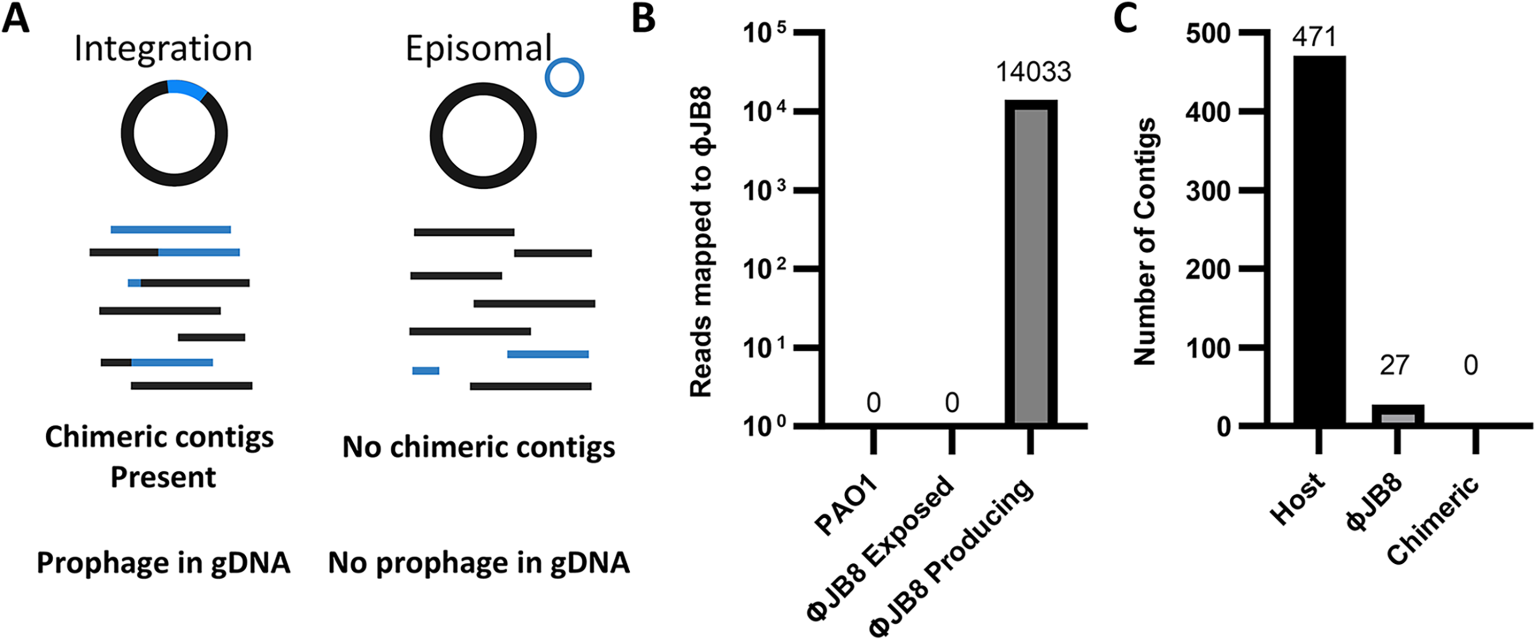
WGS shows persistence of the ΦJB8 genome in JB8-resistant cells and a lack of chimeric contigs.
We assembled contigs from each sequenced strain and in those contigs assembled from the ΦJB8 positive strains we identified 471 host contigs, 27 ΦJB8 contigs and most notably no contigs with contributions from both host and virus (Fig. 4C). This data were surprising as we had anticipated being able to identify prophage sequences that would be evidenced by contigs that are chimeras of both host and phage. Aligning reads from the ΦJB8 positive strains to a reference PAO1 genome (GenBank Accession number: NC_002516.2) showed we had achieved coverage of 2929X (±272) from end to end. This depth of coverage makes it quite likely that we would have been able to identify a prophage. In this case, our data suggest that ΦJB8 is maintained in the host, but does not integrate. This is consistent with an episomal mode of maintenance (Fig. 4A). The low ratio of ΦJB8 to PAO1 host coverage (1:68) most likely reflects the process relatively low carriage rate of the phage secondary to stochastic lysogeny, as mentioned earlier.
Discussion
Here we report ΦJB8, a novel phage discovered in a freshwater sample in Texas, USA. Upon sequencing, we calculated a crude tANI (query coverage x percent identity) of 21% compared with Pseudomonas phage Bjorn (nearest neighbor). When calculated by VIRIDIC, we found 24% intergenomic similarity, a measure analogous to tANI (Fig. 2B). 34 This meets the classification (tANI <70%) used by the ICTV to classify as a new genus. We suggest the name Junipervirus to this new genus and hence this virus would be called Junipervirus stewartii, (exemplar: Pseudomonas Phage ΦJB8).
Our characterization showed a hazy plaque suggestive of a temperate phage (Fig. 1A) and further experimentation revealed that ΦJB8-resistant colonies could continue to produce phage particles despite serial passages. This fulfilled the criteria for lysogeny that have been discussed in the literature; however, software packages for phage lifestyle prediction did not predict a temperate phage. These software packages depend on the detection of integration-associated genes with more recent systems leverage Machine Learning and Natural Language Processing. If these markers of integration are detected, then the evaluation of a phage as temperate is quite reliable. However, predicting a lytic lifestyle depends on the absence of such sequences and given the diversity of phage genomes, this is not likely to be such a robust prediction. Unfortunately, owing to the paucity of characterized episomal phages, such genomes are unlikely to make up a significant fraction of temperate phages in the training sets for these software. As such, we would speculate that their power to characterize such episomal phages as temperate is limited.
To evaluate for lysogeny, we sequenced 10 strains that had been infected with ΦJB8 and still secreted phage despite serial passaging to look for prophages. Despite excellent coverage of the phage and host genome, we did not identify any chimeric sequences that would suggest integration. Whilst phages are classically divided into lytic and lysogenic phages, passage without integration is also well-reported in the literature.46–48 We must consider two possibilities. First, that we are observing persistence of the phage and the phage genome is maintained as an episome. Second, there is the possibility that the phage does integrate but under our experimental conditions, the excision rate is very high. This would result in persistence of the phage, but we would be unable to detect a prophage as it would exist in an equilibrium that greatly favors an unintegrated state. While we cannot definitively exclude this, our high depth of coverage of the host genome (2929X) implies that the excision rate would have to be extremely high for us to have missed a prophage. Further, attempts to induce a prophage (mitomycin C, UV light exposure, ciprofloxacin, starvation) also failed. We thus favor the first explanation.
Our work has several limitations. As an initial description, we are unable to comment on any details of the molecular biology of this phage. Further, as we discussed above, we cannot exclude an integrating phage with a high excision rate. Despite these limitations, our description contributes to expanding the phage tree of life and may also contribute to better characterization of phage lifestyle as it may serve to inform bioinformaticians of refinements to their software.
Authors’ Contributions
Conceptualization, formal analysis, investigation, methodology, visualization, and writing—P.N. Conceptualization, methodology, writing, and supervision—A.W.M. Formal analysis—J.R.C. All authors have read and agreed to the published version of the article.
Footnotes
Acknowledgments
The authors wish to thank Alleigh Stewart whose local knowledge assisted in gathering the water samples that led to the isolation of this novel phage genus. We dedicate and name the phage described herein to the loving memory of our phage-hunting companion Juniper. TEM data was collected at the Baylor College of Medicine CryoEM ATC, which includes equipment purchased under the support of CPRIT Core Facility Award RP190602.
Author Disclosure Statement
No competing financial interests exist.
Funding Information
This work was supported in part by grants from NIH NIAID (T32 AI0554413 fellowship, K08 AI173452-01A1, U19 AI157981 CARBIRU Pilot sub-award) to P.N. and support from the Kleberg Foundation to A.W.M.
Disclaimer
This article was submitted solely to this journal and is not published, in press or submitted elsewhere.
Supplemental Material
Supplemental Material
References
Supplementary Material
Please find the following supplemental material available below.
For Open Access articles published under a Creative Commons License, all supplemental material carries the same license as the article it is associated with.
For non-Open Access articles published, all supplemental material carries a non-exclusive license, and permission requests for re-use of supplemental material or any part of supplemental material shall be sent directly to the copyright owner as specified in the copyright notice associated with the article.
