
Abstract
A 56-year-old man with acute myeloleukemia was hospitalized for lumbar pain. Treatment with antibiotics failed to improve the symptoms. For the diagnosis of infiltration by leukemia we performed CT-guided percutaneous needle biopsy of the L2-L3 disc and the L3 vertebral body using a left posterolateral approach. His symptoms were improved by treatment with antibiotics and he was discharged 4 days later. He again experienced lumbar pain 4 days post-discharge and was readmitted. Unenhanced CT scans of the abdomen and pelvis revealed a giant hematoma in the left psoas muscle and we suspected lumbar arterial injury. A preoperative aortography and transcatheter arterial coil embolization was then performed for the diagnosis and treatment of a lumbar artery pseudoaneurysm. On the preoperative angiography, pseudoaneurysm arising from the left lumbar artery was shown. All feeders were shown by the selective catheterization of the lumbar arteries and they were completely embolized using coils. However, contrast-enhanced CT obtained on the next day still demonstrated a pseudoaneurysm in the left psoas muscle. Thus, additional percutaneous embolization using N-butyl-2-cyanoacrylate was performed. After this procedure, complete embolization of the pseudoaneurysm was obtained and his lumbar pain was relieved.
Lumbar arterial pseudoaneurysms are commonly found as a complication after lumbar-spine trauma involving one or more fractures of lumbar transverse processes (1, 2). However, they also arise secondary to interventional percutaneous renal procedures such as biopsy (3, 4), nephrostomy (5), and nephrolithotomy (6), and more rarely after laparoscopic splenectomy (7) or during vertebral biopsy (8). As their rupture can result in rapid clinical deterioration due to retroperitoneal hemorrhage, they must be diagnosed and treated rapidly (9). We report a patient with a lumbar artery pseudoaneurysm that arose during intervertebral disc and vertebral biopsy. As the results of initial transcatheter arterial embolization were unsatisfactory we performed percutaneous embolization using N-butyl-2-cyanoacrylate (NBCA).
Case report
Patient consent for inclusion in this retrospective study was waived. A 56-year-old man with acute myeloleukemia was hospitalized for lumbar pain. Enhanced MRI showed only L2-L3 discitis and osteomyelitis. Treatment with antibiotics failed to improve his symptoms. Laboratory examination revealed mild inflammatory C-reaction protein (CRP) 1.38 (normal range <0.3); his white blood cell count was 7.3 × 10³/µL (normal range 3500–8500); there was no coagulopathy (platelet count 152 × 103/µL (normal range 145–325) and the international normalized ratio was 1.04. To assess infiltration leukemia we performed CT-guided percutaneous biopsy of the L2-L3 disc and the L3 vertebra using a left posterolateral approach and a 13-gauge bone marrow biopsy needle (Angiotech, Vancouver, BC, Canada). The histopathological diagnosis was L2-L3 discitis. Additional treatment with antibiotics improved his symptoms and resulted in a decreased CRP level; he was discharged 4 days after CT-guided percutaneous biopsy. Four days after discharge he again suffered severe left lumbar pain and numbness of the left lower extremities. Unenhanced CT of the abdomen and pelvis revealed a giant hematoma in the left psoas muscle leading us to suspect lumbar arterial injury (Fig. 1).
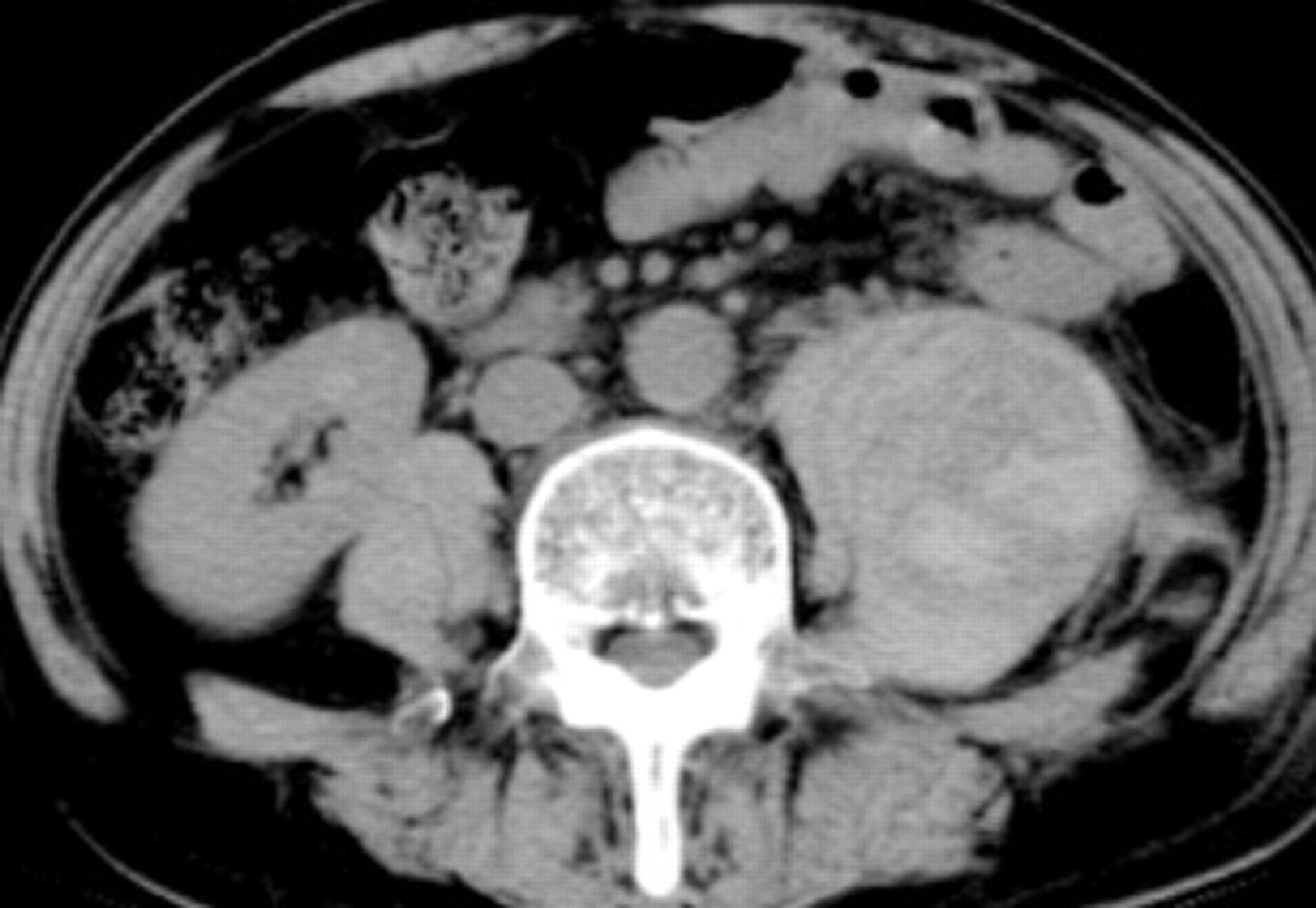
Contrast-enhanced CT scan obtained 1 week after percutaneous biopsy reveals a pseudoaneurysm medial to the left psoas muscle
Diagnostic aortography and lumbar artery angiography were performed before further treatment. These imaging studies were carried out in an interventional procedure room equipped with a commercially-available digital subtraction angiography (DSA) unit (AXIOM Artis dTA/ VB30E; Siemens, Erlangen, Germany). We used a 4-Fr pig tail catheter (Medikit Co. Ltd., Tokyo, Japan) and delivered 30 mL of iomeprol (Iopamiron 300; Bayer, Osaka, Japan) and 300 mg I/mL; the flow rate was 10 mL/s. We also performed DSA of the lumbar artery using a 4-Fr duck-head catheter (Medikit) and manually injected 10 mL of iomeprol and 300 mg I/mL as a bolus. Aortography and lumbar and CT-assisted lumbar arteriography revealed a pseudoaneurysm on the left L2 lumbar artery arising in the path of the earlier lumbar biopsy (Fig. 2).

CT-assisted lumbar arteriography revealed a pseudoaneurysm medial to the left lumbar artery
To acquire selective angiographs of the lumbar artery, 5 mL of iopamidol were manually injected as a bolus through the 2.5-Fr microcatheter (Target, Boston Scientific Corp., Watertown, MA, USA). The microcatheter was introduced with a Syncro guidewire (outside diameter 0.014 inch) featuring a free transformation tip (Boston Scientific). Selective angiography of the second left lumbar artery showed a pseudoaneurysm that was supplied directly from the second left lumbar artery. We performed transcatheter coil embolization at a site proximal to the pseudoaneurysm using four interlocking detachable coils; 4–120 mm (IDCs; Boston Scientific Corp., Watertown, MA, USA). Embolization was successful as evidenced on lumbar angiographs by contrast retension at the proximal site. Selective angiography of the third left lumbar artery was performed. Although the pseudoaneurysm was not directly supplied by this vessel, we detected an arterial anastomosis between the second and third left lumbar artery. Consequently, we performed transcatheter embolization at a site proximal to the anastomosis using six IDCs; diameter 3 mm, length 10 cm (n = 3); and diameter 4 mm, length 8 cm (n = 3) (Boston Scientific Corp., Watertown, MA, USA). Lastly, we performed selective angiography of the fourth left lumbar artery. It showed the lumbar arterial anastomosis between the third and fourth left lumbar artery, but no pseudoaneurysm.
However, contrast-enhanced CT scans obtained the next day revealed a remaining pseudoaneurysm medial to the left psoas muscle (Fig. 3). Contrast-enhanced CT scans were acquired on a 64-row multidetector scanner (Brilliance-64, Philips Medical Systems, Best, The Netherlands). We delivered 100 mL of Iopromide (Iopamiron 300; Nihon Schering, Osaka, Japan) as a bolus at a rate of 3 mL/s.

CT scan obtained on the day after coil embolization revealed a remaining pseudoaneurysm on the left psoas muscle
The pseudoaneurysm was punctured directly under CT guidance the following day. We used an 18-gauge PTCD needle (PTCD puncture set Type T; Hakko, Nagano, Japan) featuring an 18-gauge outer Teflon sheath and a 20-gauge inner stainless needle. The sclerosing agent was a 1:2 mixture of N-butyl-2-cyanoacrylate (NBCA; Histoacryl, B. Braun, Melsungen, Germany) and iodized oil (Lipiodol; Laboratoire Guerbet, Roissy, France); this yielded radiopacity. After direct puncture DSA was performed and the pseudoaneurysm was filled with a manually-injected bolus of 2 mL of iomeprol and 300 mg I/mL. After flushing the biopsy needle with 2 mL of 5% glucose solution we delivered 2 mL of NBCA slowly under fluoroscopic monitoring until the pseudoaneurysm was completely filled (Fig. 4). Follow-up MRI performed 2 days later confirmed its occlusion (Fig. 5).

Fluoroscopic image revealed that the pseudoaneurysm was completely filled with N-butyl-2-cyanoacrylate

MRI obtained 2 days after percutaneous embolization showed no enhancement in the pseudoaneurysm
Histological study revealed discitis and the patient was treated with antibiotics different from those administered initially. Subsequently his left lumbar pain and numbness of the left lower extremities improved and he was discharged after 1 month. His chronic myeloleukemia is being followed on an outpatient basis.
Discussion
Pseudoaneurysms of the lumbar arteries are infrequent; most are found incidentally after trauma to the lumbar spine (9, 10). More rarely, they are an iatrogenic complication from diagnostic or therapeutic procedures (9, 10). Different from bleeding from other intra-abdominal vascular structures, bleeding from lumbar arteries is often initially contained by surrounding retroperitoneal tissues; this acts as a tamponade to form a pseudoaneurysm (10).
Anatomically the lumbar plexus is located lateral to the intervertebral foramina and passes through the psoas major muscle. The plexus or any of its branches can be affected by an expanding mass within the muscle (10). This can induce pain and neurologic deficits in the distribution of the affected nerves. We posit that the pseudoaneurysm in our patient was due to direct trauma during lumbar disc biopsy and that exacerbation of his left lumbar pain and numbness of the left lower extremities were attributable to expansion of the mass in the psoas muscle.
The non-surgical management of pseudoaneurysms includes placement of a compression bandage and ultrasound-guided compression (11, 12). We were unable to apply ultrasound-guided compression because the aneurysm was located deep in the lumbar musculature. Maleux et al. (7) successfully treated a lumbar pseudoaneurysm and arteriovenous fistula by transcatheter embolization. We attribute our failure of proximal embolization to the presence of a complicated network of lumbar arterial collaterals within the muscles of the back and on the outside and inside of the vertebral canal. Lumbar arteries also communicate with the superior and inferior epigastric arteries, the lowest intercostal and subcostal arteries, and the iliolumbar and lateral sacral branches of the internal iliac artery (10).
The percutaneous injection of thrombin into the sac has been used to address femoral artery pseudoaneurysms. Because of its relative low number of complications and high success rate, this method has gained preference over compression or surgery to treat patients with false femoral artery aneurysms (13–15). It has also been applied to induce thrombosis in aneurysms located on the lumbar artery after renal biopsy (16). The risk of this procedure is thrombosis; as in some individuals the artery of Adamkiewicz arises from the lumbar artery, thrombosis may induce spinal cord infarction (7, 17). NBCA is a permanent embolic material and as it polymerizes immediately in blood, it provides for complete and instant vessel occlusion. The disadvantages of transcatheter embolization with NBCA are its relatively high cost, the instant occlusion of the catheter lumen, and the difficulty in predicting the exact occlusion site (18). In addition, application of the technique requires expertise; both the ratio of iodized oil to NBCA and the injection volume must be carefully determined based on the location and size of the bleeding point (18). For example; if the NBCA concentration is too high, proximal embolization may fail due to the presence of a complicated network of lumbar arterial collaterals within the muscles of the back and on the outside and inside of the vertebral canal. On the other hand, if the NBCA concentration is too low, the embolization agent may flow into communicating arteries and the lumbar venous plexus. As we have expertise in transcatheter embolization with NBCA, we chose to use this embolic agent to treat our patient.
Another disadvantage using NBCA as the transcatheter embolization agent involved the difficult evaluation on CT scans when iodized oil is added to NBCA. Because of the presence of admixed iodized oil, CT scans show high-density areas.
In conclusion, we suggest that percutaneous embolization using NBCA is appropriate for the treatment of iatrogenic pseudoaneurysms that cannot be addressed successfully by transcatheter arterial coil embolization. To avoid the migration of NBCA due to high arterial blood flow, we also recommend that transcatheter arterial coil embolization be performed as the first step in patients treated for pseudoaneurysms.