
Abstract
Administration of both streptozotocin (STZ) and nicotinamide (NA) has been proposed to induce experimental diabetes in the rat. STZ is well known to cause pancreatic B-cell damage, whereas NA is administered to rats to partially protect insulin-secreting cells against STZ. STZ is transported into B-cells via the glucose transporter GLUT2 and causes DNA damage leading to increased activity of poly(ADP-ribose) polymerase (PARP-1) to repair DNA. However, exaggerated activity of this enzyme results in depletion of intracellular NAD+ and ATP, and the insulin-secreting cells undergo necrosis. The protective action of NA is due to the inhibition of PARP-1 activity. NA inhibits this enzyme, preventing depletion of NAD+ and ATP in cells exposed to STZ. Moreover, NA serves as a precursor of NAD+ and thereby additionally increases intracellular NAD+ levels. The severity of diabetes in experimental rats strongly depends on the doses of STZ and NA given to these animals. Therefore, in diabetic rats, blood glucose may be changed in a broad range – from slight hyperglycemia to substantial hyperglycemia compared with control animals. Similarly, blood insulin may be only slightly decreased or substantial hypoinsulinemia may be induced. In vitro studies demonstrated that the insulin-secretory response to glucose is attenuated in STZ–NA-induced diabetic rats compared with control animals. This is due to reduced B-cell mass as well as metabolic defects in the insulin-secreting cells. Results of numerous experiments have demonstrated that this model of diabetes is useful in studies of different aspects of diabetes.
Introduction
Diabetes mellitus is a complex metabolic disease affecting about 5% of people all over the word. According to the classification proposed by the American Diabetes Association, diabetes is divided into different types. 1 Type 1 diabetes accounts for 5–10% of all diabetic cases and results from the autoimmune destruction of the pancreatic B-cells. This type of diabetes usually develops rapidly because of the grave destruction of the insulin-secreting cells and patients are dependent on exogenous insulin. Type 2 diabetes is more frequent, accounts for 90–95% of all diabetic cases, is characterized by insulin resistance and relative insulin deficiency, and is frequently accompanied by overweight or obesity. Hyperglycemia usually develops slowly, and at earlier stages, blood glucose is moderately elevated. In type 2 diabetes, insulin resistance is initially compensated by increased secretion of insulin; however, this prolonged overstimulation of insulin secretion leads over time to progressive exhaustion and degradation of B-cells. 2,3 In individuals with type 2 diabetes, numerous abnormalities in these cells have been described. In humans with type 2 diabetes, glucose-stimulated insulin secretion is substantially impaired. However, insulin-secretory response to amino acids or sulphonylureas is usually less affected. Moreover, the first phase of insulin secretion and the pulsatility of hormone release have also been demonstrated to be disturbed in type 2 diabetic patients. 4,5 The insulin-secretory abnormalities of B-cells are accompanied, among others, by decreased oxidation of glucose and by a lower ATP/ADP ratio. Moreover, decreased expression of insulin, glucose transporters and glucokinase have been found in pancreatic islets of humans with type 2 diabetes. It is also known that oxidative stress in pancreatic islets of type 2 diabetic humans and apoptosis of islet cells are increased compared with healthy individuals. 5–7 However, abnormalities in diabetes are still poorly elucidated.
New drugs are continually being tested and new strategies developed to prevent and treat diabetes. In these studies, different experimental animal models of diabetes have been used. One of them is streptozotocin (STZ)–nicotinamide (NA)-induced diabetes in the rat. In this model, created by Masiello et al., 8 diabetes is induced by administration of two compounds – STZ and NA. STZ is a well-known diabetogenic agent exerting cytotoxic action on pancreatic B-cells, whereas NA is given to rats to partially protect these cells against STZ. The present article focuses on the mechanisms of action of STZ and NA on B-cells and characterizes the experimental model of diabetes induced by administration of these two compounds. The information included in this study will be helpful not only in understanding the action of STZ and NA at the cellular level, but also in inducing experimental diabetes in the rat.
Cytotoxic action of STZ
STZ (2-deoxy-2(3-methyl-3-nitrosoureido)- Chemical structure of streptozotocin and nicotinamide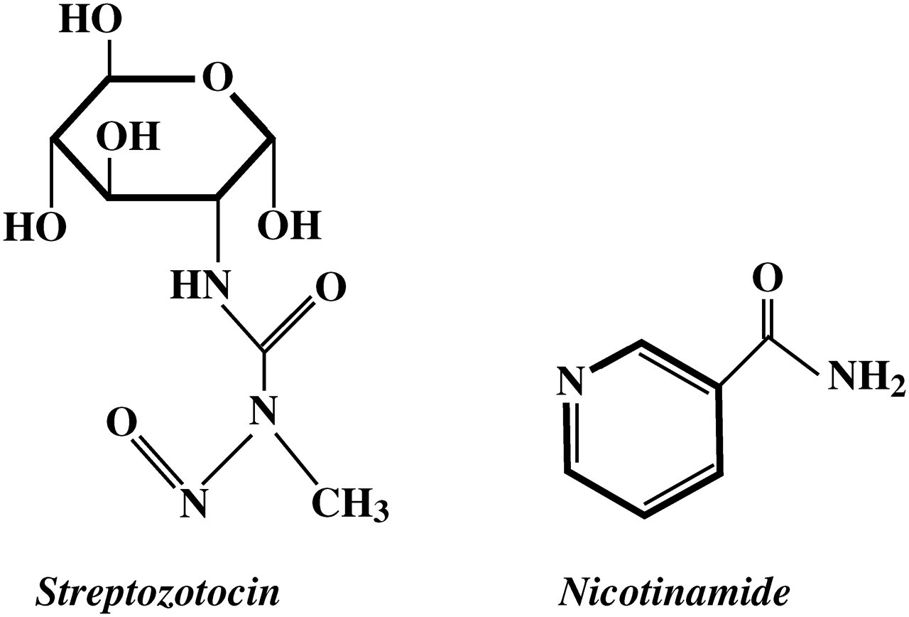
Mokhtari et al. 44 and Cheon et al. 45 revealed that c-Jun N-terminal kinase (JNK) is also involved in the cytotoxicity of STZ. Increased activity of this enzyme is observed in the case of cellular stress leading to cell death. Studies on insulin-secreting cells exposed to STZ demonstrated increased activity of JNK, whereas inhibitors of this enzyme attenuated the cytotoxic action of STZ. Activation of JNK by STZ is supposed to be preceded by increased activity of PARP-1 since PARP-1 inhibitors are able to decrease the activity of both PARP-1 and JNK. 45
Protective action of NA
NA (pyridine-3-carboxamide; Figure 1) is the amide form of vitamin B3 (niacin). The protective action of NA against cellular damage induced by different harmful agents is well established. 46–48 Results of many in vitro and in vivo studies have provided evidence that NA is also able to effectively protect B-cells against the cytotoxicity of STZ.
In vitro effects of NA
Experiments on isolated rat pancreatic islets have revealed that STZ significantly reduces proinsulin biosynthesis, and that this effect is attenuated by NA. 49 It is also known that NA ameliorates the inhibitory effect of STZ on glucose-stimulated insulin secretion by isolated rat islets. 15,50,51 STZ-induced impairment in glucose oxidation and decreased islet cell viability are also markedly improved by NA. The beneficial influence of NA on cell viability was recently confirmed in insulinoma cells (NIT-1) exposed to STZ. 45 Importantly, the protective action of NA on pancreatic islet cells also involves a decrease in DNA damage caused by STZ. 15,52
In vivo effects of NA
Administration of the diabetogenic dose of STZ to rats causes a decrease in body weight; however, this is attenuated by pretreatment of animals with NA. 8 Numerous studies have also demonstrated that the STZ-induced increase in blood glucose is significantly blunted when NA is administered prior to STZ. 8,53–56 The advantageous effect of NA on blood glucose is due to the protection of B-cells against STZ-induced injury and is accompanied by increased blood insulin. 8 According to these results, studies on perfused pancreases have revealed that glucose-induced insulin output is substantially higher in the case of pancreases obtained from rats treated with STZ and NA compared with pancreases of rats that received only STZ. 8 It is also known that STZ administered to rats diminishes pancreatic insulin content, but this effect is dose-dependently prevented by NA given prior to STZ. 8 The protective action of NA is additionally confirmed by studies demonstrating reduced damage of islets and reduced methylation of pancreatic DNA in rats pretreated with NA compared with animals that received STZ alone. 54,57
Data from the literature allows one to conclude that the mechanism of the protective action of NA against STZ-induced damage of B-cells involves two main effects: inhibition of PARP-1 and provision of NAD+ (Figure 2), whereas other effects are of minor importance.
Schematic representation of the cytotoxic action of streptozotocin and the protective action of nicotinamide on B-cells. PARP-1, poly(adenosine triphosphate [ADP]-ribose) polymerase-1; PRPP, 5-phosphoribosylpyrophosphate; NMN, nicotinamide mononucleotide; Nampt, nicotinamide phosphoribosyltransferase; Nmnat, nicotinamide/nicotinic acid mononucleotide adenylyltransferase,  , increase/activation,
  , decrease/inactivation. (A color version of this figure is available in the online journal)
Inhibition of PARP-1
NA is a well-known inhibitor of PARP-1. The inhibitory action of NA on the activity of this enzyme has been documented in different kinds of cells, including insulin-secreting cells of the pancreas. 24,28,58,59 Hyperactivity of PARP-1 in B-cells exposed to STZ is thought to be pivotal for the cytotoxicity of STZ. This was demonstrated in experiments with different PARP-1 inhibitors and in studies on PARP-1-deficient animals. 28,58,60–62 In both cases, attenuation of PARP-1 action appeared to significantly decrease the cytotoxicity of STZ. Therefore, inhibition of PARP-1 by NA plays an important role in the mechanism of the protective action of NA on B-cells. The structure of PARP-1 consists of three main domains: an N-terminal DNA-binding domain, an automodification domain and a C-terminal region. A C-terminal region catalyses the synthesis of poly(ADP-ribose) and binds to target proteins. This active site of PARP-1 is divided into an acceptor and a donor site. The donor site is occupied by NAD+ and is additionally divided into three subsites: a nicotinamide-ribose binding site, a phosphate binding site and an adenine-ribose binding site. 24 A detailed analysis performed by Pandya et al. has revealed that the inhibition of PARP-1 results from the interaction of NA with the binding site for the NA moiety of NAD+. 59 NA enters into hydrogen bound associations with Ser(904) and Gly(863) at the nicotinamide-ribose binding site of the active site of PARP-1. The inhibition of PARP-1 by NA is of a competitive nature since NA blocks the binding of NAD+ to the catalytic domain of the enzyme. 63 The inhibitory concentration of NA for 50% reduction of PARP-1 activity (IC50) was estimated to be 210 μmol/L. 59
Provision of NAD+
Apart from the inhibition of PARP-1, the protective action of NA against cellular damage induced by STZ is thought to result from provision of NAD+. Jackson et al. 64 demonstrated that administration of NA to animals increases NAD+ contents in different tissues. Moreover, NA increases intracellular NAD+ and prevents NAD+ depletion under different pathological conditions. 46,48 This effect is due to reduced utilization of NAD+ and/or its increased biosynthesis. In mammals, NAD+ is synthesized mainly from NA and its formation is catalyzed by two enzymes – nicotinamide phosphoribosyltransferase (Nampt) and nicotinamide/nicotinic acid mononucleotide adenylyltransferase (Nmnat). In the first step of NAD+ generation catalyzed by Nampt, NA and 5-phosphoribosylpyrophosphate (PRPP) are converted to nicotinamide mononucleotide (NMN). Then, NMN undergoes transformation to NAD+ in a reaction catalyzed by Nmnat. The activity of Nampt in B-cells is supposed to be low. However, Nampt acts as an intracellular (iNampt) and extracellular (eNampt) enzyme and in the case of calls with a low activity of iNampt, the majority of NMN utilized to form NAD+ is generated extracellularly. 65,66
Characteristics of STZ–NA-induced diabetic rats
Induction of diabetes and blood glucose and insulin concentrations in control and STZ–NA-induced diabetic rats
*Per kg body weight C, control rats; D, diabetic rats; iv, intravenous administration; ip, intraperitoneal administration Streptozotocin (STZ) is administered in 100 mmol/L citrate buffer, pH = 4.5, whereas nicotinamide (NA) is dissolved in saline and is administered intraperitoneally usually 15 min before STZ (with some exceptions, indicated in parenthesis)
Blood insulin and glucose
The concentrations of blood insulin in diabetic rats differ depending on doses of STZ and NA given to these animals. Blood insulin may be profoundly decreased when the dose of STZ is relatively high. Conversely, administration of a high dose of NA in relation to the amount of STZ substantially protects B-cells, and blood insulin is unchanged compared with control rats. Similarly to changes in insulinemia, diabetic rats may manifest very high hyperglycemia, moderate hyperglycemia, or blood glucose may be only slightly increased. It was found that in animals that were given low doses of NA (100–120 mg/kg body weight), administration of STZ at a dose ranging from 45 to 65 mg/kg body weight, causes a higher increase in blood glucose than that in rats receiving higher doses of NA (180–290 mg) and comparable doses of STZ (Table 1). However, in order to obtain a STZ–NA-induced diabetic model, doses of both compounds must be adjusted to avoid profound hyperglycemia, which is characteristic for STZ-induced diabetes.
The comparison of rats with STZ–NA-induced diabetes and rats with diabetes induced by STZ alone, used as a model of type 1 diabetes, demonstrates that in the latter animals, blood glucose is substantially higher, whereas blood insulin is much lower. 8,55 In obese (ob/ob) mice, representing a model of type 2 diabetes, both blood insulin and glucose are significantly elevated compared with normal mice. 69 In early stages of type 2 diabetes in humans, blood glucose is near normal or only moderately increased and blood insulin is increased. This is due to the compensatory hypersecretion of insulin. However, over time, hyperglycemia gradually develops and blood insulin is reduced. 1,70
A glucose tolerance test reveals glucose intolerance in rats with STZ–NA-induced diabetes. In healthy animals, glucose challenge causes an initial increase in blood glucose followed by a gradual decrease. However, in diabetic rats, the fall in glycemia is very slow compared with control animals. 8,71–73 The insulin-secretory response to glucose load is impaired in STZ–NA-induced diabetic rats compared with the physiological response observed in control animals. Administration of glucose to normal rats significantly increases blood insulin followed by a rapid decrease. Conversely to these dynamic changes in healthy rats, in diabetic animals, the insulin-secretory response to glucose is significantly abated and insulinemia is almost completely unchanged. 8,71,72 Therefore, glucose intolerance observed after glucose challenge in rats with STZ–NA-induced diabetes is mainly due to impaired secretion of insulin. This is in contrast to ob/ob mice, in which increased blood glucose is accompanied by increased blood insulin and hyperglycemia results from insulin resistance. 69 In humans with type 2 diabetes, glucose challenge induces a rise in blood glucose which is markedly higher than in healthy people and is comparable with that observed in rats with STZ–NA-induced diabetes. However, in type 2 diabetic humans, hyperglycemia observed after glucose load usually results from both insulin resistance and impaired function of B-cells. 70
Importantly, in rats with STZ–NA-induced diabetes, the glucose intolerance may be alleviated by pharmacological compounds, which enhance secretion of insulin such as sulfonylurea drugs. The well-known insulinotropic action of sulfonylureas results from the closure of ATP-sensitive K+ channels in the plasma membrane. In the B-cells of healthy individuals, this effect triggers insulin release and substantially potentiates insulin secretion induced by glucose. 74 In STZ–NA-induced diabetic rats, the combination of glucose and tolbutamide significantly increases blood insulin and improves glucose tolerance compared with effects caused by glucose alone. 8 Moreover, Chi et al. 56 revealed that a single administration of glibenclamide alone to rats with STZ–NA-induced diabetes reduces blood glucose with a simultaneous rise in blood insulin. Similar effects were demonstrated in diabetic animals receiving glibenclamide for 21 days or glyclazide for 30 days. 75,76
B-cells
Characteristics of the insulin-secreting cells of STZ–NA-induced diabetic rats compared with cells of control animals
STZ, streptozotocin; NA, nicotinamide; NO, nitric oxide; FAD, flavin adenine dinucleotide
In vitro studies have demonstrated that B-cells isolated from rats with type 1 diabetes induced by STZ alone, conversely to the insulin-secreting cells of STZ–NA-induced diabetic rats, are almost completely unresponsive to stimulation by glucose or sulfonylureas. 8 This is in contrast to ob/ob mice since pancreatic islets of these animals were demonstrated to secrete more insulin compared with healthy mice. Moreover, the biphasic insulin-secretory response to glucose in ob/ob mice is preserved. 69,79 Pancreatic islets of type 2 diabetic humans, conversely to islets of rats with STZ–NA-induced diabetes, are usually characterized by a loss of the first phase of insulin secretion; however, the second phase may also be deteriorated. 5,80
Interestingly, insulin release stimulated by arginine appeared to be exaggerated in the case of pancreases and islets derived from rats with STZ–NA-induced diabetes. 8,77 This hypersecretion of insulin in the presence of arginine was proposed to be due to reduced activity of constitutive NO synthase in islet cells. 77 Excessive insulin secretion induced by arginine was also revealed in Goto-Kakizaki (GK) rats, a non-obese model of type 2 diabetes. 80 In type 2 diabetic patients with reduced insulin-secretory response to glucose, B-cell response to arginine may be normal or slightly increased. 80,81
Novelli et al. 78 demonstrated that functional defects in B-cells of STZ–NA-induced diabetic rats are accompanied by metabolic abnormalities in these cells. Under physiological conditions, glucose-stimulated insulin secretion is preceded by glucose transport and oxidative metabolism to pyruvate. Then, pyruvate enters mitochondrial metabolism and the high activity of mitochondrial flavin adenine dinucleotide (FAD)-glycerolphosphate dehydrogenase (the key enzyme of the glycerophosphate shuttle) and malate–aspartate shuttle and low activities of both lactate dehydrogenase and plasma membrane lactate/monocarboxylate transporter ensure efficient mitochondrial metabolism of pyruvate. This results in increased formation of ATP and induces the sequence of events involving the increase in the ATP/ADP ratio, the closure of the ATP-sensitive potassium channels, depolarization of the plasma membrane, opening of voltage-sensitive calcium channels and the rise in cytosolic Ca2+. The increase in cytosolic Ca2+ triggers secretion of insulin. Moreover, other signals are generated to maintain the sustained secretion of insulin. 74,82 However, pancreatic islets of STZ–NA-induced diabetic rats were found to have reduced activity of mitochondrial FAD-glycerophosphate dehydrogenase compared with the activity of this enzyme in normal rats. The reduced activity of FAD-glycerophosphate dehydrogenase was also observed in pancreatic islets of type 2 diabetic humans and GK rats, but not in islets of ob/ob mice. 69,83,84 This indicates that some metabolic defects are similar in islets of both type 2 diabetic humans and STZ–NA-induced diabetic rats.
It was also revealed that the blockade of the malate–aspartate shuttle impairs insulin secretion more in islets of STZ–NA-induced diabetic rats than in islets of control rats. These abnormalities are accompanied by reduced glucose utilization by islets of diabetic rats, particularly at higher glucose concentrations. 78 Moreover, atrophy and destruction of B-cells, vacuolization, reduction of the number and size of islets, the decrease in the secretory granules of B-cells and other degenerative changes were shown in pancreatic islets isolated from STZ–NA-induced diabetic rats. 78,85,86 Induction of diabetes by STZ and NA also reduced islet insulin content. 8,78,87 However, this effect strongly depends on the doses of STZ and NA given to rats. 8
Pancreatic insulin content in rats with type 1 diabetes induced by STZ is deeply reduced compared with STZ–NA-induced diabetic animals. 8 In contrast, islets of ob/ob mice contain much more insulin than islets of normal mice and B-cell hyperplasia is observed in these animals. 69 In type 2 diabetic humans, insulin resistance is initially compensated by increased insulin secretion to maintain normoglycemia. This compensatory mechanism is thought to involve both enhanced secretory function of B-cells and increased B-cell mass. However, over time, exaggerated secretion of insulin leads to the progressive failure of B-cells, cells undergo apoptosis and B-cell mass is reduced. 70 In this context, islets of rats with STZ–NA-induced diabetes correspond to the stage of type 2 diabetes in humans when B-cell mass is reduced.
Other abnormalities in diabetic rats
Pancreatic B-cell dysfunction and changes in blood glucose and insulin in STZ–NA-induced diabetic rats are accompanied by numerous other abnormalities. Increased activities of alkaline phosphatase, aspartate transaminase and alanine transaminase and changes in the activities of different enzymes of carbohydrate metabolism were demonstrated in rats with diabetes. 76,88–90
Moreover, increased levels of tumor necrosis factor α (TNF-α), interleukin 1β (IL-1β), IL-6, nuclear factor κB (NF-κB) p65 unit and NO were found in these animals compared with control rats. 85,88 Recent studies have also demonstrated an impairment in the antioxidative defense system in STZ–NA-induced diabetic rats. The levels of lipid peroxides, hydroperoxides and protein carbonyls are significantly higher in plasma, pancreatic tissue and kidney of diabetic animals. 85,88,91 Simultaneously, the activities of superoxide dismutase, catalase, glutathione peroxidase and glutathione-S-transferase were found to be deeply reduced in erythrocytes, pancreatic tissue, liver and kidney of rats with diabetes compared with healthy animals. 85,88,92–94 However, changes observed in rats with STZ–NA-induced diabetes are less marked than in animals with diabetes induced by STZ alone. In ob/ob mice and type 2 diabetic humans, oxidative stress is also increased in different tissues and is supposed to contribute to some diabetic complications. 95,96
Utility of the STZ–NA-induced diabetic model
Rats with STZ–NA-induced diabetes are used in many studies testing the potential beneficial effects of various pharmacological and natural compounds on the course of diabetes. Experiments on these animals demonstrate that many of the tested compounds possess a real therapeutic value in diabetes. 55,56,71–73,75,76,85–94,97–104 These results indicate the high utility of the STZ–NA-induced diabetic model in this kind of studies.
Maintaining normoglycemia is one of the important problems in diabetes. Therefore, in many animal studies, the potential antihyperglycemic effects of different compounds and new drugs are tested. Rats with STZ–NA-induced diabetes manifest relatively mild hyperglycemia compared with animals with diabetes induced by STZ alone and therefore seem to be a better model for testing antihyperglycemic efficacy of various compounds. Many of these compounds, including some biologically active plant-derived compounds and sulfonylurea drugs, were demonstrated to reduce blood glucose in rats with STZ–NA-induced diabetes.
Regenerative capacity of the endocrine pancreas is also studied using different animal models. Rats with STZ–NA-induced diabetes are characterized by moderately decreased B-cell mass compared with STZ-induced diabetic animals and thus constitute a good model in these studies. 78,105
Since B-cells of STZ–NA-induced diabetic rats possess preserved insulin-secretory response to glucose and some other stimuli, these animals are often useful in studies of different aspects of B-cell dysfunction. These studies involve testing the effects of various compounds on blood insulin concentrations in diabetic animals and on insulin secretion by isolated pancreatic islets or perfused pancreas. Apart from functional investigations, pancreatic islets derived from STZ–NA-induced diabetic rats are also used to determine metabolic defects in B-cells.
Rats with diabetes induced by administration of STZ and NA are also very helpful in studies of diabetic complications, such as cardiovascular defects, effects of diabetes on exocrine pancreas and on diabetic nephropathy. 87,88,101–104 Experiments on these animals have demonstrated several symptoms of renal dysfunction – increased kidney weight, diminished creatinine clearance, renal hypertrophy and glomerular injury compared with non-diabetic rats. 88,94,100 Other diabetic complications, such as impairment in vascular dynamics, increased coronary resistance and cardiac defects were also found in diabetic rats. 101–104 Experiments on rats with STZ–NA-induced diabetes have demonstrated that some tested compounds are able to alleviate diabetic complications in these animals. Importantly, since the severity of diabetes in animals may be mild, moderate or grave, depending on doses of STZ and NA, this experimental model may be used to study diabetic complications of varying degrees of severity, from those present at the early stage of type 2 diabetic humans to complications in advanced diabetes.
Studies on STZ–NA-induced diabetic rats have demonstrated that the anti-oxidative defense system is impaired in these animals. Therefore, this model of diabetes is also a good tool for determining the anti-oxidative properties of different compounds. Some of the tested compounds were found to possess anti-oxidative activity in diabetic rats.
Other aspects of diabetes may also be elucidated using STZ–NA-induced diabetic rats and results of these experiments may be helpful in preventing and treating diabetes in humans.
Summary
Administration of STZ and NA to adult rats causes partial destruction of B-cells which leads to a decrease in blood insulin and an increase in blood glucose in these animals. Although diabetes may be easily induced using STZ and NA, doses of these compounds must be adjusted to obtain the appropriate severity of diabetes. The severity of STZ–NA-induced diabetes is much lower than that of diabetes induced by STZ alone; rats manifest moderate hyperglycemia and do not require exogenous insulin to survive.
The insulin-secretory response to glucose in STZ–NA-induced diabetes is impaired, particularly at higher glucose concentrations and the second phase of glucose- induced insulin secretion is lacking. Impairment in glucose-stimulated insulin secretion results from both a reduced mass of B-cells and some defects in existing cells. However, the insulin-secretory response to sulfonylurea drugs is preserved, and the response to arginine is exaggerated.
Diabetes induced by STZ and NA remains stable for a long time and thus this model of diabetes is suitable not only for short-term, but also for long-term, animal studies. Moreover, this model is very useful in studies of different aspects of diabetes, including diabetic complications and anti-diabetic properties of new drugs and some natural compounds.
Footnotes
ACKNOWLEDGEMENT
This study was supported by the Ministry of Science and Higher Education research project number: N N303 551439.