
Abstract
Decompression sickness (DCS) is a major concern in diving and space walk. Hyperbaric oxygen (HBO) preconditioning has been proved to enhance tolerance to DCS via nitric oxide. Heat-shock protein (HSP) 70 was also found to have protective effects against DCS. We hypothesized that the beneficial effects of HBO preconditioning on DCS was related to levels of elevated HSP70. HSPs (70, 27 and 90) expressed in tissues of spinal cord and lung in rats was detected at different time points following HBO exposure by Western blot. HSP27 and HSP90 showed a slight but not significant increase after HBO. HSP70 increased and reached highest at 18 h following exposure before decreasing. Then rats were exposed to HBO and subjected to simulated air dive and rapid decompression to induce DCS 18 h after HBO. The severity of DCS, along with levels of HSP70 expression, as well as the extent of oxidative and apoptotic parameters in the lung and spinal cord were compared among different groups of rats pretreated with HBO, HBO plus NG-nitro-L-arginine-methyl ester (l-NAME), HBO plus quercetin or normobaric air. HBO preconditioning significantly reduced the morbidity of DCS (from 66.7% to 36.7%), reduced levels of oxidation (malondialdehyde, 8-hydroxyguanine and hydrogen peroxide) and apoptosis (caspase-3 and 9 activities and the number of apoptotic cells). l-NAME or quercetin eliminated most of the beneficial effects of HBO on DCS, and counteracted the stimulation of HSP70 by HBO. Bubbles in pulmonary artery were detected using ultrasound imaging to observe the possible effect of HBO preconditioning on DCS bubble formation. The amounts of bubbles in rats pretreated with HBO or air showed no difference. These results suggest that HSP70 was involved in the beneficial effects of HBO on DCS in rats, suspected be by the antioxidation and antiapoptosis effects.
Introduction
Decompression sickness (DCS) is caused by inert gas bubbles formed in tissues and blood vessels due to inadequate decompression from one environmental pressure to a lesser pressure (in diving), or from atmospheric pressure to altitude (in aviating or performing ‘space walk’). 1 The manifestations of DCS may range from trivial symptoms such as skin itching or joint pain, to paralysis, loss of consciousness, cardiovascular collapse and even death. The widely accepted treatment is recom-pression, which requires an elaborate set of equipment, usually is not available at many sites of diving or altitude activities. Efficacious means of prevention are eagerly explored by diving or aviation physicians. 2
Hyperbaric oxygen (HBO) is a safe, clinically viable treatment that has been used as a primary therapy to DCS, carbon-monoxide poisoning and can promote wound healing.3,4 HBO preconditioning has also been reported to exert protective effects on ischemic–hypoxia injuries.5–7 In our preliminary study, we found that HBO preconditioning significantly reduced the incidence of DCS concomitant with increased levels of nitric oxide (NO) in rats. However, the increase of NO following HBO exposure did not last long and other molecules might have participated. 8 There is growing evidence to support the theory that one of the mechanisms by which bubbles induces DCS might be through the activation of platelets and endothelium, resulting in stimulation of pro-inflammatory processes.9,10 Although DCS is a systemic syndrome, the lung and spinal cord are the two frequently affected organs, especially in severe cases. 1 Empirical findings show that NO increases heat shock protein (HSP) 70 expression in the kidney, heart and liver11–13 and HBO was also found to induce the expression of HSP70 in vivo and in vitro.14,15 HSP70 has definite protective effects on the lung and spinal cord, normally through its anti-inflammatory and antiapoptosis properties.16,17 It is reported that HSP70 might be involved in heat stress-induced decrease of bubble-related injuries resulting from decompression in rats 18 and divers. 19 Furthermore, HSP70 was found to be involved in diving acclimatization to neurological DCS in rabbits.20 Besides HSP70, other HSPs such as HSP27, HSP40 and HSP90 also serve to protect cells against damage induced by physiological stress, ischemia and noxious chemicals. 21 However, no existing data have been reported on the direct mechanism of HBO precondition on DCS and HSPs’ function in the process.
In this study, we hypothesized that the prophylactic effects of HBO on DCS was related to HSPs and aimed to elucidate whether HBO facilitated acclimatization to DCS by antioxidation and antiapoptosis.
Materials and methods
Ethics statement
The experimental procedures were carried out in accordance with standard guidelines for the care of animals and were approved by the Ethics Committee for Animal Experiments of the Second Military Medical University. The approval ID for this study was the No. 20100317. All efforts were made to minimize the number of animals used as well as their suffering.
Animals
Male Sprague–Dawley (SD) rats weighing 300–310 g from the Experimental Animal Center of Second Military Medical University were housed under a controlled 12/ 12 h light/dark cycle, temperature (24 ± 1°C) and relative humidity (54 ± 2%), with free access to a pelleted rodent diet and water.
Experimental procedures
Figure 1 shows the profile of the experiments below.
The profile of the experimental procedures. In Experiment 1, Part 1 determined whether hyperbaric oxygen (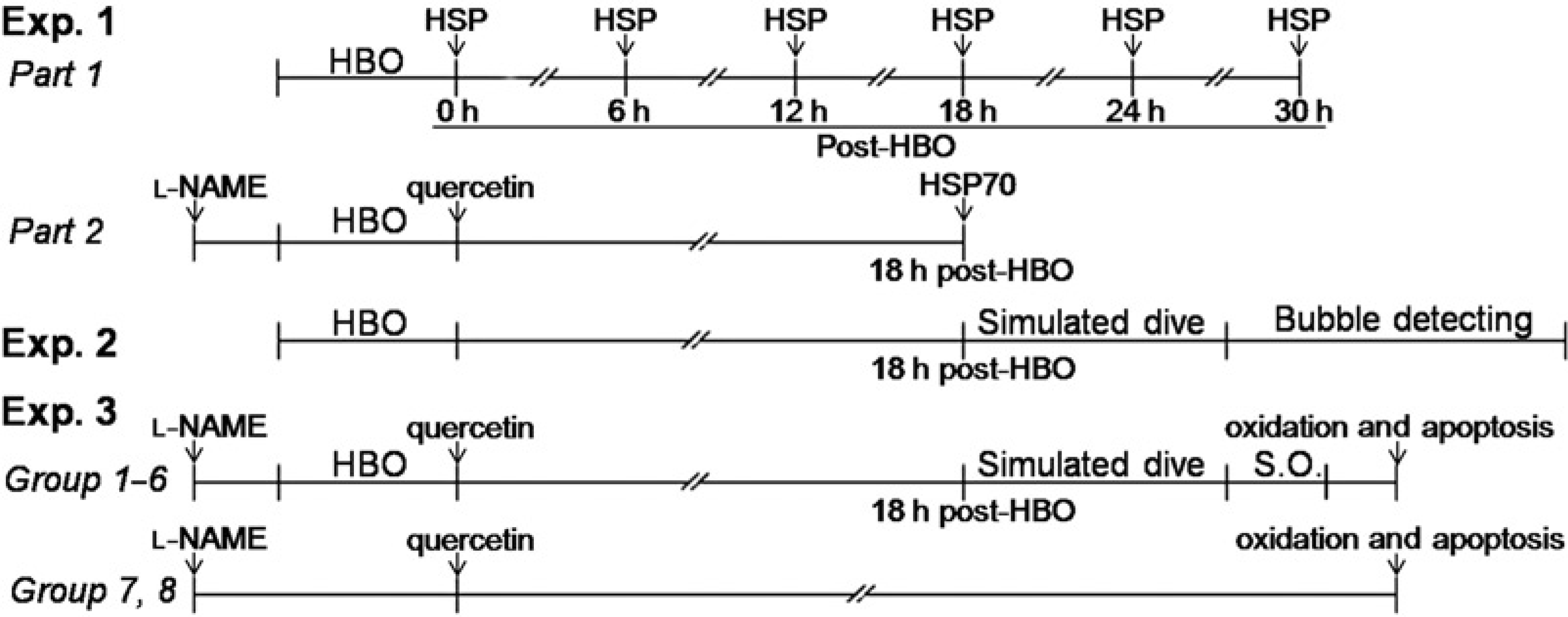
Experiment 1: HSP expression after HBO
Part 1. Rats were exposed to HBO, and levels of HSP27, 70 and 90 were determined 0, 6, 12, 18, 24 and 30 h following exposure, in the spinal cord and lung, six rats being examined for each time point. Another six rats were taken as the normal control.
Part 2. The results of Part 1 showed that only HSP70 was significantly expressed, with the greatest expression appearing at 18 h following exposure (see in Results), hence the 18 h period was selected as the time point for evaluating DCS (in Experiments 2 and 3) and HSP70 expression. Another 24 rats were used, six in each group: (1) HBO, (2) HBO + quercetin (an inhibitor of HSP70): rats received 20 mg/kg quercetin intraperitoneally instantly after HBO exposure, (3) HBO + L-NAME (an inhibitor of NOS): rats received 40 mg/kg L-NAME intraperitoneally 30 min before exposure, (4) air: sham exposure with normobaric air. HSP70 in spinal cord and lung was determined 18 h postexposure by Western blot and verified by immunohisto-chemical staining.
Experiment 2: HBO preconditioning on DCS bubble formation
Ten rats were pretreated with HBO and 10 with normobaric air as controls. Eighteen hours later, the animals were subjected to simulated air dive and rapid decompression. The bubbles flowing through the pulmonary artery were detected and analyzed.
Experiment 3: The role of HSP70 in HBO preconditioning on DCS
Rats were randomly divided into seven groups as follows: (1) air (n = 60), (2) HBO (n = 60), (3) HBO + L-NAME (n = 60), (4) HBO + quercetin (n = 60), (5) air + L-NAME (n = 60), (6) air + quercetin (n = 60), (7) L-NAME (n = 16), (8) Quercetin (n = 16), (9) normal control (n = 16). The treatments were the same as above. Eighteen hours later, the rats in Groups 1–6 were subjected to a simulated air dive and the incidence and mortality of DCS were assessed. Surviving DCS rats were anesthetized with 10% chloral hydrate (0.4 mL/100 g body weight, intraperitoneally) one hour after decompression and sacrificed for biochemical analysis. Groups 5 and 6 were used to observe the effects of L-NAME or quercetin on DCS when used alone. Groups 7 and 8 were designed to determine whether L-NAME or quercetin alone would affect the examined parameters on normal rats after the specific time intervals. Rats were injected with L-NAME (40 mg/kg, intraperitoneally, 22 h 14 min before dissection) or quercetin (20 mg/kg, intra-peritoneally, 20 h 38 min before dissection) and sacrificed for the analysis of oxidation and apoptosis. The rats were thus dissected after the same interval as the other inhibitor groups (as shown in Figure 8). Group 9 was used to determine the normal levels of oxidation and apoptosis.
HBO exposure
HBO exposurewasperformedina5.3 L transparenthyperba-ric rodent chamber (Type RDC 150-300-6, Second Military Medical University, Shanghai, China). The pressure– duration was 280 kPa–60 min, which is frequently used in animal study and the pressure 280 kPa is the current upper limit of partial pressure of oxygen (pO2) used in diving decompression or HBO therapy for humans. The compression and decompression were both carried out in three minutes. Before compression, the chamber was flushed with pure oxygen (>99.2%) for five minutes to replace the air. Carbon dioxide (CO2) absorbent was placed inside the chamber, which was continuously ventilated with oxygen during the exposure to avoid CO2 retention. The room temperature was maintained at 23–26°C for all the experiments. All the pressures described in this text are absolute pressures.
Simulated air dive
Eighteen hours after pretreatments, the rats were subjected to a simulated air dive carried out in another air chamber (the same type as above) at the pressure of 700 kPa for 90 min before decompressing linearly to the ambient pressure at the rate of 200 kPa/min. The compression was performed over five minutes which began at a low rate (50 kPa/min) to minimize possible middle ear squeeze. CO2 absorbent was used and the chamber continuously ventilated with compressed air.
Assessment of decompression sickness
Three minutes after decompression, the rats were made to walk inside an electrically controlled cylindrical cage rotating at a perimeter speed of 3 m/min for 30 min to standardize the activity level and facilitate DCS assessing. According to our experience, 30 min of observation was sufficient for all cases of DCS to become evident for the DCS model. 8 The DCS diagnosis was based on observation any of the following symptoms: abnormal breathing patterns, walking difficulties, forelimb and/or hindlimb paralysis, rolling in the rotating wheel, convulsions or death.
Determination of HSPs
Western blot
At the respective time points following HBO pretreatment, spinal cords and lungs of rats were removed and homogenized in a lysis buffer containing 1 mmol/L phenylmethylsul-fonyl fluoride, 150 mmol/L NaCl, 50 mmol/L Tris-HCl (pH 7.4), 5% sodium dodecyl sulfate and 2% Nonidet P-40. The homogenate was centrifuged at 14,000g for 20 min and the supernatant was collected. Twenty micrograms of protein were separated by 10% denaturing polyacrylamide gel electrophoresis and the gel was transferred to a polyvinylidene fluoride membrane. Membrane blocking was followed by incubation with appropriately diluted antibodies to HSP27, HSP70, HSP90 and β-actin (ab2790, ab47455, ab13492 and ab8226, respectively, all 1:1000, Abcam Ltd, Cambrige, MA, UK). There is no cross-reactivity to heat shock cognate 70 kDa in the antibody to HSP70. The intensity of each band was measured using Kodak Digital Science 1D Image Analysis System (Eastman Kodak, Rochester, NY, USA). The ratio of band intensity of HSPs to internal control (β-actin) was calculated.
Immunohistochemical staining
Rats were anesthetized and had intracardiac perfusion with phosphate-buffered saline (PBS) and 4% paraformaldehyde in PBS at 4°C. The fixed lung and spinal cord specimens were quickly removed, postfixed in 4% formaldehyde, embedded in paraffin, sectioned serially and mounted on silane-covered slides. Immunohistochemical staining was performed using the mouse anti-rat HSP70 antibody and goat anti-mouse IgG (#5470, Cell Signaling Technology, Beverly, MA, USA). Finally, a freshly prepared color development mixture containing 0.05% 3, 3'-diaminobenzidine and 0.03% H2O2 in PBS was applied to the sections for 5-10 min. The sections were then washed, dehydrated and cleared in xylene before covering. Neurons, lung alveolar cells, endothelial cells and connective tissue with brown cytoplasm were evaluated for HSP70-positive staining. Four to five sections of each specimen were observed. Sections were examined at a magnification of ×200, and 10 fields were randomly chosen to determine the expression of HSP70 using a computer image analysis system (Smart Scape, Furi Science & Technology Co., Ltd, Shanghai, China). The mean gray value scale was expressed in percent. Zero of the mean gray value represented the deepest stain and as the density of the stain became weaker the mean gray value increased.
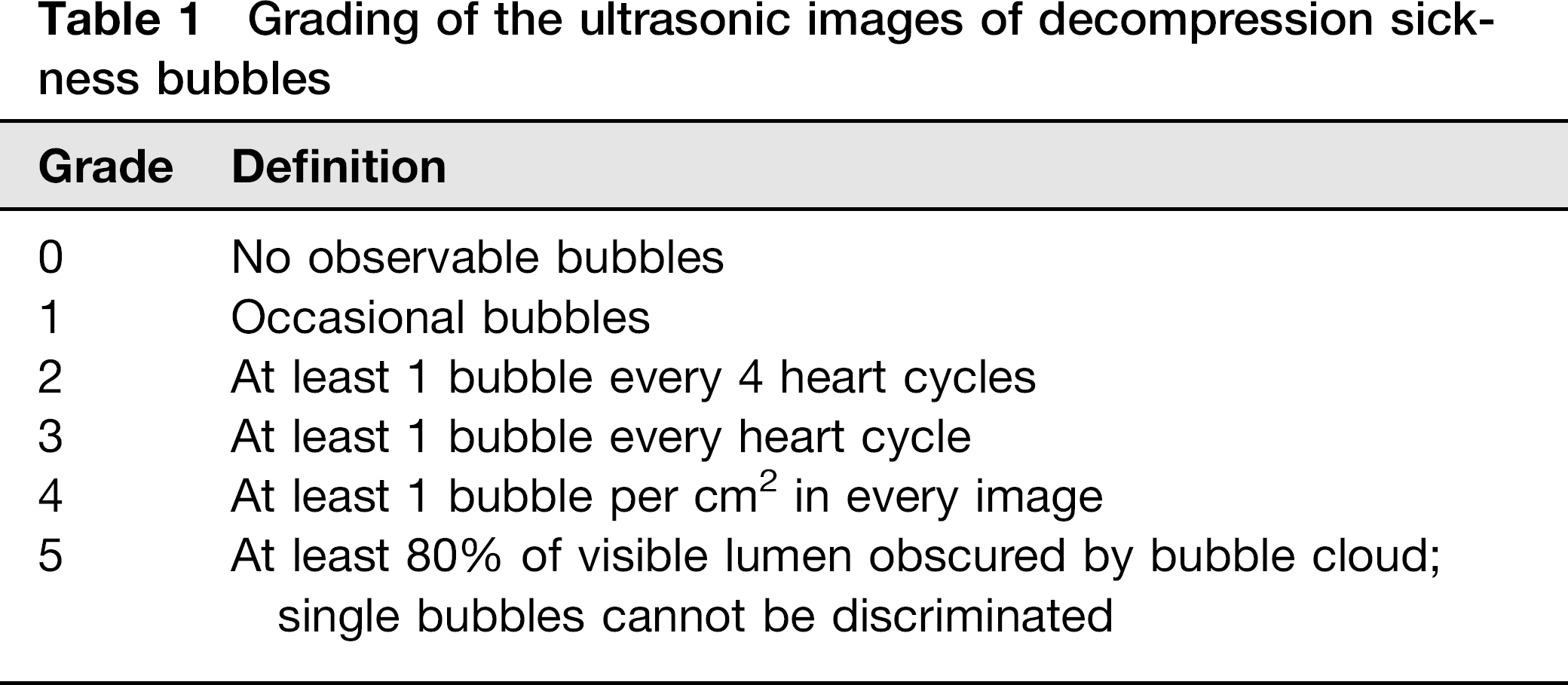
Ultrasound detecting and analysis of DCS bubbles
Immediately after fast decompression, the rats were anaesthetized with 10% chloral hydrate (3 mL/kg body weight) intraperitoneally. The hair on the chest was removed. The investigations were performed with the rats lying supine on a thermo-regulating pad (32°C). The images were obtained using a Mylab30cv ultrasonic scanner (Esaote, Italy) connected to an ultrahigh frequency (18 MHz) detector. The cross-section at the root of the pulmonary artery was insonified. Bubbles can be seen as bright spots. The detection was repeated at 5, 10, 20, 30, 45, 60, 90, 120 min after decompression, each lasted for 60 s. The ultrasound image segments were stored on the hard disk of the scanner and were copied to a personal computer for quantification of the bubbles. The bubbles can be clearly seen on slow motion playback. The number of bubbles was scored according to the grading system in Table 1. 22
Grading of the ultrasonic images of decompression sickness bubbles
Biochemical analysis
For terminal deoxynucleotidyl transferase–mediated dUTP nick-end labelling (TUNEL) staining, thoracolumbar spinal cord and left lung lobes of the rats were isolated, embedded and sectioned after perfusion fixation (n = 8 per group). For analysis of malondialdehyde (MDA), 8-hydroxyguanine (8-OHdG) and H2O2 and caspase-3 and 9, thoracolumbar spinal cord and left lung lobes (100 mg, wet weight) of other survived DCS rats in each group were isolated and homogenized immediately in 2 mL of 10 mmol/L phosphate buffer (pH 7.4) at 4°C. After centrifugation at 12,000 g for 20 min, supernatants were collected and stored at -80°C.
Measurements of oxidative parameters
MDA and H2O2 content were determined with the chemical method described as in the manufacturer's instructions (Oxis Research, Portland, OR, USA). Briefly, MDA content was measured with the thiobarbituric acid (TBA) reaction. The method was used to obtain a spectrophotometric measurement of the color produced during the reaction of TBA with MDA at 535 nm. The level of H2O2 was measured by analyzing the oxidation of ferric ions (Fe3+) by H2O2. The Fe3+ forms a complex with an indicator dye xylenol orange (3, 3-bis [N, N-di(carboxymethyl)-aminomethyl]-o-cresolsulfone-phthalein, disodium salt and XO) causing an increase in absorbance at 560–590 nm measurable as a spectrometer at a wavelength of 560 nm. 8-OHdG was measured with a commercial enzyme-linked immunosorbent assay kit following the instructions of the manufacturer (Oxis Research). Protein content in the samples was determined by Coomassie blue assay and the results were corrected per microgram of protein.
Apoptosis assay
TUNEL staining was performed on paraffin-embedded sections by using the in situ cell death detection kit (Roche Diagnostics, Rotkreuz, Switzerland). According to standard protocols, the sections were dewaxed and rehydrated by heating the slides at 60°C. Then these sections were incubated in a 20 μg/mL proteinase K working solution for 15 min at room temperature. The slides were rinsed three times with PBS before being incubated in TUNEL reaction mixture for one hour at 37°C. Sections were dried by filter paper and added Converter-AP on samples for one hour at 37°C. After rinsing with PBS (5 min, 3 times), sections were colored in dark with nitroblue tetrazolium (NBT) and 5-bromo-4-chloro-3-indolylphosphate (BCIP). Six visual fields (0.6 mm2) of the lung and spinal cord were photographed in each section. The number of staining cells in each field was counted at higher magnification (×400). The data were represented as the number of cells per high-power field.
Caspase-3 and -9 activities were determined using fluoro-metric assay kits (Biovision Research Products, Mountain View, CA, USA), according to the manufacturer's instructions. In brief, 20-200μg lysates were incubated in a 96-well plate with 2 × Reaction Buffer (50 μL). The reaction was started by adding 1 mmol/L DEVD-APC substrate (5 μL). After incubation in the dark at 37°C, the plate was read in a fluorometer equipped with a 400-nm excitation filter and a 505-nm emission filter. Fold-increase in caspase-3 or -9 activity was determined by comparing these results with the levels of the uninduced controls.
Statistical analysis
Incidences of DCS of different groups were compared by means of χ2 test. One-way analysis of variance (ANOVA) followed by the Student Newman-Keuls tests were used to compare the ratio of HSP70 to β-actin, bubble scores, MDA, 8-OHdG, H2O2, caspase 3 and -9 levels among groups. For analyzing the results of cell counting, a non-parametric Kruskal-Wallis ANOVA was used followed by Dunn's test. P < 0.05 was considered statistically significant. Except for the incidence and mortality, all results were expressed as means ± SEM.
Results
HSPs expression after HBO exposure
The results of Western blot showed that HSP27 and HSP90 had a slight and non-significant increase after HBO exposure (P > 0.05, Figure 1), while for HSP70, it increased and reached at a peak level at 18 h after HBO exposure both in the lung and the spinal cord (P< 0.05) (Figure 2). The interval of 18 h was then selected to observe the effects of HBO following exposure. Western blot and immunohisto-chemistry further verified that at 18 h following HBO, the expression of HSP70 increased significantly (P < 0.05), mainly in neurons, alveolar epithelia and endothelia. Quercetin (an inhibitor of HSPs) or L-NAME (NG-nitro-L-arginine methyl ester, a non-selective nitric oxide synthase [NOS] inhibitor) significantly inhibited the effects of HBO (P < 0.05). (Figure 3).
Heat-shock protein (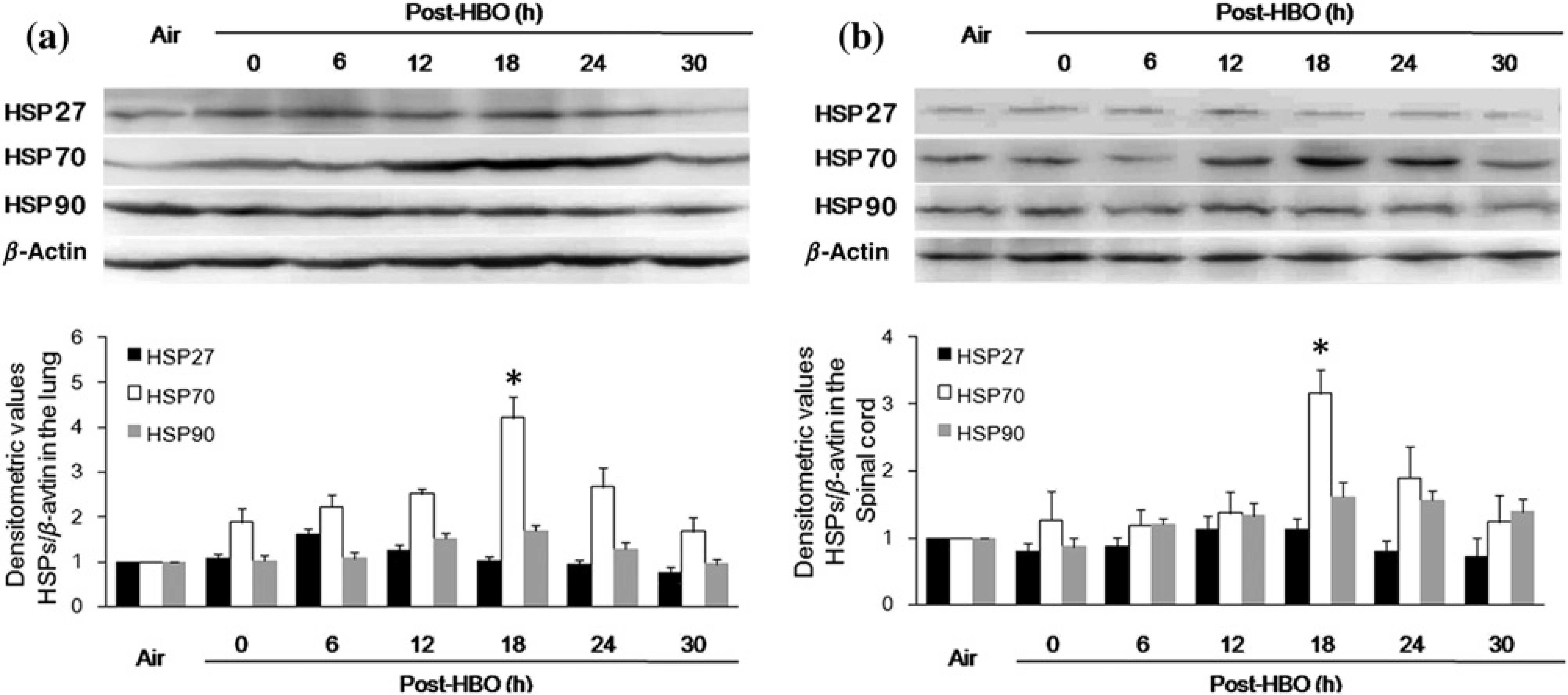
Heat-shock protein 70 (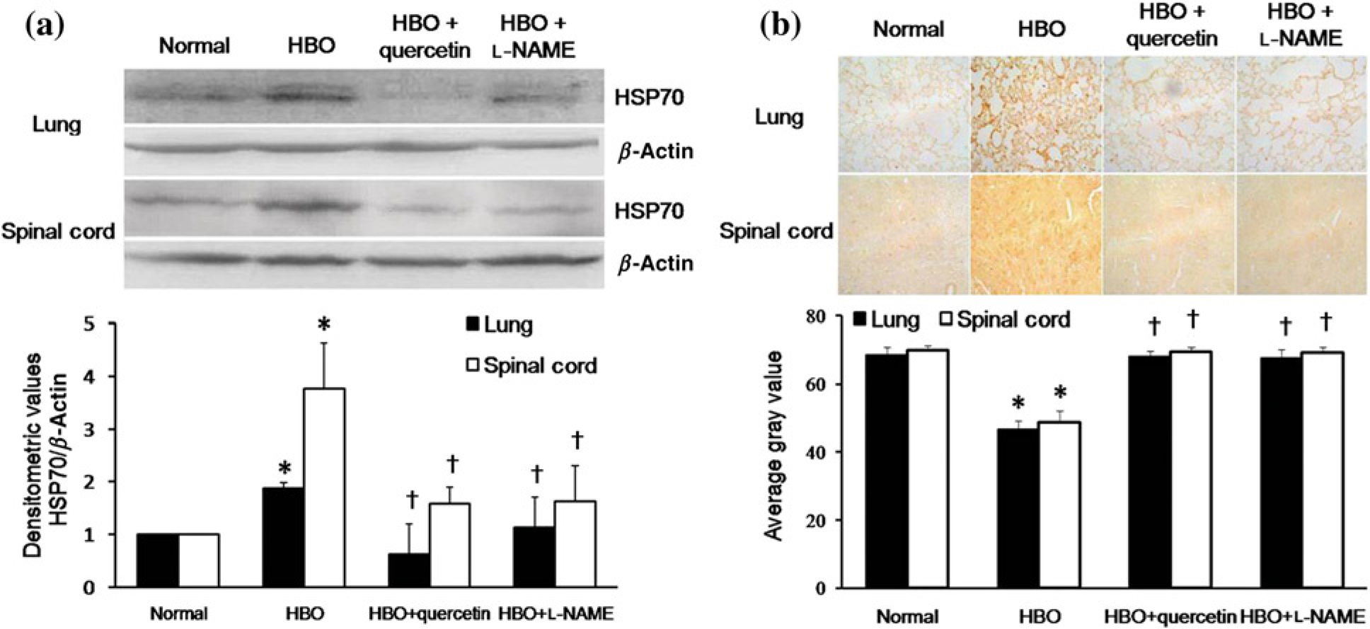
Bubble formation in DCS rat models with different pretreatments
Gas bubbles could be clearly seen (moving bright spots) in the pulmonary artery in ultrasound images. The frame frequency was 40 per second, so each video of 60 s contains 2400 frames of images. The relative bubble count increased gradually after decompression, to reach a maximum at around 31 min (95% confidence interval [CI] 22–40 min). There was no difference in the bubble scores at each time point or the total scores, or in the time to reach a maximum value between the two groups (Figure 4).
Bubble scores in rat decompression sickness models pretreated with hyperbaric oxygen or Air. Eighteen hours after 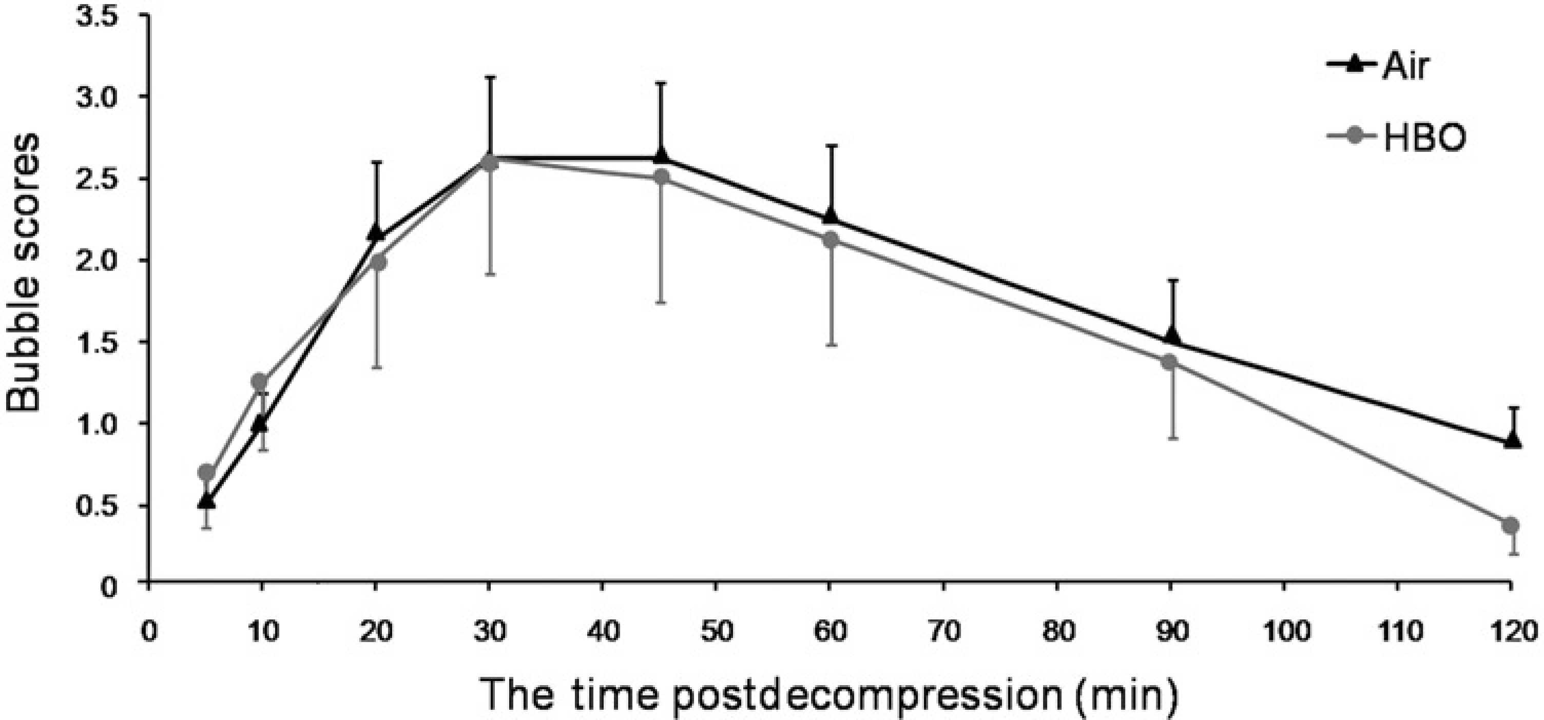
Effects of HBO preconditioning on the incidence of DCS
Based on the outcomes after a simulated dive and fast decompression, the rats in Experiment 3 were divided into three subgroups: no DCS, DCS and death. HBO significantly decreased the incidence and mortality of DCS (P < 0.05). L-NAME or quercetin nullified the protective effects of HBO. They did not affect DCS incidence when used alone (Air + L-NAME, air + quercetin). (Figure 5).
Effect of hyperbaric oxygen (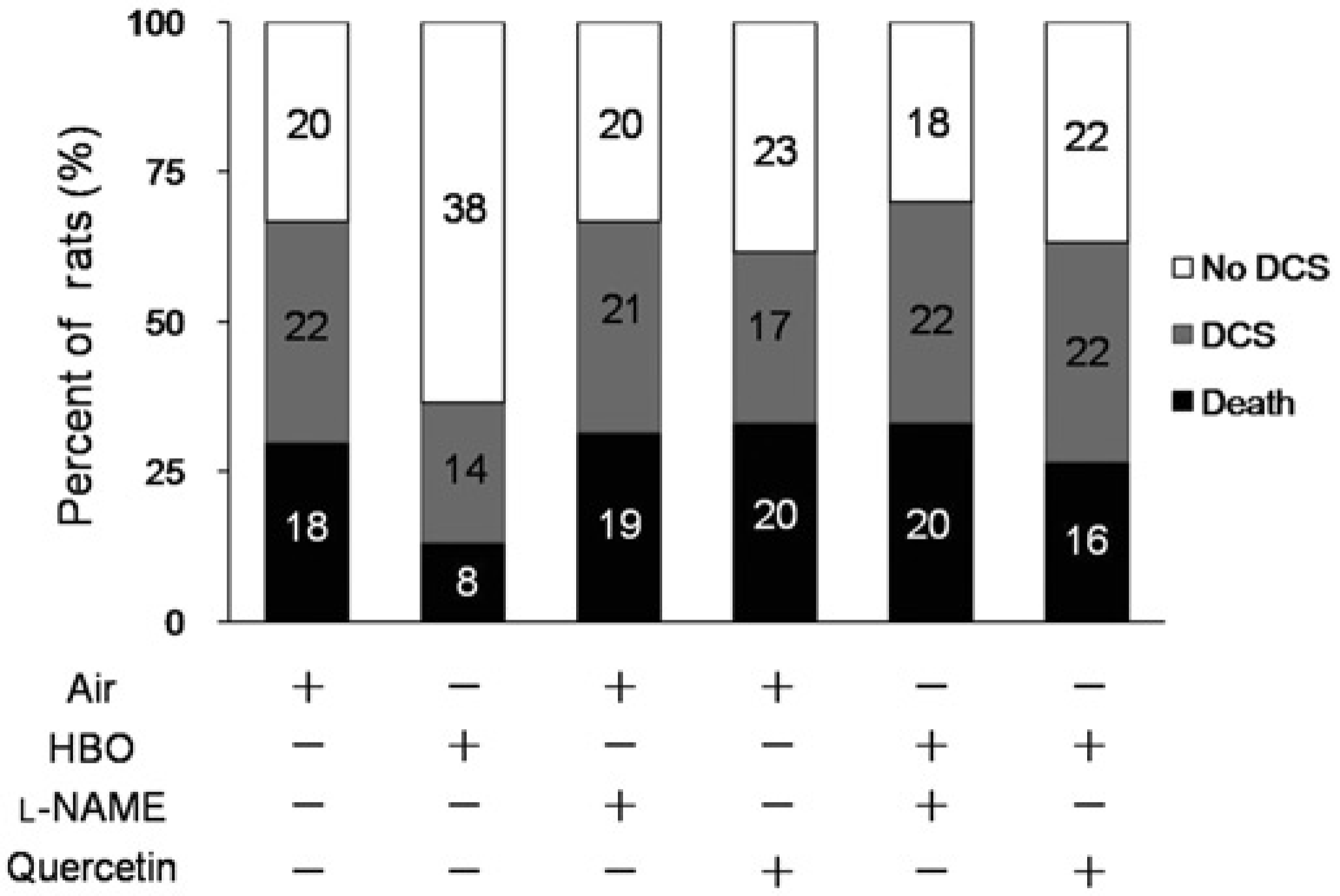
Changes of oxidation in DCS rats with different pretreatments
Compared with the normal controls, the levels of MDA, 8-OHdG, hydrogen peroxide (H2O2) in the lung and spinal cord increased significantly (P < 0.01) in DCS rats (Air Group). HBO preconditioning significantly lowered the levels (P < 0.01), which could be partially counteracted by L-NAME or quercetin (P < 0.05 or P < 0.01). L-NAME or quercetin did not affect the oxidation parameters both in normal or DCS rats when used around 20 h before. (Figure 6).
Malondialdehyde (MDA), 8-hydroxydeoxyguanosine (8-OHdG) and hydrogen peroxide (H2O2) levels in DCS rats with different pretreatments. Tissues were obtained from rats one hour after decompression. Data are expressed as mean ± SEM. The numbers of rats in each group were shown in the figure. *P < 0.05, **P < 0.01 compared with the normal control; †P < 0.05, ††P < 0.01 compared with the Group Air + dive; ‡P < 0.05, ‡‡P < 0.01 compared with the Group HBO + dive. There is no significant difference between the inhibitor-treated groups and the controls, with (Column 6 or 7 compared with Column 3) or without (compared between the last 2 columns and the first column) simulated dive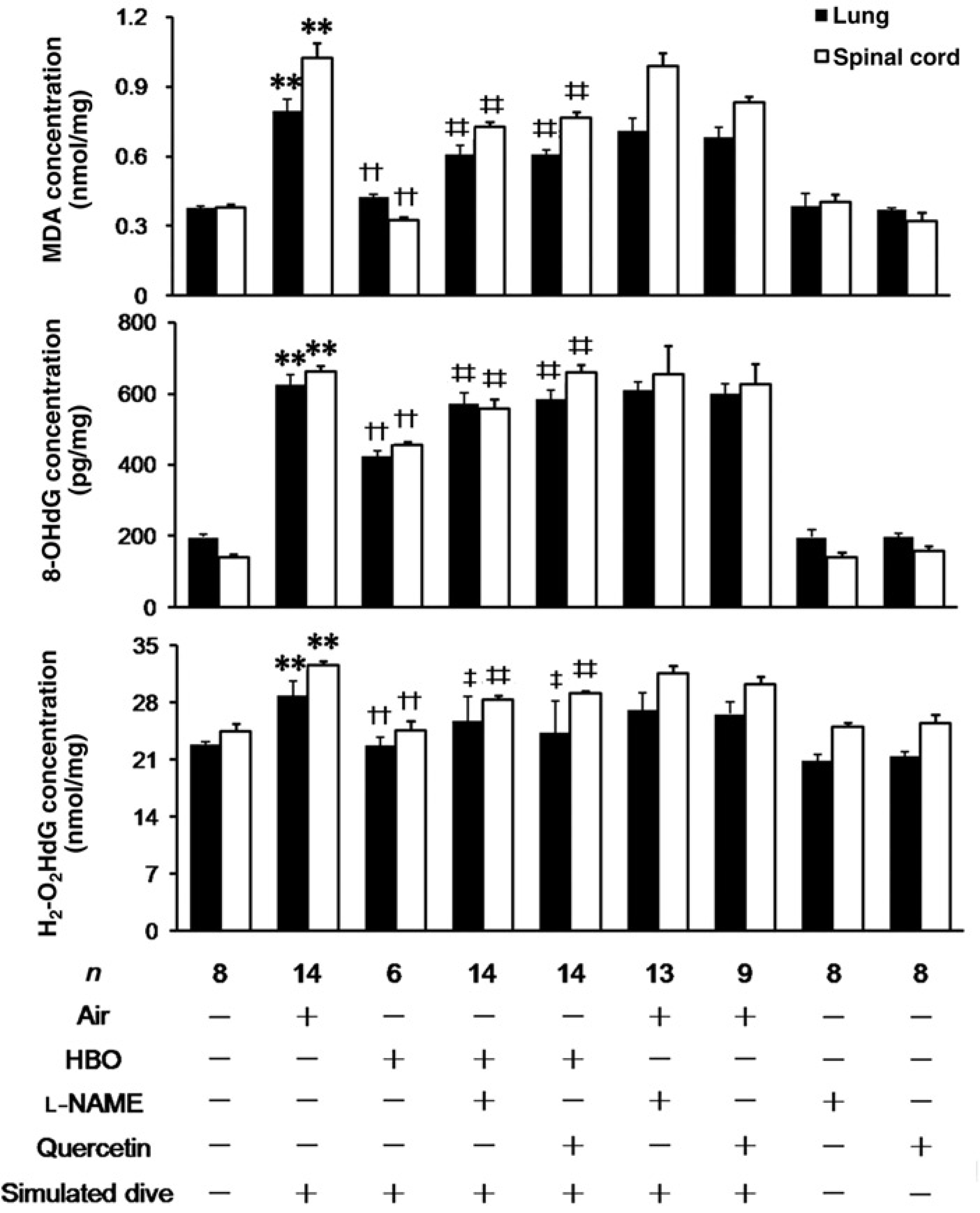
Changes of apoptosis in DCS rats with different pretreatments
Photos in Figure 7 were representative graphs with different magnifications of TUNEL staining of lung and spinal cord in rats with different pretreatments. Cells with double-strand breaks in DNA were stained in brown color. A few TUNEL-positive cells were identified in samples from normal control (A1–4). TUNEL-positive cells were markedly increased in DCS rats (B1–4) (P < 0.05). HBO preconditioning dramatically reduced the number of positive cells (C1–4), the effects were inhibited by L-NAME (D1–4) or quercetin (E1–4).
Terminal deoxynucleotidyl transferase-mediated dUTP nick-end labelling (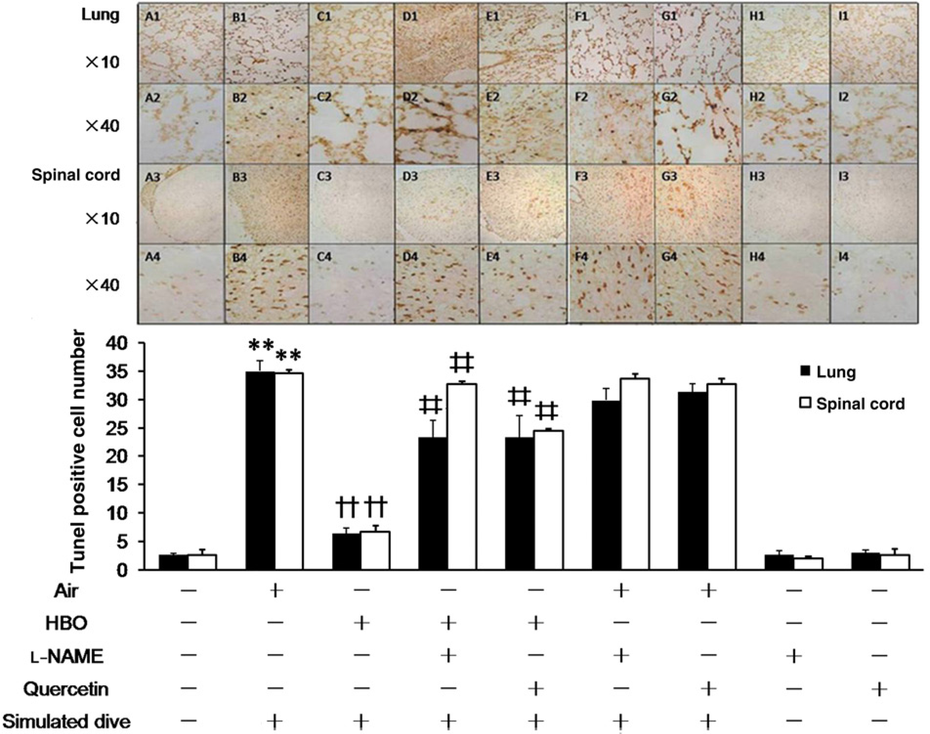
The activities of caspase 3 and -9 increased significantly in DCS rats, and were decreased in rats pretreated with HBO (P < 0.05). L-NAME or quercetin inhibited the effects of HBO (Figure 8).
Caspase-3 and -9 levels in spinal cord and lung in rats with different pretreatments. Tissues were obtained from rats one hour after decompression. Data are expressed as mean ± SEM. The numbers in each group were shown in the figure. *P < 0.05, **P < 0.01 compared with the normal control; †P < 0.05, ††P < 0.01 compared with the Group Air + dive; ‡P < 0.05, ‡‡P < 0.01 compared with the Group 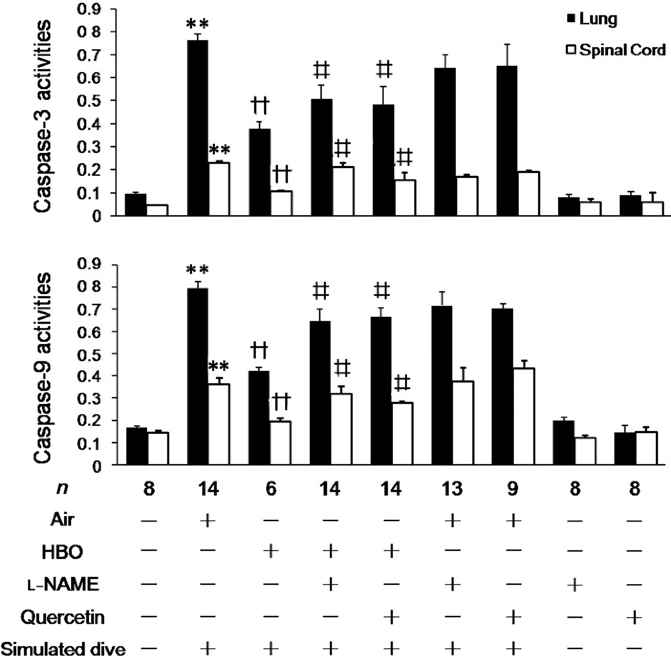
L-NAME or quercetin administered around 20 h before had no effects on TUNEL-positive cell numbers and caspase activities either in normal or DCS rats.
Discussion
DCS is an environmental injury which occurs during hyper-or hypo-baric exposures, and is a key problem in diving related activities. 1 Recently, our laboratory revealed that HBO had beneficial effects on many ischemic injuries.6,7,23 This study further verified our preliminary findings that a single exposure of HBO 18 h before significantly decreased the incidence of DCS in rats via NO. 8 The routinely applied pressures of oxygen in diving decompression or clinical hyperbaric therapy are 200–280 kPa, and so pressures of 250 and 280 kPa are frequently used in experimental studies of hyperbaric medicine. With the expectation of acquiring a more pronounced effect, we selected 280 kPa rather than the 250 kPa level which was used in our preliminary study. 8 The results of incidence and mortality showed no significant difference between the two preconditioning profiles. Furthermore, in order to acquire sufficient cases of survived DCS rats for biochemical assessment, 60 rats were assigned for each of the groups in Groups 1–6, Experiment 3. This big sample provides a convincing support of the conclusion that HBO preconditioning is effective on ameliorating DCS in rats. Bubble formation is the precipitating cause of DCS, but the signs and symptoms may result from a variety of subsequent mechanisms. 1 The protective mechanisms underlying HBO on DCS was speculated to be related to the reduction of the formation of gas bubbles, which could be the results of the decrease of the number of gas nuclei, or the acceleration of expulsion of inert gas. HBO before diving could replace the resident gas in the micronuclei by oxygen, which could be consumed by mitochondria and thus resulted in a reduced volume and number of micronuclei. 24 Furthermore, HBO increased NO production,8,25 and NO may influence the degree of DCS bubble formation.26,27
In the tissues well supplied of blood, microbubbles flow from capillary vessels into veins, circulate to the right heart and sequestered in the lung through pulmonary vas-culature. 28 Right heart ventricule and pulmonary artery conduct DCS intravascular bubbles before they are sequestered in the lung and these are the target sites of detecting DCS bubbles. 22 Ultrasonic detection of intravascular bubbles is by far the most objective and stable methods of detecting realtime decompression-induced bubbles. 22 By using ultrasound imaging in this study, bubbles flowing through the pulmonary artery were detected. The results revealed that HBO exposure 18 h before did not influence the generation of bubbles in rat DCS model. This indicates that HBO reduced the risk of DCS by ways other than decreasing bubble formation. HSPs play a crucial role in maintaining cell homeostasis and survival against various injuries. 29 The inducible members including HSP 27, 32, 40 and 70 families are associated with cellular protection. HSP27 correlates with increased survival in cells exposed to cytotoxic stimuli in several disease model systems. 30 HSP32 has been implicated as a protective mediator in numerous experimental disease models. 31 And HSP40 serves as the co-chaperone of HSP70 to regulate in cellular signal transduction and apoptosis. 32 HSP90 is a constitutive molecular chaperone that regulates numerous client proteins and protects cells against various injuries. 33 HBO has been found to enhance the expression of some of the HSPs.15,34 Quercetin, a widely distributed bioflavonoid, has been shown to inhibit the synthesis of HSP27, 70 and 90 by changing the tertiary structure of heat shock factor 1, which was required for the transcription of most HSP genes except HSP32.35,36 So, the expression of HSP27, 70 and 90 after HBO exposure were detected in this study. The results showed that HSP27 were not significantly induced by HBO (less than 1.5 fold of the control at 6 h time point). And the expression of HSP90 was also not altered significantly though an increasing trend occurred at 18 h after HBO exposure. For HSP70, the expression in rat lung and spinal cord were both increased, with the greatest expression appeared at 18 h following exposure. These results indicate that HSP70 might be the main HSP enhanced by a single exposure of HBO. Because the interval of detection was six hours, and the second peak time point was at 24 h, the actual time point of highest expression of HSP70 was most probable around 20 h following HBO exposure. Thus, the interval of 18 h between HBO exposure and simulated dive was selected, so as to the stress induced by rapid decompression occurred around 20 h after HBO exposure. While in our preliminary study, 18 h was selected as the interval merely because this spacing is convenient for the experiment and for the diving practice. 8
HSP70 is the most widely studied proteins in the highly conserved HSP family in mammals. 37 Much evidence indicate that HSP70 confers injury tolerance in the lung and spinal cord.38,39 Heat stress prior to simulated dive conferred protection against DCS and HSP70 is involved in endothelial protection during DCS process. 40 The protective effects of HBO against DCS may be relative to HSP70.
In this study, either quercetin or L-NAME counteracted the increase of HSP70 induced by HBO and did not affect the pathophysiological process in DCS. They also diminished the beneficial effects on the incidence and mortality of DCS (HBO + L-NAME, HBO + quercetin). Neither of the inhibitors influences DCS outcomes when administered more than 20 h before (Air + L-NAME, air + quercetin). The results suggested that HBO induced HSP70 expression via NO, and HSP70 was involved in the prophylactic effect of HBO on DCS in rats.
HBO induces the production of NO instantly during the exposure. 41 So, as in many other related studies, L-NAME was administered 30 min prior to HBO exposure. Quercetin also influences the synthesis of HSP90, 36 which is important in NOS function. 42 So, quercetin was given instantly after HBO exposure to avoid the possible influence of NO synthesis. From the current results, quercetin effectively inhibited the increase of HSP70 and nullified the protective effect on DCS 18 h after HBO exposure. However, L-NAME was reported to increase bubble formation in DCS rats when chronic oral administered, 43 and quercetin was proved to act as an antioxidant in some diseases. 44 Therefore, they may affect the incidence, mortality, oxidation and apoptosis of DCS by the effects other than the inhibition of HSP or NO induction. Accordingly, we determined the effects of quercetin or L-NAME on the above variables in DCS rats when administered at the respective time periods (20 h 38 min or 22 h 14 min, respectively) ahead. The results showed that they had no influence on the measures detected more than 20 h later.
Much work has shown that HSP70 directly interfered with oxidative injury and ischemia-like insults, which nduces significant alterations in cellular redox status that plays a critical role in cell survival/death path-ways.45–47 HSP70 can exert cytoprotective effects by modulating the activities of glutathione peroxidase and glutathione reductase. 48 The oxidative damage following hypoxia–ischemia caused by DCS bubbles plays an important role in the course of DCS, especially in severe cases. 15 Our previous study revealed that selective antioxidant hydrogen protected rats against DCS by alleviating the inflammation. 49 H2O2 is a direct reflection of the degree of oxidative stress, and MDA and 8-OHdG represent the oxidative injury to cell membrane and DNA, all serve as sensitive biomarkers of intracellular oxidative stress. 50 HSP70 is also capable of antiapoptosis, and can inhibit caspase activation by interfering with Apaf-1 and prevent the recruitment of procaspase-9 to the apoptosome.47,51 Cell apoptosis was found participating in the process of decompression injury after simulated diving in mice, and was postulated to be an important pathophysiology mechanism in DCS process. 52
In the present study, the oxidation and apoptosis parameters significantly increased in DCS rats, and HBO provided significant protection against the injuries. The beneficial effects of HBO were mostly inhibited by quercetin or L-NAME, suggesting that the protective effects were related to the antioxidation and antiapoptosis effects of HSP70.
The mechanism by which HBO induces HSP70 expression has not been clearly elucidated. In mouse neuroblastoma cell lines, HSP70 expression may be regulated by HBO through the activation of c-Jun N-terminal kinase (JNK). 11 HBO increases the production of reactive oxygen species and NO, which were found playing pivotal role in activating HSP70 expression, possibly through JNK, protein kinase C/A or p38/MAPK pathways in cultured MDA231 and T84 cells.53–55 How HBO induces HSP70 expression in pulmonary and spinal cord tissues in vivo warrants further studies.
This is the first study to explore the role of HSPs in HBO on DCS. We suggest that a single exposure to HBO exerted significant protection against DCS in rats mostly via antiox-idative and antiapoptotic function of HSP70 induced by NO. Whether a double or multi-exposure of HBO could further enhance the expression of HSP70 and the beneficial effects against DCS deserves further study. We conclude that, as a mild oxidative stress, HBO exposure is a promising modality to induce HSP70, with little or no adverse effects. HSP70 induced by HBO could not only prevent DCS, but also could contribute to the prevention of a great deal of prospective injuries such as surgical operations. We look forward to more experimental or clinical studies in the topic of HBO and HSP70.
Footnotes
Acknowledgements
This work was supported by the Chinese National Natural Science Fund No. 81171873.