




Abstract
Objective
To find out variations in the deep forearm flexors that are associated with anomalous origin of the first lumbrical muscle.
Introduction
Muscular anomalies of the upper extremity could cause peripheral nerve disorders. Although variations in the attachments of lumbrical muscles have been commonly studied, muscle with an additional origin in the proximal aspect of the forearm from the deep flexors is not much discussed.
Methods
Fifty-three formalin-fixed upper limbs were dissected to assess the aberrant origin of the first lumbrical from the deep forearm flexors. The measurement, shape, origin, insertion and innervation of aberrant deep forearm flexors were noted and photographed.
Results
In two (3.77%) out of 53 upper limbs unilateral aberrant deep forearm flexors were found giving accessory anomalous origin to the first lumbrical muscle. In 11 cases (20.75%) the presence of accessory head of flexor pollicis longus was seen, and found co-existing with the aberrant deep forearm flexor in one (1.89%) limb.
Discussion
The clinical implication of these muscular anomalies, such as aberrant origins and hypertrophy of the first lumbrical, in nerve entrapments, especially in the aetiology of carpal tunnel syndrome, is considered. The awareness and identification of the muscular variations of the antebrachial and carpal regions will facilitate safe surgery of cases of median nerve entrapment syndromes.
Keywords
Introduction
Abnormal and additional muscles are not an uncommon occurrence in the forearm and hand. Understanding the anatomical variations and abnormalities of the muscles of this region is important in order to define the anatomical features of each in relation to clinical diagnosis and for surgical procedures. 1
Anatomical variations in muscles in the superficial flexor compartment of the forearm, especially those of flexor digitorum superficialis (FDS), are quite common, among which many are associated with anomalous accessory first lumbricals. 2–4 Variations of flexor digitorum profundus (FDP) in the deep flexor compartment are not rare. But, rarely, a lumbrical muscle may arise from the deep flexors in the forearm 5 even though variations in their attachments are common. 5–7 Among the accessory forearm flexor muscles, Gantzer's muscles 8 are prevalent; the incidence of accessory head of flexor pollicis longus (FPLah) being more common than that of FDP. 9–12
An anomalous origin of the lumbrical from muscles in the forearm has the potential to cause compression of the median nerve in the carpal tunnel. Although lumbricals show frequent variations (20% of individuals), rarely their number is increased by accessory slips. In the case of the first lumbrical, an accessory slip may arise from the tendon of flexor pollicis longus (FPL), FDS, the first metacarpal, opponens pollicis or the palmar carpal ligament. There were cases in which a fasciculus arose from the muscular belly of the superficial or deep flexor and joined the first lumbrical. 7 A rare case of a unilateral bipennate first lumbrical muscle with an additional origin arising in the distal quarter of the forearm but from the same FDP tendon was reported. 13 However, bilateral presence of an additional distal forearm flexor that formed a conjoint tendon with the index FDP tendon but had normal lumbrical was also reported in the literature. 1
Materials and methods
During routine dissections we had come across a peculiar contribution to the first lumbrical muscle from an anomalous forearm flexor. Fifty-two formalin-fixed upper limbs were further dissected to assess the aberrant origin of first lumbrical from the deep forearm flexors. Their measurement, shape, origin, insertion and innervation were noted and photographed.
Results
In two (3.77%) out of 53 upper limbs, unilateral aberrant deep forearm flexors (AFM) were found giving accessory anomalous origin to the first lumbrical muscles. FPLah was also present in 11 cases (20.75%); all of them presented a fusiform muscle belly that arose from the deep aspect of origin of FDS to get inserted to the ulnar margin of FPL. In one (1.89%) limb, FPLah was found co-existing with AFM. In both cases of AFM the origin was mainly from the interrosseus membrane, between FPL and FDP, and their contribution to first lumbrical muscle mass was significant.
Case 1
The fusiform muscle belly of AFM arose from the anterior surface of radius and the adjoining interrosseus membrane. Beneath the tendons of FDP it became tendinous, passed through the carpal tunnel and terminated by giving muscle fibres that merged with the hypertrophied first lumbrical muscle (Figure 1). The length and width of the AFM muscle belly was 5.3 and 0.9 cm, respectively, and the tendinous part was 7.1 cm long. The nerve supply was from the branches of the median nerve.
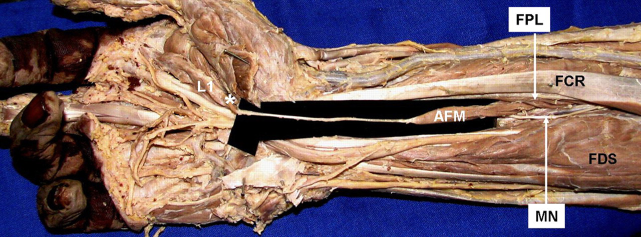
Anterior view of the right forearm and hand. FPL, flexor pollicis longus; FCR, flexor carpi radialis; FDS, flexor digitorum superficialis; MN, median nerve; AFM, aberrant deep forearm flexor; L1, first lumbrical. *Denotes the accessory contribution to lumbrical muscle mass from the tendon of AFM
Case 2
The origin of AFM was in the form of a sheet of muscle fibres from both the interosseus membrane and the fascial sheath of pronator quadratus. Beneath the flexor retinaculum the tendon of AFM joined the FDP tendon of the index finger to form a conjoint tendon to give origin to the muscle belly of first lumbrical that was bulky. The incursion of second lumbrical into the carpal tunnel was also observed (Figure 2). Besides, FPLah was present as a fusiform additional muscle belly arising beneath the origin of FDS to the ulnar margin of FPL. Both FPLah and AFM were supplied by the anterior interosseus nerve (AIN) that passed posterior to the FPLah to occupy the plane between the FPL and the AFM (Figure 3). The length of the AFM muscle belly was 13.3 cm and that of the tendon, which formed within the anterior margin of this unipennate muscle, was 10.6 cm.

Anterior view of the left forearm and palm. Flexor digitorum superficialis tendons are pulled medially to expose the aberrant deep forearm flexor (AFM) and flexor digitorum profundus (FDP). FCR, flexor carpi radialis; FPL, flexor pollicis longus; PQ, pronator quadratus; FDPi, flexor digitorum profundus indicis tendon; AIN, anterior interosseus nerve; L1, first lumbrical; L2, second lumbrical; †, AFM tendon; ‡, conjoint tendon. *Denotes the accessory contribution to lumbrical muscle mass from the conjoint tendon

Anterior view of the left forearm. Flexor digitorum superficialis (FDS) tendons are pulled medially to expose the accessory head of flexor pollicis longus (FPLah), aberrant deep forearm flexor (AFM) and flexor digitorum profundus (FDP). FCR, flexor carpi radialis; FPL, flexor pollicis longus; AIN, anterior interosseus nerve; FDP, flexor digitorum profundus; †, AFM tendon
Discussion
Although rare, lumbrical origins in the forearm from the deep flexors were reported. 1,13,14 Kopuz et al. reported a distal and complete additional FDP muscle to the index finger bilaterally from the distal part of the forearm, that shared the same synovial compartment with a normal separate FDP tendon for the index finger and ended as a conjoint FDP tendon; however, the FDP for the index finger and its lumbrical muscle showed no abnormalities. 1 But in our study the conjoint FDP tendon contributed to the lumbrical muscle mass and even in the other case of direct accessory origin of lumbrical from AFM-tendon, the first lumbrical was hypertrophied. Unlike those reported distal forearm muscles, the origins of AFM were extending proximally from the upper or middle one-third of the forearm in ours. A proximal origin, under FDS, of an aberrant deep flexor contributing to the second lumbrical but supplied by the ulnar nerve could also be found in the literature. 14 While describing anomalous origin of first lumbrical from the index FDS tendon, it was suggested that the anomalous muscles that have a proximal origin in the forearm can be identified as accessory first lumbrical muscles, if both are supplied by the median nerve. 3 In our study, the AFM was supplied by the median nerve in one case whereas in the other by the AIN.
Apart from its anatomical interest, FPLah as a causative factor of anterior interosseous nerve syndrome 10–12,15 needs to be considered as significant when it is present between the median nerve anteriorly and the AIN posteriorly 9,11,12 as in our study. It is implicated in the causation of Kiloh–Nevin syndrome characterized by paralysis of AIN because of compression in the forearm. 15,16
Causation of carpal tunnel syndrome (CTS) by compression of the radial branch of the median nerve due to anomalous hypertrophied muscle belly of the first lumbrical at the distal edge of the transverse carpal ligament is a relatively new diagnostic entity. 17 But anomalous and additional lumbrical muscle as a cause of CTS had been reported. 2,4,13,14,18,19 Causes of CTS with respect to the lumbricals include incursion of the lumbrical muscles within the tunnel during finger movements, 20,21 hypertrophy of the lumbricals, 18 anatomic variants such as abnormally long lumbrical muscles 19 and aberrant tendinous origin of the first lumbrical. 2,4,13,14
The present findings will supplement our knowledge of muscle variations in the antebrachial and carpal region and should also be considered in the aetiology of CTS, the awareness of which will facilitate safe surgery of median nerve entrapment syndromes. The identification of muscular variations in the carpal region is important for chiropractors as well, who use manoeuvres to alleviate pain resulting from CTS.