




Abstract
A 56-year-old man presented complaining of urinary frequency, passing urine eight times per day, urethral irritation and dysuria. Investigations showed no evidence of urinary tract infection or sexually transmitted infections. Three months later he presented, again complaining of increased urinary frequency and urethral irritation. He brought with him a urine specimen containing a small ‘worm’, 2 mm in length, identified as a drain fly (or moth fly) larva, of the genus Psychoda (dipterous flies). Psychoda lay eggs in organically polluted water such as sewage plants, sink drains or on decaying vegetables and fruits. Urogenital myiasis may arise from hatching of larvae near the urethral opening and ascending migration along the urethra with consequent urethritis. Following larval identification, ivermectin was prescribed and the man's symptoms improved after six weeks.
Keywords
INTRODUCTION
Myiases, or fly infections, were first described by Hope in 1840 and consist of invasions of living or necrotic tissue by larvae (maggots) of a number of genera of dipterous (two-winged) flies. The invasive process is part of the normal life cycle. Invasion can be of tissues such as skin, cavities such as urogenital, oral and pulmonary or other systems such as intestinal and optic infections. Accidental ingestion may lead to migration into the gastrointestinal system. Episodes of urogenital myiases are rarely reported, especially in the English language literature. 1
CASE HISTORY
A 56-year-old UK-born man who has sex with men presented complaining of increased urinary frequency, urethral irritation and dysuria. His past medical history revealed chronic hepatitis B infection (treated with interferon), recurrent non-specific urethritis (NSU) and urethral gonorrhoea. A urine specimen was cloudy, but microscopy and swabs showed no evidence of NSU or sexually transmitted infections (STIs). He was reassured and no treatment was given. He presented two months later again complaining of increased urinary frequency (from 2 to 8 times per day), cloudy urine, burning and discomfort in the last one-third of his urethra. As his symptoms had continued since his last visit, he had obtained clean, but unsterilized, specimen containers and collected urine specimens, one of which he presented containing a small ‘worm’, 2 mm in length, shown in Figure 1.
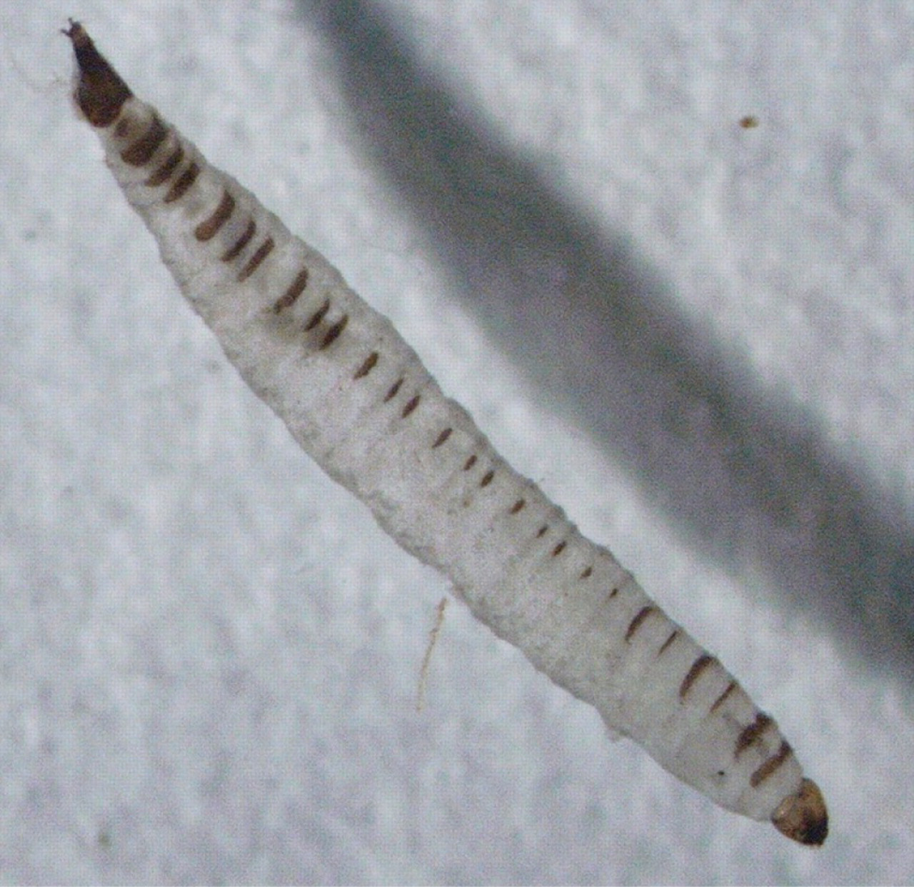
Found in urine specimen, a 2 mm worm identified as a Psychoda (drain fly) larva
INVESTIGATION AND TREATMENT
There was microscopic evidence of non-gonococcal urethritis, but all tests for STIs were negative. The London School of Hygiene and Tropical Medicine Reference Laboratory identified the ‘worm’ as the larva of a drain fly, family Psychodidae, genus Psychoda. A computed tomography scan, done to exclude urogenital abnormalities, showed a 4.5 mm calculus within the collecting system of the right kidney. There was no ureteric calculus, but relative under-filling of both collecting systems.
Initial treatment was with doxycycline and metronidazole followed by azithromycin because of persistent symptoms and pending results of STI tests and ‘worm’ identification. There was no symptomatic relief. Following identification of the ‘worm’, ivermectin 2 was prescribed 12 mg once daily for two days. The man's symptoms improved after six weeks.
DISCUSSION
Psychoda's natural substrate for egg laying is wet, decaying vegetation in aquatic or subaquatic environments. The flies are commonly found in sewage plants, cesspools and sink drains in bathrooms and kitchens, hence the common name ‘drain flies’. The larvae may opportunistically enter human orifices and give rise to rarely described cases of intestinal, nasopharyngeal and urogenital myiasis. 3 There are 15 British species of Psychoda, the most common being P. alternata and P. albipennis.
Indwelling urethral appliances such as stents or catheters may provide avenues for infection including myiases. Hyun et al. 1 reported a case where they believed that ureteric stent placement coupled with poor hygiene and exposure to contaminated water increased the likelihood of urinary myiasis by providing an entry means for psychodid larvae. Our patient reported no history of catheter use and there was no past history of urological surgery.
A case report by Taylan-Ozkan et al. concluded that lack of personal hygiene and partial obstruction of the urinary flow due to a 2 cm calculus in the right kidney were probably contributing factors for the myiasis caused by P. albipennis 4 in their patient. Our patient's renal calculus may have caused some obstruction in urinary flow thus contributing to possible entry by the psychodid larva. He also had a past history of urethral gonococcal infection and, possibly, scarring or stricture formation could have affected urinary flow. A cystourethrogram was not performed to investigate this.
In both cases cited poor personal hygiene or contact with infected water were mentioned as possible contributory factors. Our patient had mentioned drainage problems in his block of flats, which might have provided a site for the breeding of drain flies. Laying females could, therefore, have oviposited near his urethral opening or in urine-soiled underwear that he may have used without washing. There was no evidence to suggest an extra-urethral source or whether multiple infections had occurred.
The case presents a combination of unusual factors. Urogenital myiasis is unusual. Psychoda do not have a life cycle involving invasion of living, dead or necrotic tissue. However this case has a number of similar features to those described in the literature. Urogenital myiasis is often connected with poor environmental conditions and personal hygiene, while poor urinary flow may facilitate larval invasion. In our case the diagnosis was made due to the diligence of the patient in the face of persistent urogenital symptoms.