




Abstract
Here we provide an up-to-date review of research that explains why uncircumcised men are at higher risk of HIV infection. The inner foreskin is a mucosal epithelium deficient in protective keratin, yet rich in HIV target cells. Soon after sexual exposure to infected mucosal secretions of a HIV-positive partner, infected T-cells from the latter form viral synapses with keratinocytes and transfer HIV to Langerhans cells via dendrites that extend to just under the surface of the inner foreskin. The Langerhans cells with internalized HIV migrate to the basal epidermis and then pass HIV on to T-cells, thus leading to the systemic infection that ensues. Infection is exacerbated in inflammatory states associated with balanoposthitis, the presence of smegma and ulceration – including that caused by infection with herpes simplex virus type 2 and some other sexually transmitted infections (STIs). A high foreskin surface area and tearing of the foreskin or associated frenulum during sexual intercourse also facilitate HIV entry. Thus, by various means, the foreskin is the primary biological weak point that permits HIV infection during heterosexual intercourse. The biological findings could explain why male circumcision protects against HIV infection.
INTRODUCTION
It is now well established that male circumcision (MC) provides men with over 60%, and possibly up to 75%, protection against HIV infection during heterosexual intercourse. 1–9 MC also protects ‘insertive only’ men who have sex with other men. 10–13 These studies, based on observational data, systematic reviews, meta-analysis and randomized controlled trials (RCTs), do not address the biological mechanisms involved. Why is it then that the presence of a foreskin increases the risk of HIV infection? Older research on the biological mechanism has been supplemented by newer and emerging analyses making the picture clearer. In the present review, we examine the research that has accumulated over recent years that now shows the likely biological mechanisms involved.
ERECTION EXPOSES THE VULNERABLE INNER FORESKIN
We begin with the anatomical changes in the foreskin during an erection. When the penis becomes erect in an uncircumcised man, the anatomical arrangement of the foreskin changes somewhat as the glans becomes exposed (Figure 1). When the man then inserts his erect penis into the vagina or any other orifice, such as the anorectum, the whole of its inner surface becomes exposed to the biological fluids and other contents of that cavity.
14
The thrusting that ensues causes movement of the foreskin so increasing exposure of the inner surface during each inward stroke, with the foreskin then retreating during each outward stroke.
Foreskin-associated factors that have been proposed to explain the higher HIV infection observed in uncircumcised men. The diagram of an uncircumcised penis in the erect state (centre) depicts a typical appearance of the foreskin during thrusting. It should be noted, however, that the length of the foreskin of different men varies across a wide range from very short to very long. Thus the extent to which the glans is bared during intercourse varies greatly between different uncircumcised individuals. The foreskin is pulled forward during the outward motion and backwards during the forward motion involved in the thrusting that takes place during intercourse. When inserted into the vagina of an infected woman the vulnerable inner lining becomes exposed to the infectious inoculum. Thrusting exacerbates exposure of the inner lining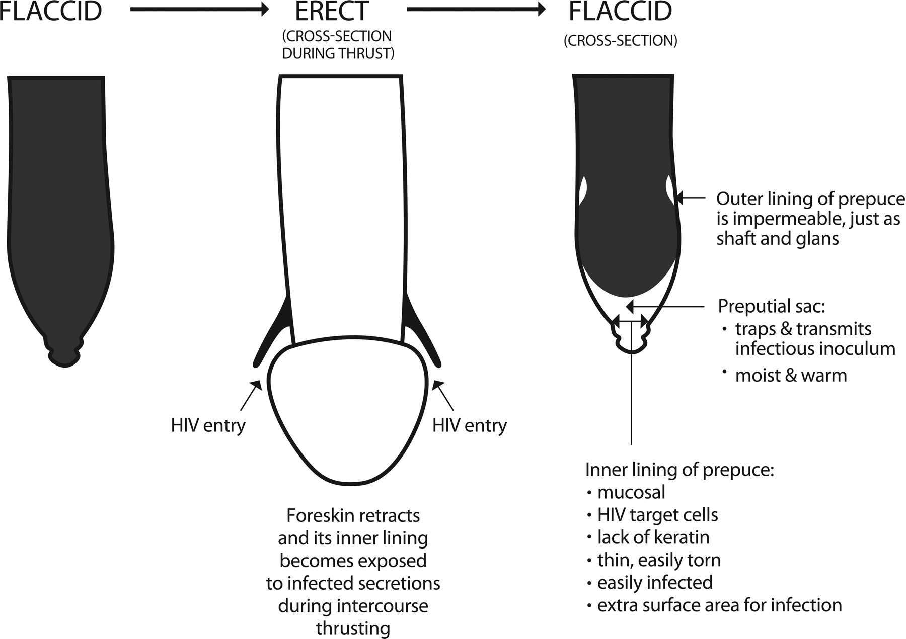
So how does this increase the risk of HIV passing from the sexual partner to the man?
INFECTIOUS INOCULUM TRAPPED UNDER THE FORESKIN
An early suggestion that attempted to explain the higher risk of HIV infection in uncircumcised men was that after insertive sexual intercourse with an infected partner and the return of the penis to its flaccid state, any infectious inoculum acquired would become physically trapped within the warm, moist preputial space, which would provide a hospitable environment for infection to occur. 15 The preputial space was termed a ‘cesspool for infection’. 16 It was also suggested at the time that the increased surface area, traumatic physical disruption during intercourse and inflammation of the glans penis (balanitis) could aid in recruitment of target cells for HIV-1. 15
THINNER KERATIN LAYER OF INNER FORESKIN
Early thoughts were that the portal of entry could potentially be the glans, sub-prepuce and/or urethra. It was suggested that in a circumcised penis the drier, more keratinized skin covering the penis could prevent entry. 15 However, subsequent studies showed that the glans of the circumcised and uncircumcised penis were in fact identical in histological appearance and had the same amount of protective keratin. 14 In contrast, the inner lining of the foreskin was shown to be a mucosal epithelium in which keratin layers were less numerous than in the outer foreskin. 17 The lining of the nasal passages and vagina are also mucosal surfaces and are targets for infection by microorganisms.
So did this also apply to the inner foreskin?
Later data led to equivocation on the keratin issue. Keratin in the foreskin of 60 Chinese preschool boys and 20 healthy men was reported to be much greater in the inner than in the outer layer. 18 Then a study of 16 adult male donors undergoing elective circumcision in Chicago reported no difference in keratinization of the inner and outer layers of foreskins. 19 At the time we criticized the latter findings pointing out that the foreskins were from men circumcised for foreskin pathologies that could have increased keratinization; only one sample was assessed per patient and this sample could have been from the distal end of the foreskin that has thicker keratin than the proximal foreskin near the coronal sulcus. 20 For Kenyan RCT samples collected 1 cm from the distal end of the intact foreskin no difference in keratin thickness was reported between the inner and outer foreskin. 21
In recent work, two novel models were established that more closely resemble the physiological state of the foreskin in vivo. 22,23 Based on their examinations the authors rejected as artefacts the earlier claims that there is no difference 19 or greater 18 keratin thickness in the inner as compared with the outer foreskin. They also showed that the thick keratin layers in the outer foreskin prevented infection by HIV, whereas HIV readily crossed the inner, mucosal epithelium, 23 as will be discussed in detail below.
CELL TARGETS FOR HIV
The mucosal inner lining of the adult foreskin is rich in potential HIV-1 target cells and receptors. Antigen presenting cells in the inner foreskin 24 were very early recognized as a primary target for HIV infection in men. 14 Both the inner and outer foreskin contain Langerhans cells and T-cells expressing CD4, the principle receptor for HIV-1, as well as the co-receptors CCR5 and CXCR4; and also present are other possible HIV-1 target cells (macrophages and dendritic cells), residing below the epithelium. 17,21,24–28 The proportions of CD4+ T-cells, Langerhans cells and macrophages were estimated as 22.4%, 11.5% and 2.4% of the total cell population, respectively. 17 This contrasts with the neonate, where the foreskin is deficient in such cells; 17,29 the proportion in a 22-month-old infant being instead 4.9%, 6.2% and 0.3%, respectively. 17 In the cervical mucosa of women, the respective percentages for these immune system cells were 6.2%, 1.5% and 1.4%, respectively. 17 The external layer of the foreskin has, like the rest of the penis, very much lower proportions of these cells: 2.1%, 1.3% and 0.7%, respectively. 17 Although the urethra is also a mucosal surface, Langerhans cells are rarer, 24 and until recent preliminary in vitro findings were reported 30 the urethra had not been regarded as a common site of HIV infection. 25,31–33
UPTAKE OF HIV IN TISSUE CULTURE
In 1998 it was shown that application of simian immunodeficiency virus (SIV) to the foreskin of male monkeys resulted in them becoming infected. 34 In this work, infected Langerhans cells were observed.
The selective entry of HIV via the inner surface of the human foreskin was first demonstrated in 2002 by Patterson et al. 17 This study used punch biopsies taken from fresh foreskin within three hours of circumcision and that had been kept at 4°C post excision. Upon infection of agarose-sealed foreskin tissue explants, cell-free HIV-1 entered the Langerhans cells of the inner foreskin. 17 No uptake occurred for the outer foreskin. The mean number of HIV copies per 1000 cells (determined by quantitative polymerase chain reaction) one day after infection was 301 for the mucosal inner foreskin, but was undetectable in the outer, external, foreskin. 17 These workers performed a comparison with cervical biopsies, finding mean HIV copy number to be 30, thus suggesting that the inner foreskin was 10 times more susceptible to HIV-1 infection than the cervix. 17 The HIV co-receptor CCR5 was, moreover, especially prevalent in cells in the foreskin tissue. 17 This study tested only high doses of HIV-1 at time points exceeding 24 hours.
Others have questioned the sealing efficiency and thereby polarization of the infection in the system used. 28 Using non-polarized foreskin tissue explants, thus allowing HIV-1 access to both apical and basal tissue surfaces, they reported that foreskin, glans, meatus and urethra were all equally susceptible to infection by cell-free HIV-1. 28 They then simply noted that ‘circumcision would remove two out of three of the exposed surface areas of the penis, reducing the chance of the virus coming into contact with susceptible target cells’. 28
Such explant models have been criticized because the functional integrity of the mucosal barrier is maintained for only a few hours, the surgical procedure itself activates migratory immune cells, and efficient sealing of the edges of the explant tissue to ensure polarization of the infection (i.e. from the apical to the basal compartment as takes place in vivo) is not always met. 23 The drawback of these models is that unlike the in situ situation, neither the stratified architecture nor the presence of immune cells occurs. Therefore two novel models were developed. 22,23 One involved inner or outer foreskin being placed on top of a permeable membrane in a two-chamber system with the apical side facing up, and with hollow plastic cloning ring cylinders glued tightly to the epidermal surface, thus creating a highly sealed apical chamber for polarized inoculation of HIV-1. The other involved seeding primary inner and outer foreskin fibroblasts and keratinocytes in the apical compartment of a two-chamber system, together with immature Langerhans cells and dendritic cells. Culture conditions were optimized to allow keratinocytes to mimic the low-keratin or high-keratin structure of the inner or outer foreskin, respectively. The result was in vitro models resembling the typical natural structural and morphological characteristics of the inner and outer foreskin. Using these, efficient HIV-1 transmission was shown following one hour of polarized exposure to the inner, but not the outer, foreskin epithelium. 22 By fluorescent and confocal microscopy, the authors showed that HIV-1 particles became trapped within the thick apical keratin layer of the outer foreskin, but readily penetrated the epidermis of the inner foreskin.
MECHANISMS INVOLVED IN UPTAKE OF HIV BY CELLS
The immune cells of the inner lining of the foreskin help fight bacteria and viruses that accumulate under it. However, in the case of HIV, they act as a ‘Trojan horse’, serving as portals for uptake of HIV. 35 In studies of various stratified mucosal epithelia, HIV ‘hijacks’ the physiological processes of pathogen recognition by Langerhans cells in order to invade the body. 35 Confocal imaging microscopy and mRNA quantification has demonstrated abundant and superficially present potential HIV target cells (CD3+ and CD4+ T-cells, Langerhans cells, macrophages and submucosal dendritic cells) in the inner foreskin. 21 In this study, there was no difference in the findings when foreskins from men with positive and negative herpes simplex virus type 2 (HSV-2) serostatus were compared. HIV-1-infected cells can form tight, although transient, interactions with the mucosal surface of epithelial cells, using a mechanism referred to as viral synapse formation. This contact between the two cells leads to local budding of HIV particles in the synaptic cleft. The fully mature HIV particles are then endocytosed and transcytosed across epithelial cells and released, still infectious, into the submucosa. The heparan sulphate proteoglycan agrin and adhesion molecules are key players in viral synapse establishment, as occurs in immunological and neurological synapses. Agrin is differentially expressed at the apical epithelial surface and serves as an attachment receptor. 36 The envelope glycoprotein subunit gp41 binds specifically to agrin, reinforcing the interaction of gp41 with its epithelial receptor galactosyl ceramide. 36 HIV entry into T-cells requires CD4 receptors and co-factors such as chemokine receptors CCR5 and CXCR4, present in high density on the surface of Langerhans cells. 37 After cell–cell contact is made between an infected cell and CD4 receptors on the target cell, CCR5, CXCR4 and integrin counter-receptors (ICAMs) interact with env, activated LFA-1 and other b2 integrins to ensure a firm connection. 35 Although cells with HIV receptors CD1a, CD4, CCR5, CXCR4, HLA-DR and DC-SIGN are present throughout the epithelia of the inner lining, HIV can only infect those cells to which it can gain access. 17 Moreover, CCR5/CXCR4 chemokines can inactivate cell-free HIV, but not HIV inside infected cells. 35 Most Langerhans cells are in the epithelium closest to the surface of the inner foreskin lining (1.2% versus 0.3% of cells in surface epithelium versus submucosa, respectively), whereas macrophages are mainly in the submucosa (0.04% versus 0.02% in submucosa versus surface epithelium, respectively). 26 CD4+ T-cells are present in each region. Furthermore, Langerhans cells send dendritic projections up between the keratinocytes, and these are particularly superficial in the inner foreskin (4.8 µm) compared with the outer foreskin (20 µm). 25
Several mechanisms appear to mediate internalization of HIV. 38,39 During one hour of exposure to HIV-1, epidermal Langerhans cells modify their spatial distribution within the foreskin by migrating towards the apical surface of the epidermis, sample HIV-1, rapidly internalize it and then transfer it to T-cells across Langerhans cell–T-cell conjugates. 22,23 Chemokines induced by HIV-1 may mediate changes in the spatial distribution of Langerhans cells and T-cells. 23 The process involves the presence on Langerhans cells of the c-type lectin, Langerin, that can bind HIV. 38 In the inner foreskin, but not the outer, tumour necrosis factor-α can activate Langerhans cells and stimulatory cytokines cause an influx of CD4+ T-cells into the epithelial layer. 40 The higher permeability of the inner foreskin is associated with increased interaction of HIV target cells with HIV and other external factors. HIV can, moreover, infect T-cells independently of Langerhans cells. 39,41 The success of HIV in establishing a systemic infection might nevertheless depend on its early interaction with Langerhans cells. 39,41 At low viral levels Langerin is able to clear HIV, shunting it to intracellular granules for degradation, but this mechanism becomes overwhelmed at higher viral loads. 42,43
It is now known that HIV-1-infected cells form viral synapses in the region of apical foreskin keratinocytes.
23,35
This leads to polarized budding of HIV-1 and rapid internalization by dendrites of Langerhans cells within one hour
22,23
(Figure 2). In contrast, cell-free HIV-1 does not translocate efficiently and the small amounts that are taken up become degraded.
22,23
Inoculation with HIV-1-infected cells induces a 1.6-fold increase in the known T-cell chemokine CCL5/RANTES and a 0.6-fold decrease in CCL20/MIP-3-alpha secretion.
44
The increase in CCL5/RANTES recruits T-cells from the dermis to the epidermis. While at one hour Langerhans cells are attracted to the apical surface to sample HIV-1 from infected cells, by four hours they have migrated back towards the basal membrane. The latter process is correlated with the decrease in CCL20/MIP-3-alpha observed. The T-cells recruited form conjugates with Langerhans cells that then transfer their cargo of HIV-1 to the T-cells.
44
The ability of HIV to activate C-type lectin DC-SIGN not only promotes HIV replication, but also HIV transmission to T-cells.
45
This is but one of a multitude of effects involving various pattern recognition receptors on dendritic cells that facilitate infection and transmission of HIV to T-cells.
45
The thick keratin layers in the outer foreskin prevented infection.
23
In one study, seminal plasma and cervico-vaginal secretions alone were found to have no effect on HIV-1 translocation, whereas a mixture of each reduced entry of HIV-1 into the inner foreskin.
23
Other studies found that seminal plasma can block the capture and transmission of HIV to CD4+ T-cells.
46,47
Current understanding of the foreskin-related mechanism of HIV-1 infection.
23
This involves the formation of apical viral synapses between cells highly infected with HIV and dendrites of Langerhans cells. Local HIV budding and HIV capture ensues, resulting in cell-to-cell transfer of HIV (black dots) from infected cells to dendrites of Langerhans cells, a process that takes one hour. A reduction in CCL20/MIP-3-alpha secretion takes place as Langerhans cells then migrate to the basal layers of the epidermis within four hours, where they transfer their HIV cargo to T-cells. At four hours, T-cells are recruited from the dermis into the epidermis as a result of increased CCL5/RANTES secretion, so fuelling the formation of Langerhans cell–T-cell conjugates.
44
The T-cells can then also infect dendritic cells. In contrast, cell-free HIV particles or HIV in cells with a low viral load are taken up by Langerhans cells and degraded
These researchers then used similar techniques to develop two explant models of the human urethra, 30 suggesting that urethra is an important site for entry of HIV in circumcised as well as uncircumcised men. One hour of exposure to HIV-1-infected mononuclear cells led to penetration of the urethral epithelium, but not the urethral meatus or glans, with HIV-1 and infection of urethral macrophages, but not urethral T-cells, was observed. 30
In the vaginal mucosa HIV enters CD4+ T-cells almost exclusively by CD4 and CCR5 receptor-mediated direct fusion, leading to overt productive infection. 41 In contrast, entry of HIV into CD1a+ Langerhans cells occurs primarily by endocytosis, involving multiple receptors, and the virions persist intact within the cytoplasm for several days. 41 Human ectocervical epithelial cells in culture capture HIV on their surfaces and maintain it in a fully infectious state for at least six days. 48 Although the infectivity of HIV in vitro is low in the absence of seminal fluid, the 248–286 peptide fragment of prostatic acid phosphatase (a major constituent of semen, being present in semen at approx. 35 µg/mL) forms fibrils known as ‘semen-derived enhancer of viral infection’, and these increase infectivity 100,000-fold by enhancing the attachment of HIV to target cells. 49,50 This may enhance the transmission of HIV from the male to a sexual partner.
TRAUMA
The uncircumcised penis is more susceptible to minor trauma, with tearing of the frenulum and thin tissue of the prepuce not uncommon in uncircumcised men. 37 HIV would then more readily infect through the breaks in the epithelial barrier. A large two-year RCT found significantly lower penile coital injuries among men in the circumcised arm of the trial, adjusted odds ratio being 0.71 for soreness, 0.52 for scratches/abrasions/cuts and 0.62 for bleeding. 51 Although minor trauma to mucosal tissues can occur during a sexual encounter to permit entry, this is not a requirement for HIV infection to occur. 52
FORESKIN AREA
For uncircumcised men, those with a higher foreskin surface area are more likely to be infected with HIV, 53 so adding to the evidence that the foreskin is an important factor in acquisition of HIV. Area was 43 cm2 in those who acquired HIV and 37 cm2 in those who did not (P = 0.01). HIV incidence was 0.80 per 100 person-years for men whose foreskin area was in the lowest quartile (<26 cm2), and was 2.5 per 100 person-years in men whose foreskin area was in the highest quartile (>46 cm2), a four-fold difference.
ULCERATIVE DISEASE, OTHER SEXUALLY TRANSMITTED INFECTIONS AND INFLAMMATION
Genital ulcerative disease is more common in uncircumcised men. 37,54 This may be caused by HSV-2, syphilis, chancroid or other causes, each of which are associated with higher risk of HIV acquisition. 54–56 Genital ulcers also increase HIV shedding. 35 Urethritis caused by Neisseria gonorrhoeae is associated with a 10-fold increase in HIV in semen. 57 A meta-analysis showed, however, that MC offers little protection against sexually transmitted urethritis. 58 The foreskin is also more vulnerable to infections and inflammation, so that disruptions to the epithelial barrier caused by ulcerative diseases and balanoposthitis could cause the epithelium to be more susceptible to HIV infection. The preputial sac can harbour pathogenic organisms in a pool of smegma. 37 Infected cells, such as peripheral blood mononuclear cells in vaginal fluids or semen, can adhere to mucosal surfaces or can migrate through abrasions, so serving as a continuing source of budding virions. 52
HSV-2 infection was found to increase HIV risk in men and women by three-fold in one study 59 and three- to five-fold in another. 60 A further study that found individuals with HSV-2 to have twice the risk of acquiring HIV than those without noted that those infected with both viruses were more likely to transmit HIV than if they just had HIV. 61 Interventions that treat sexually transmitted infections (STIs), such as giving co-infected patients aciclovir, were suggested to be cost-effective in HIV prevention in populations that engage in high-risk behaviours or have low circumcision rates. 62 Models based on the Kenyan RCT data suggested, however, that no more than 10–20% of the HIV infections prevented by circumcision were due to efficacy against STIs. 63,64 It has nevertheless been suggested that circumcision, by lowering HSV-2, should contribute to a lowering of HIV infection. 65 Despite modelling projections regarding the effect on HIV prevalence of vaccination against HSV-2, 66 an efficacious HSV-2 vaccine does not exist. There could be a synergy between HIV and HSV-2 infections, with mathematical modelling showing that HSV-2 in either partner increased female-to-male HIV transmission 3.0-fold. 67 While good in theory, HSV-2 suppressive therapy has, however, failed to decrease HIV acquisition in men, as seen in two RCTs. 68 A RCT in women similarly found that HSV-2 suppressive therapy had no effect on them becoming infected with HIV. 69 Further analysis of RCT data from the Orange Farm trial in South Africa revealed that while HSV-2 infection increases HIV acquisition significantly, by 28% (95% CI 18–37), the protective effect of MC was similar in men with and without HSV-2. 70 Moreover, the Ugandan RCT has shown that only 11.2% of HIV infections are mediated by a reduction in genital ulcer disease after circumcision, and only 8.6% by a reduction in HSV-2 incidence. 55 Ulcers prevented by circumcision were mostly not from herpes, but rather from foreskin tearing during intercourse. 55 It has been suggested, moreover, that the persistence and enrichment of HIV receptor-positive inflammatory cells in biopsies from healed genital lesions after HSV-2 infection would help explain the inability of anti-HSV-2 therapy to reduce HIV acquisition. 71
In the HIV RCT in South Africa, being positive for high-risk human papillomavirus (HPV) in urethral swabs was associated with a 3.8-fold higher incidence of HIV. 72 There was no association with low-risk HPV. High-risk HPV may be simply a marker for increased sexual activity (which would increase risk of both HIV and HPV), although it is too early to say whether there may be a causal explanation; thus the association observed may be coincidental. Circumcised men infected with high-risk HPV clear the HPV faster than uncircumcised men. 73–75
Inflammation of the epithelium of the foreskin has been noted in 4.2% of men with neither HIV nor HSV-2, 7.8% of men with HSV-2 only, 19% of men with just HIV, and 32% of men with both. 76 For stromal inflammation, the figures were 14%, 30%, 33% and 61%, respectively. Both epithelial and stromal inflammation were more common in men with smegma. Even in the absence of visible lesions the mucosal tissue can show histological signs of inflammation. 21 Although inflammation can increase the risk of infection, one study showed that it is not a requirement for infection to occur. 52
In addition, HIV and HSV-2 co-infection synergistically increases CD8+ T-cell densities. 77 The increase in CD4+ cell density in the foreskin of HSV-2-positive men should increase their susceptibility to HIV infection. 77
Wetness under the foreskin is an indicator of poor hygiene and is associated with a 40% increase in risk of HIV infection. 78 A wet penis may enhance attachment of infectious virions for longer, reduce healing after trauma, or may lead to balanitis under the foreskin and consequent micro-ulcerations. 78
MICROBIOME OF THE PENIS
A report in 2010 identified the entire microbiome of the penis of 12 men before and after circumcision. 79 Among the 42 unique bacterial families identified, Pseudomonadaceae and Oxalobacteraceae were the most abundant irrespective of circumcision status. Circumcision was associated with a significant change in the overall microbiota (P = 0.007) and with a significant decrease in putative anaerobic bacterial families (P = 0.014). Two families in particular – Clostridiales Family XI (P = 0.006) and Prevotellaceae (P = 0.006) – were uniquely abundant before circumcision. Within these families the authors identified a number of anaerobic genera previously associated with bacterial vaginosis, including: Anaerococcus spp., Finegoldia spp., Peptoniphilus spp. and Prevotella spp. The researchers concluded ‘the anoxic microenvironment under the foreskin may support pro-inflammatory anaerobes that can activate Langerhans cells to present HIV to CD4 cells in draining lymph nodes’. They suggested that the reduction in putative anaerobic bacteria after circumcision might play a role in protection from HIV and other STIs.
ALLOIMMUNIZATION
Mucosal alloimmunization has also been suggested as a protective factor against HIV. 33 Counterintuitively, HIV risk is lower in circumcised men who have more frequent exposure than it is in circumcised men with less frequent exposure. 32 This led to the hypothesis that repeated contact of the small area of exposed urethral mucosa, or more likely the meatus, which unlike the urethra does contain a small number of HIV receptors, 25 with subinfectious inoculums may induce an immune response having a protective effect over and above that afforded by removal of the vulnerable foreskin. 32 The small area exposed may mean that the infectious inoculum per act of insertive intercourse could be less likely to overwhelm the effects of partial protection as compared with the mucosal area exposed in a foreskin or vagina. 32 This hypothesis remains to be tested.
WHY THEN ARE CIRCUMCISED MEN NOT COMPLETELY PROTECTED AGAINST HIV?
The fact that circumcised men can nevertheless still become infected with HIV means that mechanisms must exist for this to occur. Based on the evidence to date we speculate that likely reasons may include situations in which the epithelial barrier of the circumcised penis becomes compromised. This may occur as a result of abrasions incurred during vigorous intercourse, loss of epithelial integrity as a result of genital ulcer disease and epithelial changes caused by the particular STIs that weaken the epithelial barrier. While each of these factors is less common in circumcised men, they are far from abrogated completely, so offering a likely explanation for the residual vulnerability of circumcised men to HIV infection during penetrative sexual intercourse with an infected partner. Infection via the urethra is an additional possibility. 30
CONCLUSION
The present comprehensive review highlights the reasons why the foreskin, in particular the inner mucosal lining, is the weak point that allows HIV to infect men during unprotected vaginal or anal intercourse with an infected partner. 80 HIV-laden T-cells of the partner adhere to keratinocytes and transmit HIV to Langerhans cells via dendrites residing just below the epithelial surface. Langerhans cells migrate to the dermis and transfer HIV to T-cells and thence to dendritic cells. Disruption of the penile epithelium, especially the thinner foreskin, by tearing or infections results in direct transfer into the bloodstream. After having acquired HIV, the preputial cavity serves as a hospitable environment for an infectious inoculum that can then be transmitted to subsequent sexual partners. MC, by removing a major portal of HIV entry, greatly reduces the risk to men of HIV infection. 81,82