




Abstract
We report the successful endovascular repair of a ruptured abdominal aortic aneurysm (AAA) in a multimorbid patient 8 months after endovascular abdominal aortic aneurysm repair (EVAR). A 74-year-old man with a history of EVAR 8 months earlier presented with hypotension, severe back pain, and tenderness on abdominal palpation. A contrast-enhanced computed tomographic scan showed a large retroperitoneal hematoma and confirmed the diagnosis of secondary abdominal aortic rupture. Because the patient had severe comorbidities, the endovascular method was chosen for further management. Two stent grafts were placed appropriately to eliminate a type 1a and a type 3 endoleak owing to modular separation of the left iliac graft limb from the main body stent graft. An additional self-expanding stent was deployed in the solitary right renal artery to open its origin, which was partially overlapped by the proximal cuff. The patient was discharged on the tenth postoperative day and is alive and well 1 year postoperatively. This case indicates that endovascular repair is feasible not only in cases of primarily ruptured AAAs but also in secondarily ruptured AAAs after failure of EVAR.
Keywords
Although rare, rupture after endovascular abdominal aortic aneurysm repair (EVAR) represents a major life-threatening complication that requires prompt emergent diagnosis and treatment. To date, no specific guidelines are available for the optimum management of this entity and only limited data based on small case series exist. More often, such patients are managed with open surgery. However, endovascular repair may be a feasible, safe, and advantageous alternative for selected patients. To our knowledge, only a few cases of endovascular reintervention for the repair of secondarily ruptured abdominal aortic aneurysm (AAA) after EVAR have been reported. 1,2 To increase interest in such an alternative approach, we present herein a case of successful endovascular repair of a rupture after EVAR.
Case Report
A 74-year-old man was admitted to our emergency department complaining of severe back pain of sudden onset. His medical history was remarkable for hypertension, severe coronary artery disease, peripheral vascular disease, chronic obstructive pulmonary disease, left nephrectomy, and endovascular repair of an AAA that was 5.5 cm in maximum diameter. The latter had been performed 8 months before at another vascular center with the use of a bifurcated stent graft system (Lifepath, Edwards Lifesciences, Irvine, CA). Although at the 1-month follow-up after EVAR no signs of endoleak had been detected, at 6 months, abdominal color duplex scanning revealed a small endoleak (Figure 1A). However, as this finding was not confirmed by contrast-enhanced computed tomography (CT) (Figure 1B) and no sac enlargement was demonstrated, no further investigation or reintervention was considered necessary at that time.

A, Color duplex scan demonstrating a small endoleak 6 months after endovascular abdominal aortic aneurysm repair (EVAR). B, Contrast-enhanced computed tomographic scan with no signs of endoleak 6 months after EVAR.
At admission to our department, the patient's arterial blood pressure was 80/40 mm Hg and the heart rate was 110 beats/min. At physical examination, a pulsatile abdominal mass, as well as tenderness on abdominal palpation, was found. Laboratory investigation revealed severe anemia (hematocrit 22%). An abdominal radiograph demonstrated a disconnection of the left graft limb from the main body of the stent graft (Figure 2A), whereas on an abdominal CT scan, a large retroperitoneal hematoma around the aneurysm sac was detected (Figure 2B). Consequently, the patient was transferred to the operating room with the diagnosis of secondary rupture after EVAR. Initial angiography was performed to assess the suitability for endovascular repair as the patient was multimorbid and in poor medical condition. Angiography revealed a type 3 endoleak owing to modular separation of the left iliac graft limb from the main graft body (Figure 2C); thus, an endoluminal intervention under local anesthesia was decided. During the procedure, the technique of hypotensive hemostasis was employed by limiting resuscitation while maintaining a detectable blood pressure. In addition, an aortic occlusion balloon was available and ready for use; however, it was not used since our patient remained hemodynamically stable during the whole operation.
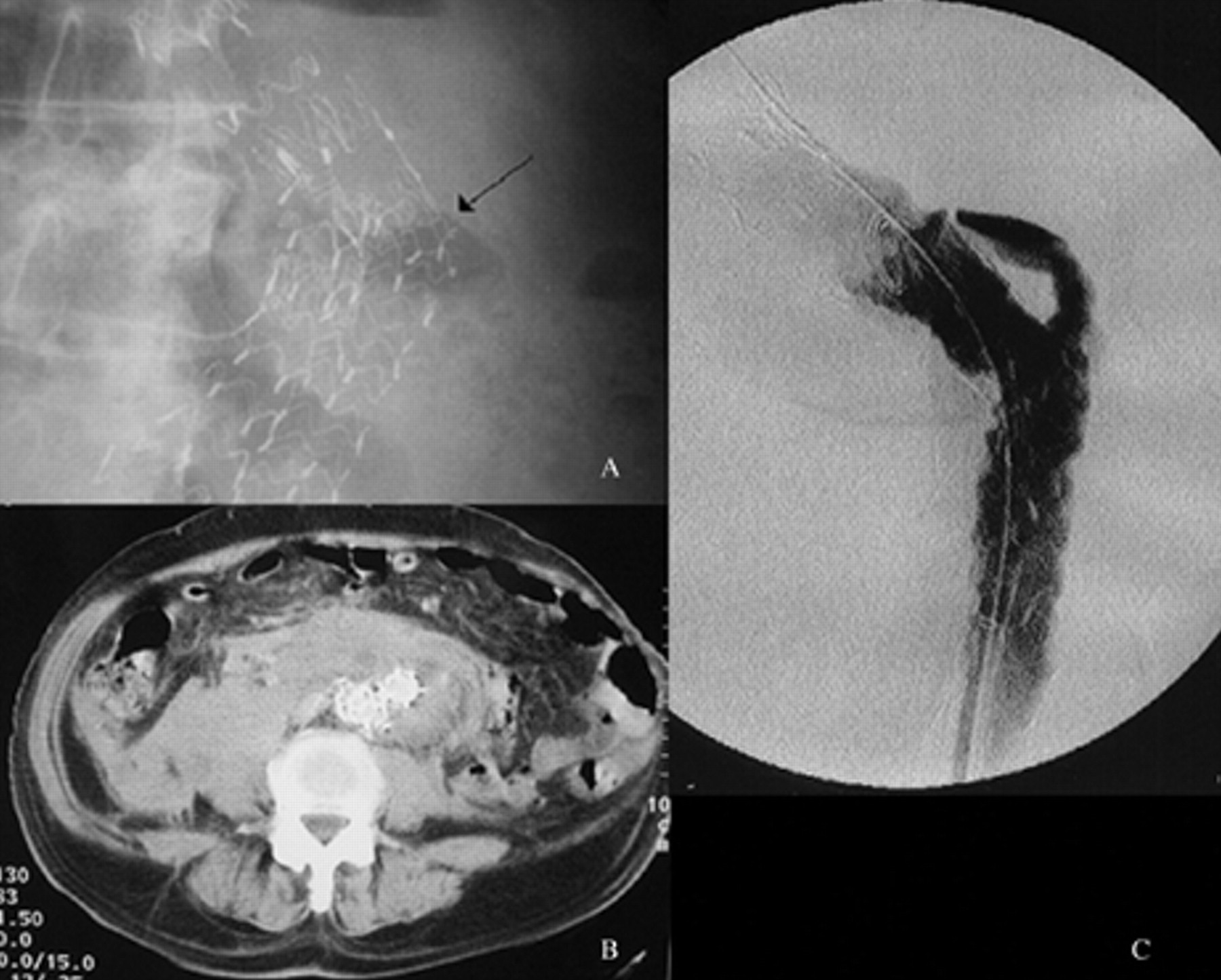
A, Abdominal radiograph demonstrating modular disconnection of the left graft limb from the main body of the stent graft (arrow). B, Contrast-enhanced abdominal computed tomographic scan showing a large retroperitoneal hematoma around the aneurysm sac. C, Angiogram demonstrating a type 3 endoleak owing to modular separation of the left iliac graft limb from the main graft body.
The left common femoral artery was exposed, and a 6F sheath was introduced. A hydrophilic Terumo guidewire was then used to pass the limb and main body of the endoprosthesis. The right position of the wire was verified using a “pigtail”-shaped catheter that was advanced at the level of the proximal edge of the main body stent graft. At this location, free rotation with maintenance of the catheter shape indicated its correct position inside the main graft. Subsequently, the wire and the sheath were exchanged for a 0.035-inch Landerqwist superstiff guidewire and a long 18F sheath, respectively. An 18 mm × 10 cm Excluder stent graft (W.L. Gore & Associates, Flagstaff, AZ) was used to bridge the site of leakage. To achieve secure “sealing,” the proximal edge of the device was placed 1 cm proximal to the bifurcation of the main stent graft. Subsequent angiography demonstrated successful elimination of the type 3 endoleak but revealed a second, type 1a, small endoleak. The latter could have been due to a low deployment at the primary procedure or a migration of the proximal stent graft component and probably preexisted but was obscured on an angiogram by the larger type 3 component. An explanation could be that device migration associated with type 1a endoleak led with time to modular stent graft separation, resulting in a second type 3 endoleak.
To eliminate the type 1a endoleak, a 32 mm × 4 cm proximal trunk extension cuff (W.L. Gore & Associates) was used. The latter had to be deployed within the limited space between the right renal artery origin and the proximal edge of the “bridging” graft to avoid overlapping and occlusion of either the solitary right renal artery or the left limb. After deployment, the type 1a endoleak component was successfully eliminated, but stenosis of the renal artery origin was noticed. Therefore, and because the patient had a solitary kidney, an additional bare stent was placed to open the orifice of the right renal artery. Completion angiography showed successful exclusion of the ruptured aneurysm and no remaining stenosis in the renal artery.
Following an uneventful hospital stay, the patient was discharged 10 days postoperatively. At the 6- and 12-month follow-ups, no signs of endoleak were observed on abdominal CT scans (Figure 3A); the retroperitoneal hematoma had shrunk, the aneurysm sac diameter was reduced from 5.6 to 5.0 cm, and the right renal artery remained patent (Figure 3B).
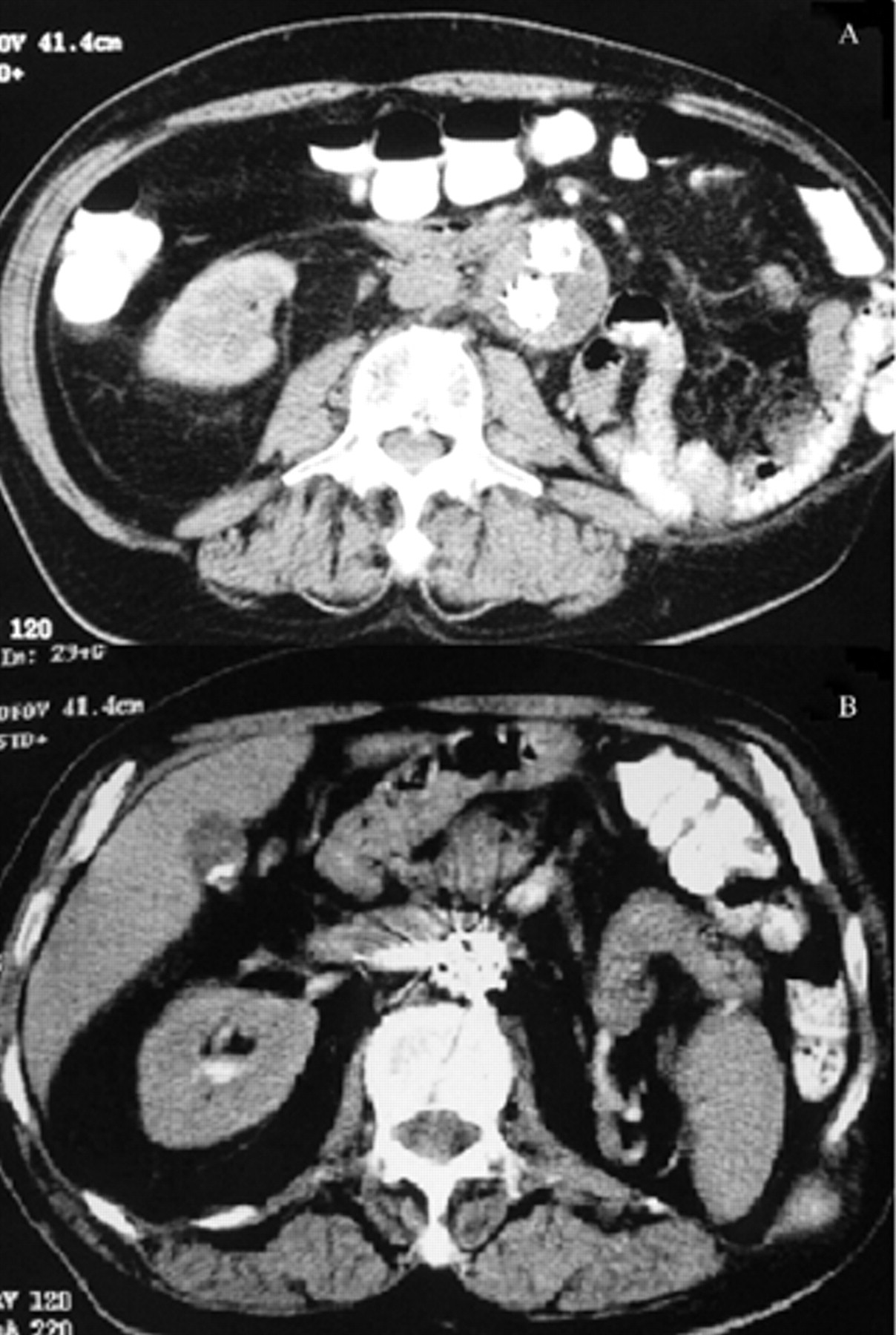
A, Contrast-enhanced computed tomographic (CT) scan at the 12-month follow-up demonstrating aneurysm sac diameter reduction, with no signs of endoleak. B, Contrast-enhanced CT scan at the 12-month follow-up demonstrating a patent stented right renal artery.
Discussion
Although rupture after EVAR represents a major life-threatening complication that requires prompt diagnosis and emergent intervention, no specific guidelines are available to date for the most appropriate management of this situation; limited data from small case series indicate that more often these patients are treated with open surgery. 1 However, the operative risk of secondarily ruptured AAAs following EVAR is high 3 as such patients usually suffer from significant comorbidities, owing to which the initial endoluminal approach was chosen. In addition, open surgery can be very challenging owing to technical difficulties related to the existing endoluminal prosthesis. Endovascular repair has been used for the management of primarily ruptured AAAs, with promising results 4–8 ; however, such an approach has been infrequently reported in cases of secondarily ruptured AAAs after EVAR. 2,9
Indeed, in emergent situations, issues such as the availability of experienced staff or appropriate stent graft dimensions may limit its feasibility. Difficulties during endovascular repair of secondarily ruptured AAAs after EVAR can also arise owing to the incompatibility of different device characteristics. In our case, the first endograft was a bifurcated Lifepath system with a standard diameter of 15 mm in both limbs, whereas the iliac graft proximal diameter was 16 mm. Consequently, the bridging stent graft had to be of the appropriate size to fit with the endografts already in place. This potential incompatibility highlights the need for accurate measurement of the devices already placed. In addition, the endovascular specialist should be aware of the dimension characteristics of all commercially available endografts. To overcome such difficulties, especially in an emergency, it is thought that all patients treated with EVAR should carry a personal electronic card created at the time of the initial procedure and updated at follow-up consultations with precise information about the characteristics of their endograft. More specifically, this card should include preoperative anatomic details of the AAA (eg, neck length and diameter, neck angulation), the exact type and dimensions of the device placed at primary EVAR, and information relevant to the follow-up of these patients concerning endoleak detection, aneurysm expansion rate, device migration, etc. The patient's preoperative and follow-up imaging studies should also be included.
The exact positioning of a stent graft when another one is already in place can be challenging owing to interactions between the devices. In the case presented herein, the proximal cuff had to be deployed within the limited space between the origin of the solitary renal artery and the “bridging” graft. This deployment, however, led to a stenosis of the renal artery origin. This complication could have been avoided if the proximal cuff was placed first and the bridging stent afterward. However, the fact that the two endoleaks were not noticed from the beginning prevented us from applying such a sequence.
Although type 1 and type 3 endoleaks are common enough and associated with a higher risk of aneurysm rupture, 10 the mixed-type endoleak-types 1a and 3-is rare. 11 Whether this pattern of endoleak is related to an even higher risk of rupture remains undetermined. In the present case, a relatively small 5.5 cm AAA ruptured within 8 months from its initial endovascular repair owing to a mixed-type endoleak, types 1a and 3. This event highlights the fact that such endoleaks may be associated with high morbidity, making their reliable detection mandatory. Especially for secondary late endoleaks, it has been reported that successful endovascular repair leads to weakening of the aneurysm wall, decreased resistance to dilatation, and therefore increased susceptibility to rupture in the event of secondary pressurization. 12
In the present case, an endoleak was revealed on a duplex sonogram 6 months after endovascular repair but was ignored since this finding was not confirmed on CT scan and there was no evidence of aneurysm expansion. Although contrast-enhanced CT is currently the most common imaging modality used for endoleak detection, with acceptable sensitivity and specificity, 13 it may not be ideal or superior to color duplex ultrasonography. Duplex ultrasonography as a dynamic investigation may be more likely to reveal a low-flow endoleak than CT, and it may also be a better method of localizing the source of a low-flow endoleak. 14 Indeed, a recent study supports the efficacy of contrast-enhanced ultrasonography for endoleak detection after EVAR, particularly when depiction fails with other imaging modalities. 15 This case accentuates the fact that ultrasonographic signs of even low-flow endoleak should not be ignored, despite the absence of endoleak findings on the CT scan. Contrast-enhanced angiography is also valuable 16 as it allows determination of the ingress and egress channels, which is obviously very important for endoleak classification and different treatment strategies. Finally, plain abdominal radiography should be routinely included in follow-up protocols since it may demonstrate impending dislocation, migration, or modular disconnection of the device.
Although in our patient, a proximal aortic cuff was used to treat the type 1a endoleak, other management options could have been followed, including additional balloon dilatation of the proximal edge of the stent graft with optional deployment of a giant Palmaz stent or the use of an aortouni-iliac stent graft and conversion of the bifurcated graft into an aortouni-iliac configuration with femorofemoral bypass.
A basic minimum stock is necessary in vascular centers dealing with endovascular treatment of ruptured AAAs. Stent graft devices, especially with suprarenal fixation, large diameters, and short lengths, combined with a variety of iliac extensions and proximal cuffs, should be available to match as many AAA anatomic configurations as possible that may arise in emergencies. It should be emphasized that aortouni-iliac devices are extremely useful in these scenarios, especially if patients are hemodynamically compromised. Additionally, physicians should be familiar with techniques such as the safe and effective use of aortic occlusion balloons and bifurcated stent graft conversion to aortouni-iliac configuration.
In conclusion, this case report highlights the potential role of endovascular repair for secondarily ruptured AAAs following EVAR. Such an approach is not only feasible, it also offers theoretical advantages against conventional open surgery. Furthermore, in this report, it is indicated that for the management of patients with rupture after EVAR, details of their primary repair should be easily available. Thus, better planning of their treatment could be achieved, and additional technical difficulties owing to the already placed devices could be eliminated.