




Abstract
Hypothenar hammer syndrome (HHS) is a rather rare condition and is a term used to describe an aneurysm or thrombosis of the ulnar artery. It is considered an occupational or recreational injury and is usually a result of a repetitive trauma to the hypothenar region where the unique anatomy of the Guyon canal allows arterial injury. HHS is one of the less common causes of symptomatic ischemia of the upper extremity. The aim of this article is to describe our experience with six patients. Presenting signs, symptoms, differential diagnoses, and literature review of this probably underdiagnosed syndrome are described. There are two pathologic entities: the aneurysmal type and the thrombotic type. Although most authors recommend watchful observation for the thrombotic type, reconstructive surgery is the treatment of choice for the aneurysmal type.
Hypothenar hammer syndrome (HHS) is a term used to describe thrombosis or aneurysm of the ulnar artery at the Guyon canal secondary to a repetitive injury to the artery. The term was first coined by Conn and colleagues in 1970. 1 HHS is considered an occupational or a recreational injury. 2,3 HHS is a rather rare condition, with an incidence of up to 14% in the population at risk, 4 and is considered one of the less common etiologies of ischemia of the upper extremity. The predisposing factor in the development of HHS is a repetitive or even a single trauma to the region of the hypothenar eminence. HHS mostly occurs in young males using the palm of the hand for pounding or pushing, 1,5 resulting in an aneurysm or thrombosis, leading, in turn, to digital ischemia. The anatomic structure of the Guyon canal makes the ulnar artery especially vulnerable to injury.
This article presents six patients with HHS of different etiologies. Presenting signs, symptoms, diagnoses, and treatment modalities are discussed, and the pertinent literature is reviewed.
Patients
Over the last 10 years, we have treated six patients with HHS. All patients were male, with an average age of 28 years (range 20 to 43 years). All patients had HHS in their dominant hand with no evidence or signs of HHS in the other hand. Four patients had ulnar artery aneurysms, whereas the other two had thrombosis. In the four patients with an ulnar artery aneurysm, the presenting symptom was a growing pulsatile mass of 1 to 3 cm at the hypothenar area. Only one patient had moderate to severe local pain. None of them had suffered digital ischemia. All four patients had a history of trauma to the hand either while using it as a hammer (three cases) or from acute sport injury (one). The two remaining patients had thrombosis of the distal ulnar artery accompanied by digital artery emboli; one patient presented with numbness of the fourth and fifth fingers, and the other had a nonhealing ulcer in his fourth finger.
The Allen test was positive in only two of six patients, those with thrombosis of the distal ulnar artery.
All patients with the aneurysmal type of HHS were operated on. In two, the aneurysm was resected, but no reconstruction was performed. The decision not to reconstruct was based on good backflow, as determined intraoperatively by the surgeon, and a large gap unsuitable for reanastomosis in the face of patients' refusal for an additional cut or extended procedure. An ipsilateral cephalic vein interposition graft (for the ulnar artery) was carried out in the other two. The two patients with the thrombosis type of HHS were initially treated conservatively. However, the patient with the ulcer had suffered intractable pain, and cervical sympathectomy was performed, after which he had an uneventful recovery.
On follow-up visits, all patients did well. The ulcer was healed after sympathectomy in the above-mentioned sixth patient. He still complained of some sensitivity, especially in cold weather. The patency of the grafts was demonstrated on subsequent duplex ultrasonographic examinations.
Demonstrative Case Report
A 31-year-old television producer was referred to our clinic with a tender mass at the hypothenar area of his left hand. The patient reported acute trauma to his hand during fast boat sailing in a stormy sea 6 months earlier. Initial radiographs of the wrist and hand were evaluated by an orthopaedic surgeon as normal. A bone scan showed a moderate increase in the uptake at the left wrist. The patient was treated by physical therapy and analgesics, without improvement. On physical examination, the patient was not distressed; his blood pressure was 125/75 mmHg, and his heart rate was 68 beats per minute and regular. Pulses were intact bilaterally, without bruits. Examination revealed a soft pulsatile and nontender mass with poorly defined borders in the hypothenar area of the left hand. There were no signs of digital ischemia or emboli. Blood coagulation tests were normal, and an electrocardiogram revealed normal sinus rhythm. A duplex scan suggested a pseudoaneurysm of the ulnar artery. Angiography of the left upper extremity showed a 1 cm aneurysm of the ulnar artery at its bifurcation extending into the superficial branch (Figure 1). There was no filling of the deep branch of the ulnar artery. No other anomalies were found. Approximately 7 months after the injury, the patient was operated on. At surgery, a 15 mm long by 10 mm wide ulnar aneurysm was found at the Guyon canal (Figures 2 and 3). The aneurysm was resected and, owing to a rather long gap after débridement, a reversed segment of the ipsilateral cephalic vein harvested from the distal forearm was used as an interposition graft. A postoperative electromyographic and nerve conduction velocity test revealed partial sensory axonal damage of the ulnar nerve at the wrist. This damage is probably a combination of both the disease process and the surgical insult. After 6 months, the patient had resumed his previous level of sport activity. Duplex scans done 3, 12, and 20 months following surgery demonstrated a patent graft with a normal flow.
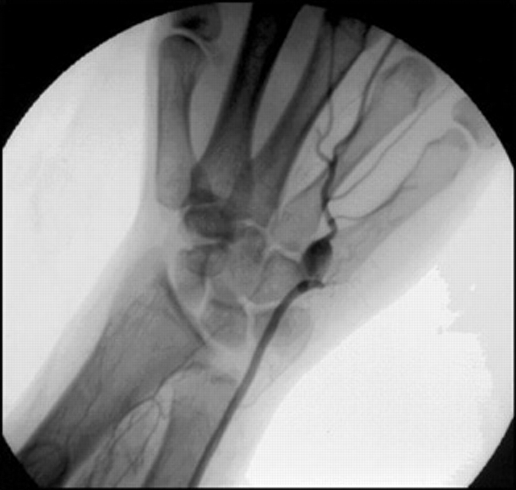
Angiography of the left upper extremity demonstrating a 1 cm aneurysm of the ulnar artery at its bifurcation extending into the superficial branch. There is no filling of the deep branch of the ulnar artery.

Operative view of the aneurysm after its dissection.
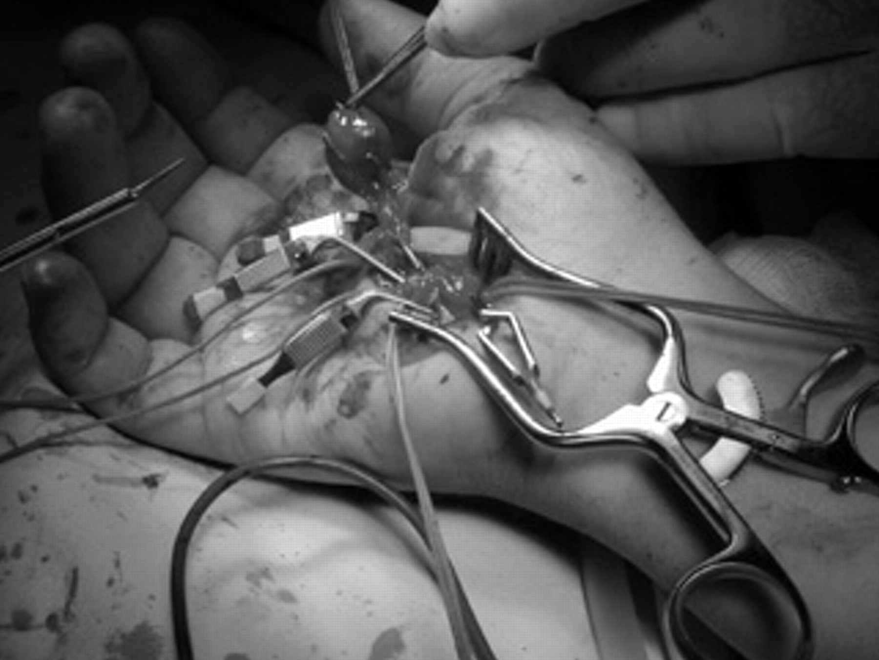
Removal of the aneurysm. Vessel loops and delicate vascular clamps are applied to enable vascular reconstruction.
Discussion
The first descriptive report of digital ischemia as a result of ulnar artery thrombosis was by von Rosen in 1934. 6 It was recognized as an occupational disease 2,3 occurring mainly in automobile repair workers and mechanics habitually using their hands for pounding, pushing, and twisting. 3–5 Conn and colleagues, in 1970, coined the term hypothenar hammer syndrome for the post-traumatic digital ischemia. 1
HHS is most commonly seen in males younger than 50 years of age and is also seen in athletes (especially in those practicing volleyball and karate); thus, it is first seen by an orthopaedic surgeon and a sport physician. Although considered a rarity, HHS must have a greater incidence than the literature reports (fewer than 150 cases). Little and Ferguson screened a population at risk (automobile repairmen) and found that 14% (11 of 79) were digital ischemia cases. 4 A positive correlation with duration of work was noted. No evidence of HHS was found in workers who did not use their hands as a hammer.
The pathophysiology is based on the unique vascular anatomy of the hand. 7 The ulnar artery and nerve enter the hand, passing the Guyon canal. The two terminal branches of the ulnar artery, deep palmar branch, and superficial palmar arch supplying blood to the fingers arise in the Guyon tunnel bounded medially by the pisiform and hamate bones, dorsally by the transverse carpal ligament, and superficially by the volar carpal ligament. Over a short segment of approximately 2 cm, the artery lies just anterior to the hook of the hamate bone covered only by the slight fibers of palmaris brevis muscle, subcutaneous tissue, and skin. The hook of the hamate may function as an anvil, and the fixation of the artery by the course of the deep branch allows little movement to escape the trauma. The nature of the vessel damage results in thrombosis, aneurysm, or a combination thereof. Intimal tear or flap causes thrombosis, whereas medial injury results in an aneurysm and distal embolization. 6–9 The diagnosis of thrombosis alone implies a better prognosis and allows a different clinical approach. 9
Recently, however, the traumatic etiology has been challenged by Ferris and colleagues, suggesting a fibromuscular dysplasia mechanism. 10 Intimal and medial hyperplasia and disruption of the internal elastic lamina were seen in many of their histopathologic specimens. In addition, there were angiographic findings of alternating areas of stenosis and dilatation. Also, the striking incidence of bilateral angiographic abnormalities in patients with unilateral symptoms led the authors to suggest the a priori presence of an underlying arteriopathy. This theory may explain the relative rarity of HHS.
Several signs and symptoms may be present. At the time of injury, some patients report on severe lacerating pain over the hypothenar eminence. Later, a typical dull pain and tenderness are present. HHS has a slow, insidious progression, and ischemic symptoms develop weeks or even months following the injury. 1,3,4,9,10 Ischemia can appear as a unilateral cold sensation, paresthesia, blanching, cyanosis, and even mottling of the digits and atrophic ulceration. The ring finger is most frequently symptomatic, whereas the thumb has not been reported to be involved and is spared owing to its radial blood supply. 1,5,7,9 This may be a distinguishing sign from Raynaud phenomenon, which is symmetric and bilateral and includes the thumb. In addition, in HHS, there is a male predominance (rather than female in Raynaud phenomenon). On physical examination, ischemic changes are seen, and localized tenderness, a prominent callus, or a pulsatile mass may be present. The Allen test should be suggestive for ulnar artery occlusion but is negative in the presence of an aneurysm. There is absence of hyperemic phase in patients with HHS because the artery is occluded, not spastic. Signs of frank necrosis of the fingers are less common.
The differential diagnosis includes orthopedic trauma, Raynaud disease or phenomenon with connective tissue disease (scleroderma, systemic lupus erythematosus, rheumatoid arthritis, hand-arm vibration syndrome), vasculitis, Buerger disease, thoracic outlet syndrome, cardiac-derived emboli, and proximal upper extremity aneurysms or atherosclerotic lesions. 10–13
Digital plethysmography, Doppler-derived digital pressure, and duplex scan are helpful noninvasive studies. Angiography, computed tomographic angiography, or magnetic resonance angiography is mandatory for the diagnosis because of the almost pathognomonic findings. 9–15 Nevertheless, angiography still remains the gold standard for diagnosis. Among its advantages, arteriography can evaluate distal emboli, identify thrombosis, define the precise anatomic location, and diagnose a proximal pathology causing upper limb ischemia.
The treatment of thrombosis of the ulnar artery is mostly symptomatic. Intra-arterial catheter-directed thrombolytic treatment is beneficial only within 2 weeks of the occurrence of acute occlusion. Rest, local warmth, smoking cessation, and calcium channel blockers may be helpful. Cervical sympathectomy is suggested for vasodilatation and may reduce the pain associated with ischemia. Ulnar artery aneurysms should be resected to eliminate the source of emboli and to remove a painful aneurysmal mass. 1,9,16 In case the outflow arteries are patent, one should also reconstruct the vessels by either end-to-end anastomosis or mostly by a vein interposition graft. 5,9,10 This approach improves the digital perfusion and adds the vasodilatory effect of periarterial sympathectomy. 9,17 Patients without symptoms and no threat of digital loss can be left under watchful observation. 18,19
Conclusion
HHS is a rare but underdiagnosed syndrome. It presents itself by pain in the hypothenar region, sometimes accompanied by a pulsatile mass or digital ischemia. The diagnosis may be established by the use of ultrasonography. Arteriography is used for the precise definition of the pathology and planning the operative treatment. The approach to HHS should consider two separate pathologic entities: thrombosis and aneurysm. Most authors recommend conservative treatment for thrombotic HHS. A reconstructive surgery is the treatment of choice for aneurysmal HHS as well as in all cases with ischemia. Further studies comparing conservative treatment with surgical repair are indicated in the thrombotic type to establish a more uniform and evidence-based approach to this rare entity.