




Abstract
Ti-based bulk metallic glasses are reported with high strength, low Young modulus and high corrosion resistance, suggesting their potentials in biomedical applications. However a thorough in vivo evaluation of its biocompatibilities has not been conducted yet. In this study, we implanted bars of Ti-based bulk metallic glass in the femoral bone of rats, followed up local tissue reaction as well as its component ions’ diffusion in local area and whole body. The Ti-based BMG (Ti40Zr10Cu34Pd14Sn2) alloy exhibited favorable features of both high strength and high elasticity. In vivo implant evaluation showed that it has a good tissue compatibility, equivalent bone integration and bonding ability with Ti sample. No component ion diffusion was detected up to 3 months post implantation. The possibility and efficacy of its use for bone implant is confirmed. Thus further long term implant study is recommended.
Introduction
“Bulk metallic glass (BMG) alloys, i.e. amorphous alloys, are materials without any long-range atomic order that are prepared by solidification of a liquid melt at a sufficient high speed to suppress the nucleation of crystals. BMG alloys have a random atomic structure and do not contain any segregations and defects. In BMG alloys, slip planes are not generated and elastic deformation continues even under considerable stresses because such deformation is caused by mass movement of the constituent atoms. Accordingly, BMG alloys have a higher strength and a lower Young’s modulus than crystalline alloys” [1]. “Due to absence of long range atomic order, BMG alloys present unique mechanical properties such as superior strength, high elastic strain limit, relatively low Young’s modulus, excellent corrosion resistance, and good wear resistance” [2,3]. “Because of these characteristics, BMG alloys could constitute a new generation of metallic materials for orthopaedic implants” [2,4].
“Ti-based bulk metallic glasses have comparative advantages for quick application in biomedical area, mostly because Ti alloys have a very long history of being used as orthopedic implants and also are considered as the ultimate choice in biomedical fields” [5,6]. Recently we developed a Ti-based BMG (Ti40Zr10Cu34Pd14Sn2: in nominal atomic percentages), it exhibited high activation energy for crystallization (334.3 kJ/mol) and high compressive strength about 2000 MPa [7]. In this study, we evaluated it in vivo as bone implant with rats. Biocompatibility, osteointegration, as well as local and whole body ion diffusion were evaluated.
Materials and methods
Sample preparation
According to the method described in our preview study [7], Ti-based BMG rod samples at the size of 1.5 mm in diameter and 4 mm in length were prepared as follows: “The master ingot of Ti40Zr10Cu34Pd14Sn2 alloy was prepared by arc-melting the pure elements with purities above 99.9% in a vacuum arc furnace with argon atmosphere. The alloy composition represents the nominal atomic percentage of the mixture. The ribbon sample with a cross section of 0.02 × 2 mm2 was prepared by melting the master ingot, and ejecting through a nozzle onto a Cu wheel rotating with a surface velocity of 40 m/s. The rod sample was prepared from melting master ingot by casting into a copper mold with proper diameter”. Titanium samples with the same shape for control were commercially obtained from the Nilaco Corporation, Japan.
Material characterization
“Glassy structure was examined by X-ray diffraction (XRD, Rigaku RINT-Ultima, monochromatic Cu Kα radiation). Compressive tests were conducted by a mechanical testing machine (Shimadzu AG-X) under a strain rate of 5 × 10⁻⁴/s. The compression sample was 1.5 mm in diameter and 4 mm in length” [7]. Chemical analysis of sample elements ratio was carried out by using inductively coupled plasma emission spectroscopy (ICP; ICP10P, ARL, Valencia, CA). Sample surface was observed with a scanning electron microscope at a magnification of ×80 (SEM, model S-4500, Hitachi). Surface roughness (Ra) was measured with a Laser Microscope at a magnification of ×100 (LM, VK-9500, Keyence).
In vivo evaluation
All animal experiments were carried out in compliance with and approved by the institutional review board of The Tokyo Medical and Dental University. All possible steps were taken to avoid animal suffering in the experiment. Fifteen-week-old male Sprague-Dawley rats weighing 400 to 450 g were used in the experiment. Twenty rats were randomly assigned to two groups. After anesthesia, 20 Ti-based BMG alloy bars were implanted into both sides of the center of femoral diaphysis in 10 animals (BMG group), while 20 control Titanium bars were implanted in the other 10 animals (Ti group).
Twelve weeks later, after sacrifice with lethal dose anesthesia, blood samples were taken from 5 rats of each group respectively and sent to Mitsubishi Chemical Medience Corporation (Tokyo, Japan) for measurement of serum copper level. Ten samples including surrounding bone tissue from 5 rats of each group were harvested, followed by dehydration, embedding in Osteoresin (Wako, Tokyo, Japan), cross-sectioning and staining with toluidine blue. Histologic images of implant and its surrounding tissue were taken by a light microscope (BX51, Olympus, Tokyo, Japan). An image analyzing software, Image Pro Plus 6.0 (Media Cybernetics, Silver Spring, MD, USA) was employed for the measurement of following parameters: (1) Bone attachment ratio: length of bone directly attaching on the implant surface/length of implant surface. (2) Surface bone formation: area of bone tissue newly formed on the implant surface within a distance of 200 µm. Elemental analysis was carried out linearly at the border area of implant to bone on the exposed stump surface by energy dispersive X-ray spectroscopy (EDX, SEM, model S-4500, Hitachi). For the left 5 rats of each group, bone bonding strength was evaluated with 10 samples respectively by Pull-out test on a mechanical testing machine (Autograph AG-X, Shimadzu, Japan). The load speed was 1 mm/min, maximum bonding strength was recorded.

A typical XRD pattern of the Ti-based bulk metallic glass alloy sample. A diffuse diffraction pattern typical for a glassy phase is confirmed.
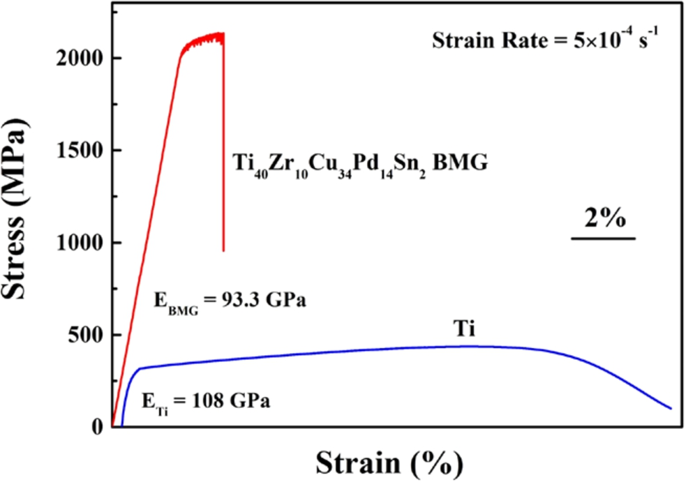
The compressive stress-strain curves of the Ti-based BMG and Ti samples. High yield strength, plasticity and relatively low Young’s modulus are displayed for the BMG sample. Ti sample shows much lower strength, elasticity and higher Young’s modulus for the Ti sample. (Colors are visible in the online version of the article;
Statistical analysis was executed with the software Statcel 3. A non-parametric ANOVA, Mann-Whitney U test was applied to calculate the significance of differences between mean values for each parameter. A p value less than 0.05 was considered statistically significant.
Results
Material characterization
Inductively coupled plasma emission spectroscopy result of the BMG sample
Inductively coupled plasma emission spectroscopy result of the BMG sample
Note: Elements composition is confirmed as designed.
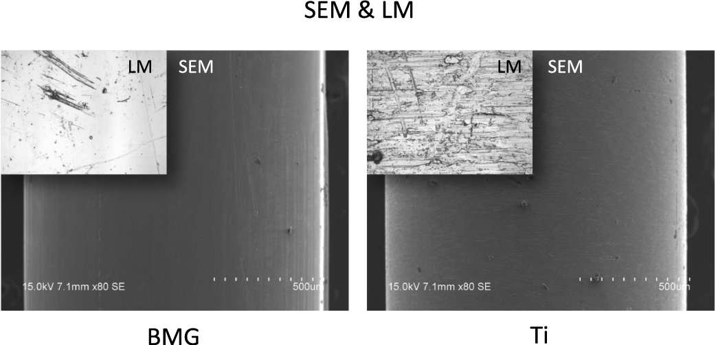
SEM and LM images of BMG and Ti sample surfaces. Approximate surface roughness is shown.
A typical macroscopic view of the Ti-based BMG sample at 12 weeks after implantation is shown in Fig. 4. Inflammatory reaction was totally not observed, implant dislocation or loosing is not observed in all cases, indicating excellent biocompatibility and integration to bone tissue (Fig. 4). Histological images is shown in Fig. 5, revealed that both BMG sample and Ti sample were well covered by surrounding bone tissue, and there were no abnormal findings in surrounding bone tissue.
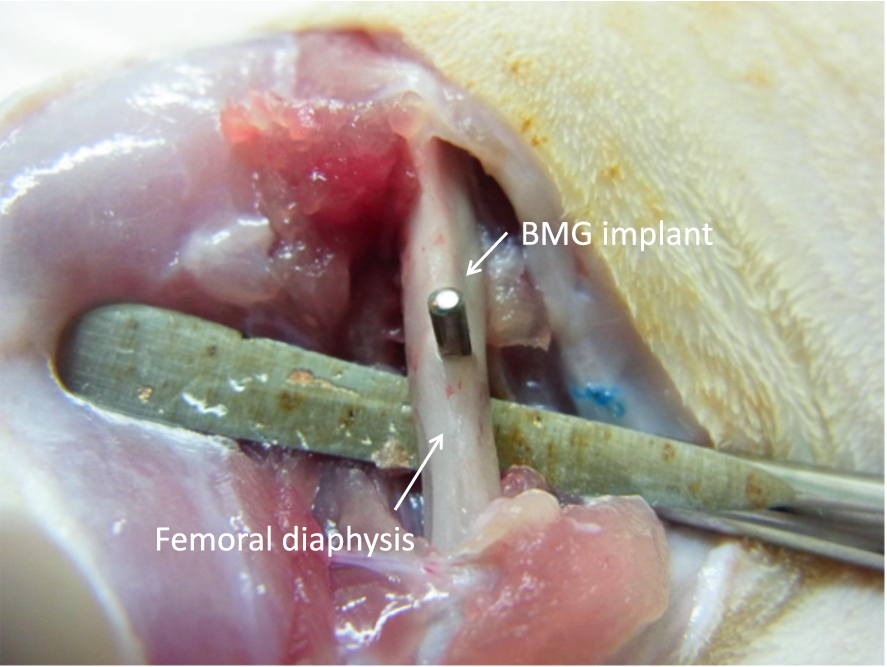
Macroscopic view of the Ti-based BMG sample at 12 weeks after implantation in femoral diaphysis. No inflammatory signs or implant loosing is observed. (Colors are visible in the online version of the article;
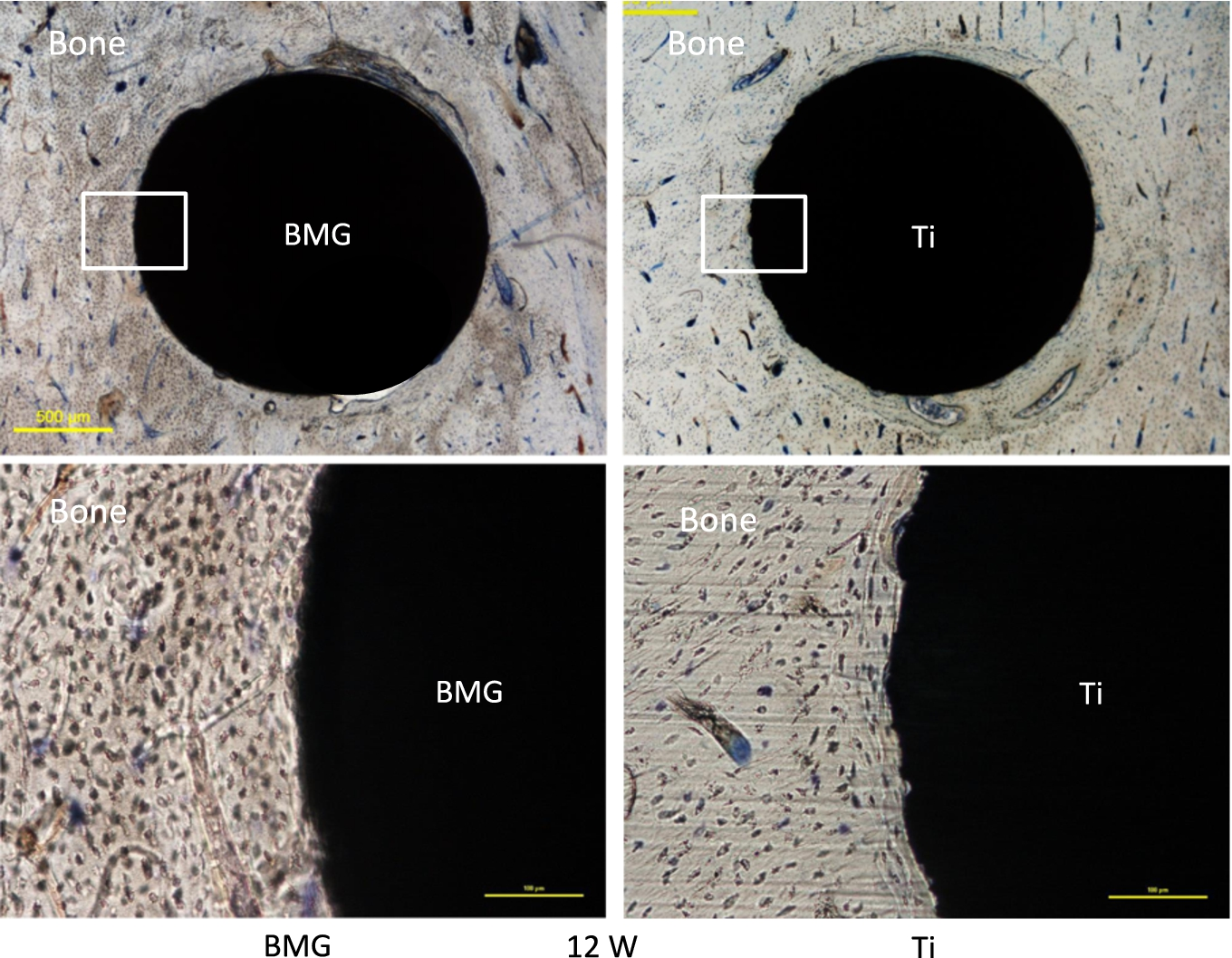
Histological views of Ti-based BMG implant and Ti implant. Both samples are well covered by surrounding bone tissue. There are no abnormal findings in surrounding bone tissue. Objective magnification of upper images is ×4 and lower images is ×20. (Colors are visible in the online version of the article;
Bone attachment ratio was shown in Fig. 6. Although the average value in BMG group is a little bit higher than that in Ti group, significant difference was not detected, suggesting same level of bone attachment to implant. Also with bone formation area on surface (Fig. 7), no significant difference was found between the two groups, indicating that their influence to surrounding bone tissue were equivalent. With bone bonding strength (Fig. 8), a similar pattern with that of bone attachment ratio was displayed with no significance detected. It is not hard to comprehend the correlation of bone attaching area with bone bonding strength.
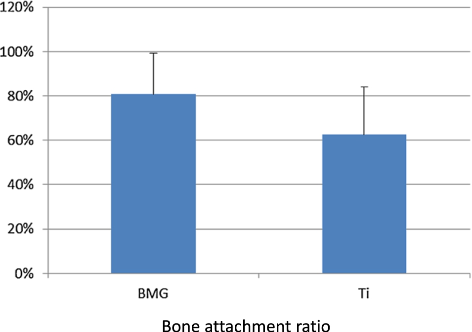
Result of bone attachment ratio. No significant difference is detected between BMG and Ti groups, suggesting they have same level of bone attachment to implant. (Colors are visible in the online version of the article;
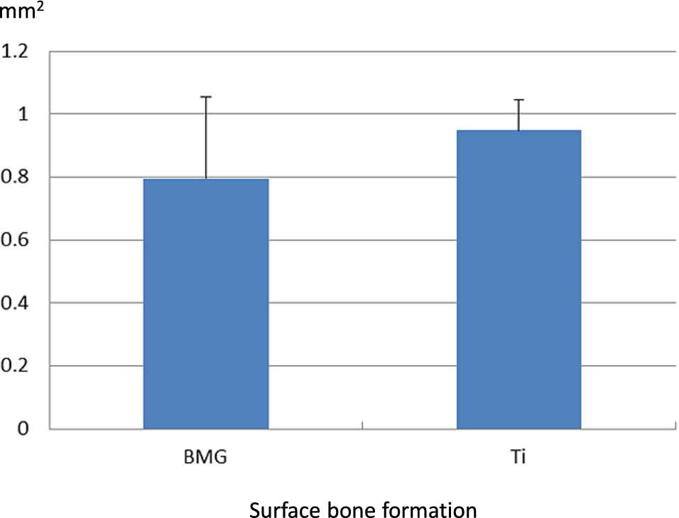
Result of bone formation area on surface. No significant difference is found between the two groups, indicating that their influence to surrounding bone tissue is equivalent. (Colors are visible in the online version of the article;
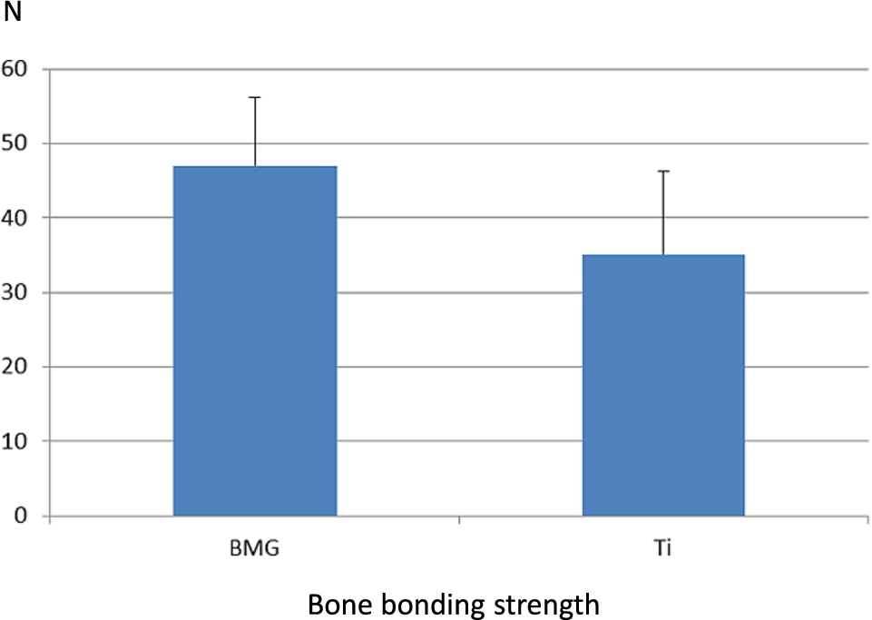
Result of bone bonding strength. A similar pattern with that of bone attachment ratio is shown, suggesting that two groups have comparable bone bonding ability. (Colors are visible in the online version of the article;
EDX elemental analysis of implant border area found no diffusion of any metallic ions in the BMG sample (Fig. 9). Assay of serum Cu level showed same level of serum copper that is just normal [8] (Fig. 10).
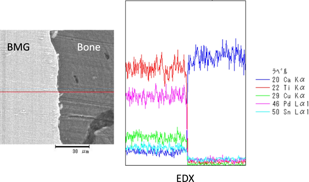
EDX elemental analysis of implant border area. No diffusion of any metallic ions in the BMG sample is found. (Colors are visible in the online version of the article;
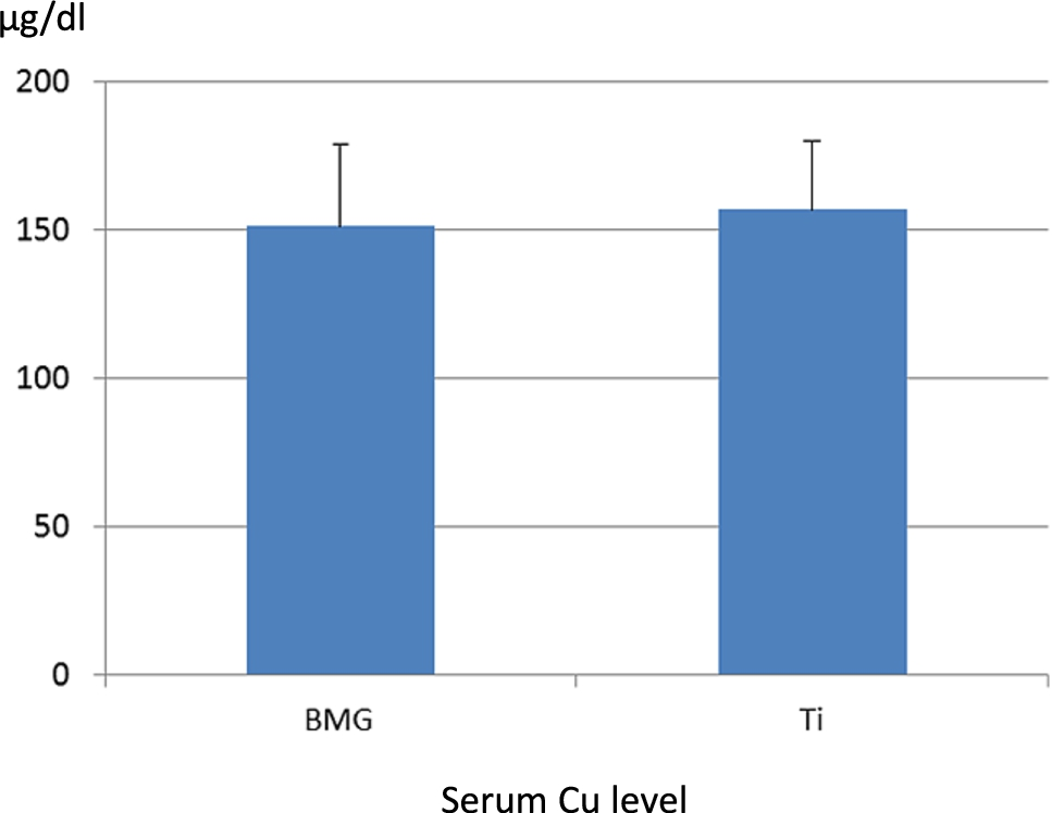
Result of serum Cu level measurement. The two groups show normal level of serum copper. (Colors are visible in the online version of the article;
“BMG alloys have a higher strength and a lower Young’s modulus than crystalline alloys” [1]. “Due to absence of long range atomic order, BMG alloys present unique mechanical properties such as superior strength, high elastic strain limit, relatively low Young’s modulus, excellent corrosion resistance, and good wear resistance” [2]. In this study, compressive test of the Ti-based BMG rod sample revealed that its yield strength was above 2000 MPa, elastic deformation limit was 2.2% and Young’s modulus was calculated at 93.3 GPa. While relative data of Ti sample are reported at 800 ± 50 MPa, <1% and 100 ± 7 GPa, as well as those of cortical bone are 200 ± 100 MPa, <2% and 40 ± 10 GPa [6]. Our Ti-based BMG sample presented favorable features of both high strength and high elasticity, making it superior to pure Ti for bone implant applications. Furthermore, BMG’s good corrosion resistance and wear resistance are especially favorable for bone implant too.
The in vivo results of the Ti-based BMG bone implant showed that it has a very nice biocompatibility, equivalent bone integration and bonding ability with Ti sample. Considering its main constituent is Ti, these data are not surprising. No component ion diffusion was detected up to 3 months post implantation, indicating BMG’s good corrosion resistance. We think the possibility and effect of its use for bone implant is confirmed.
The Ti–Zr–Cu–Pd bulk glassy alloy was developed by Xie’s group in 2007, it exhibited high glass-forming ability, higher strength and lower Young’s modulus [9]. The Sn added Ti-based BMG exhibited higher activation energy for crystallization (334.3 KJ/mol), addition of 2–4% Sn can enlarge the supercooled liquid region, indicating good thermal stability [7].
However concerning the potential toxicity of diffusion of Cu, Pd and Sn ions, long term in vivo implant experiment is required, as well as comparison of other Ti-base BMG or alloys.
Conclusions
The Ti-based BMG (Ti40Zr10Cu34Pd14Sn2) alloy exhibited favorable features of both high strength and high elasticity, making it superior to pure Ti for bone implant applications. In vivo implant evaluation showed that it has a very nice biocompatibility, equivalent bone integration and bonding ability with Ti sample. No component ion diffusion was detected up to 3 months post implantation. The possibility and efficacy of its use for bone implant is confirmed. Further long term implant study is recommended.