
Abstract
In this paper, magnesium matrix hydroxyapatite composite material was prepared by electrophoretic deposition method. The optimal process parameters of electrophoretic deposition were HA suspension concentration of 0.02 kg/L, aging time of 10 days and voltage of 60 V. Animal experiment and SBF immersion experiment were used to test the biocompatibility and bioactivity of this material respectively. The SD rats were divided into control group and implant group. The implant surrounding tissue was taken to do tissue biopsy, HE dyed and organizational analysis after a certain amount of time in the SD rat body. The biological composite material was soaked in SBF solution under homeothermic condition. After 40 days, the bioactivity of the biological composite material was evaluated by testing the growth ability of apatite on composite material. The experiment results showed that magnesium matrix hydroxyapatite biological composite material was successfully prepared by electrophoretic deposition method. Tissue hyperplasia, connective tissue and new blood vessels appeared in the implant surrounding soft tissue. No infiltration of inflammatory cells of lymphocytes and megakaryocytes around the implant was found. After soaked in SBF solution, a layer bone-like apatite was found on the surface of magnesium matrix hydroxyapatite biological composite material. The magnesium matrix hydroxyapatite biological composite material could promot calcium deposition and induce bone-like apatite formation with no cytotoxicity and good biocompatibility and bioactivity.
Keywords
Introduction
With the development of science and technology, the requirements for artificial bone with high properties were exigent [1]. Titanium alloy was mainly metallic biomaterial currently. However, due to high elasticity modulus, nondegradation and nos toxic effect of metal, the wide application in medical field were limited. With excellent mechanical strength, low elastic modulus and biodegradable properties, magnesium alloy was a kind of potential metallic biomaterials [2]. But the magnesium alloy rapid degradation in body made it fail during the reserve service period and hinder its clinical application [3,4]. Through surface modification, the problem of the poor corrosion resistance of magnesium alloy was solved. A good biological material with appropriate degradation rate, excellent mechanical strength and lower elastic modulus was got. With good biocompatibility, bioactivity, bone induced ability and corrosion resistance, hydroxyapatite was an ideal coating material of artificial bone [5]. The hydroxyapatite was coated on magnesium alloy to prepare magnesium matrix hydroxyapatite biological composite material which had appropriate degradation rate, excellent mechanical strength, low elastic modulus of magnesium alloy, good biocompatibility, bioactivity and bone induced ability of hydroxyapatite [6,7].
There were chemical method, physical method and physicochemical method for the preparation of magnesium matrix hydroxyapatite biological composite material at present. Specifically, there were laser cladding method, micro-arc oxidation method, electrochemical deposition method, sol-gel method, biomimetic solution method, hydrothermal synthesis method, plasma spraying method, etc. [8]. The electrophoretic deposition method had some notable advantages [9,10]. As a kind of mild conditions of surface coated method, electrophoretic deposition could avoid the phase change of the coating and brittle caused by high temperature, and could improve the bonding strength between coating and matrix after heat treatment. Furthermore, with the non-straight process of electrophoretic deposition, the HA coating could deposit on all kinds of surface of complex shape. In this paper, the electrophoretic deposition method was used to prepare HA coating on magnesium alloy AZ31B. And the biocompatibility [11] and bioactivity [12,13] of magnesium matrix hydroxyapatite biological composite material were key factors that kept the composite material stay in body to play a role and function for a long time [14]. Therefore, it was necessary to verify the biocompatibility and bioactivity of magnesium matrix hydroxyapatite biological composite material.
Experimental procedure
Apparatus and materials of experiment
Electrophoretic deposition experiment
Electrophoretic deposition method was used to prepare magnesium matrix hydroxyapatite composite material.
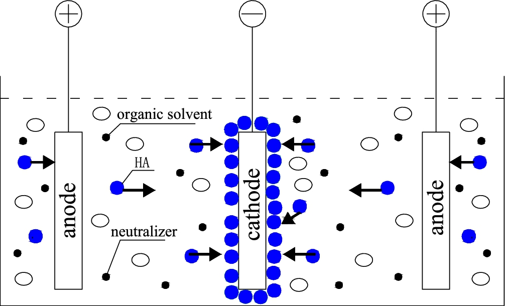
Electrophoretic deposition device.
The compatibility of magnesium matrix hydroxyapatite biological composite material was tested by animal implantation experiment.
SBF immersion test was used to inspect biological activity of magnesium matrix hydroxyapatite biological composite material. Magnesium matrix hydroxyapatite biological composite material prepared by electrophoretic deposition method was trimmed into experimental samples with the size of 9 mm ∗ 9 mm ∗ 2 mm. As a result of the electrophoretic deposition, the surface of experimental sample had coating on both sides. In order to avoid confusion, one side was used as experimental surface, and the other side whose coating was removed as the control surface. Before immersion test, the sample was soaked in anhydrous ethanol about 10 min, and then was cleaned twice in deionized water. After cleaning, the experimental sample was upright placed in plastic bottles which was filled with SBF solution (simulated body fluid). The scale between SBF solution volume and the plate area of sample is about 100 ml/cm2. Then the experimental samples and plastic bottles of SBF solution were put in constant temperature box of 37 ± 1°C for 4 weeks. The experimental samples were taken out from plastic bottles to clean twice in deionized water, then to dry naturally for XRD analysis.
Results and discussion
Preparation and characterization of mg/HA composite material
HA powder, magnesium alloy base and deposition coating were used respectively to do diffraction experiments on X-ray diffractometer. The phase composition of the coating was determined through contrast analysis of the diffraction pattern. XRD diffraction pattern of magnesium alloy, hydroxyapatite (HA) and HA/Mg composites were shown in Fig. 2. The major diffraction peaks of deposition coating could be well corresponded with that of HA powder. It meant that the ingredient of electrophoretic deposition coating was HA. Electrophoretic deposition did not cause HA crystal phase change. At the same time, the diffraction peaks of magnesium alloys of HA/Mg composite material were different from that of the original magnesium alloy. Compared to the XRD diffraction pattern of original HA powder, the diffraction peak shape of HA in HA/Mg composites was quite narrow, which showed that the crystallinity of HA was relatively low and there was less HA coating in HA/Mg composites. Compared with the diffraction peaks of original magnesium alloy base, the diffraction peaks of magnesium alloys in HA/Mg composites were relatively low, which indicated that the content of magnesium alloy was relatively lower.

XRD diffraction pattern of magnesium alloy, HA and HA/Mg composite.
The scanning electron microscope (Japanese JMS-6490LA) was used to scan the deposition coating which prepared by electrophoretic deposition. As shown in Fig. 3. The coating was tightly arranged by HA particles which look like the needle, and it had uniform density, no crack.
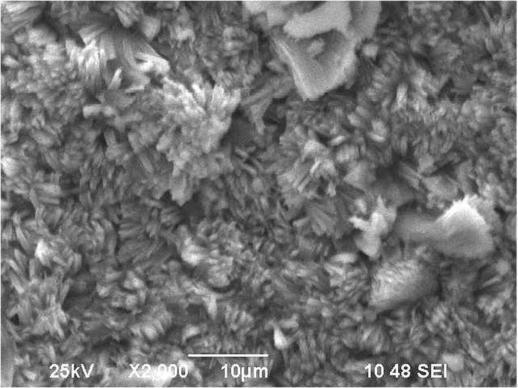
Scanning electron microscope morphology of the coating (×2000).
Surgical wound of SD rat showed minor red swelling and had no osmosis after the surgery. The wound healed after about 2 weeks. The experimental rats were killed and dissected at 2 weeks, 4 weeks and 8 weeks after the surgery respectively. The implant surrounding tissue was taken to do tissue section, HE dyed and organizational analysis by biomicroscope. There were some tissue section pictures of implant surrounding at 2 weeks, 4 weeks and 8 weeks after HA/Mg composite material implant, as shown in Fig. 4. Figure 4(a) acted as the control group. Figure 4(b), (c) and (d) showed that obvious hyperemia, effusion, suppuration, degeneration and necrosis did not appeared in the implant surrounding soft tissue. Compared with the control group, the Fig. 4(b) showed that connective tissue hyperplasia and capillary proliferation appeared, and a few inflammatory cells of lymphocytes and megakaryocytes in connective tissues and softer tissues infiltrated. Compared with the control group shown in Fig. 4(b), (c) showed that more connective tissue and less inflammatory cells appeared around the implant. As shown in Fig. 4(d), less inflammatory cells and a amount of connective tissue hyperplasia forming fibrous capsule around the implant appeared in the tissue section picture. The fibrous capsule was not completed and the connective tissue was torn for the destroy during the implant material removing. After implantation experiment, the electronic analytical balance was used to measure the weight of implant. The results showed that the implant was not degraded, HA of implant coating, to a certain extent, could reduce the rate of corrosion of magnesium alloy. Through above analysis, magnesium matrix hydroxyapatite biological composite material possessed excellent biocompatibility.

The implants section figure (∗100): (a) the control group section figure; (b) the section figure after 2 weeks; (c) after 4 weeks; (d) after 8 weeks (∗40).
After immersed in SBF solution for four weeks, magnesium matrix hydroxyapatite biological composite material has not been corroded. Moreover, the thickness of HA coating increased and a layer of white covering deposited on the HA coating surface. As shown in Fig. 5, there was apatite phase in the white covering. The diffraction peak intensity of apatite phase of HA coating increased, and the diffraction peak intensity of magnesium alloy decreased after immersed in SBF solution. The results showed that the apatite of magnesium matrix hydroxyapatite biological composite material increased, while the contrast surface without HA coating was badly corroded. Atom absorption spectrum analysis was used to analyze the concentration of calcium ions of SBF solution. The experimental results showed that the concentration of calcium ions of SBF solution was reduced from 105.23 mg/L to 67.35 mg/L. Therefore, magnesium matrix hydroxyapatite biological composite material could promote deposition of calcium and induce bone-like apatite.

The XRD diffraction pattern before and after soaked HA/Mg composite.
Magnesium matrix hydroxyapatite biological composite material was successfully prepared by electrophoretic deposition method. The animal implantation experiment showed that the body’s immune response caused by magnesium matrix hydroxyapatite biological composite material was not obvious after implantation for 8 weeks. The composite material had no inflammatory reaction, or cytotoxicity but excellent biocompatibility. The SBF immersion test showed that magnesium matrix hydroxyapatite biological composite material could promote deposition of calcium, and induce bone-like apatite with excellent bioactivity. The magnesium matrix hydroxyapatite biological composite material showed good corrosive resistance through animal implantation experiment and SBF immersion test. HA could slow down the corrosion rate of magnesium alloy, and make magnesium alloy meet the requirements of mechanical properties in service period.
Conflict of interest
The authors have no conflict of interest to report.