
Abstract
A homogeneous and uniform array of nanotubes with a diameter of about 70 nm was produced on titanium (Ti) surface by anodic oxidation. The wall thickness of the nanotubes was around 20 nm, and the depth was about 200 nm. The biological properties of the anodized Ti surface were investigated by simulated body fluid (SBF) soaking test and in vitro cell culture test. The mechanical properties were evaluated by instrumented nanoindentation test and friction-wear test. The results showed that the anodized Ti surface can induce the formation of bone-like apatite after immersion in SBF for four weeks, enhance cell adhesion, proliferation and gene expression, it also showed decreased friction coefficient, similar stiffness and Young’s modulus to those of the cortical bone. Based on these results, it can be concluded that anodic oxidation endowed the Ti surface with improved biological and mechanical properties, which was attributed to the formation of nanostructured surface.
Introduction
Titanium (Ti) not only has excellent biocompatibility, but also provides many favorable characteristics such as high corrosion resistance, good ductility, and non-toxicity, which made it the most popular implant material as artificial bone tissues in clinic to cure bone fracture or bone defect. However, the mismatch of bioactivity and mechanical properties between Ti and bone tissues usually leads to poor osseointegration of the implant material, raising the potential risk of loosening in the long-term usage [1]. Therefore, it is necessary to modify the Ti implant surface to achieve improved biological and mechanical properties.
Many methods have been applied to improve the bioactivity of the Ti surface, such as mechanical methods (e.g., sand-blasting [2]), chemical methods (e.g., acid etching [3]), and the use of various coatings (e.g., hydroxyapatite, wollastonite [4]). Through these conventional approaches, improved biological properties have been achieved due to the creation of favorable surface features and/or surface chemistries preferred by osteoblasts. However, a disadvantage of these methods is that they cannot produce highly controllable surface structure. Moreover, some of these methods have the potential to form surface residuals fallen off from the matrix in complex physiological environment, which are harmful to the surrounding tissue [5]. Thus, alternative methods to modify Ti surface to produce well-defined topography having good bioactivity are desirable.
The mechanical force interaction at the cell-implant interface is a critical indicator for implant stability and longevity. The mechanical mismatch (e.g. high Young’s modulus of Ti) cannot provide proper mechanical stimulus for sensor cells such as bone lining cells of osteoblastic origin and osteocytes, resulting in that the cells cannot generate sufficient biochemical signals to transduce the obtained mechanical signal and modulate bone formation and resorption [6,7]. During the last two decades, a variety of implant materials with porous coatings have been applied to approach the mechanical properties of bone tissues [8]. It has been found that implants with porous surface usually have lower stiffness and Young’s modulus than those with dense surface [1]. The methods used to achieve porous surface are diverse, such as the electron beam melting process, creep expansion of argon-filled pores, laser-engineered net shaping, electric current activated/assisted sintering technique, powder metallurgy (PM), and space-holder technique, etc. [1,9]. However, these methods are limited by their difficult implementation and/or costly instruments. There is necessity to develop new methods to obtain the porous structure on Ti surface.
One method that may create controllable porous surface on Ti is anodic oxidation. Anodic oxidation is an economical, simple, and versatile technique to produce uniform and controllable nanoporous surfaces on metals (e.g. Alumina, Ti-based alloys) [10]. Although there have been several researchers investigating the influence of anodic oxidation induced nanopatterns on biological properties of Ti surface, their direct influence on mechanical properties of Ti has not been studied till now. Therefore, this study aimed to fabricate the controllable porous surface on Ti by anodic oxidation, and then systematically investigate the influence of surface topography on biological and mechanical properties.
Materials and methods
Preparation of samples
Pure titanium plates in dimensions of 10 mm × 10 mm × 1 mm were firstly polished consecutively with #360, #600, #1000 and #2000 carborundum sandpaper, and then cleaned in acetone, ethanol and deionized water for 5 minutes, respectively. Anodic oxidation experiments were carried out in 1M NaF solution using a direct current (dc) voltage source (WYK-150, Yangzhou, China) under a constant voltage of 10 V for 1 h at room temperature. The Ti plate was used as anodic electrode while graphite (40 mm × 40 mm × 5 mm) was used as cathodic electrode. The distance between anodic and cathodic electrodes was 40 mm. During the experiment, the solution was stirred using a magnetic stirrer. After the anodic oxidation treatment, the samples were rinsed with deionized water, dried and characterized. For comparison, the as-polished Ti plates without anodic oxidation were used as the control group.
Scanning electron microscopy (SEM, HITACHI S-4800) was used to observe the surface morphology of the Ti samples. Image processing software of Adobe Photoshop was utilized to measure the pore density on Ti surface. The crystal structure was examined by X-ray diffraction (XRD) analysis on a RIGAKUD/MAX2500 diffractometer with Cu Kα radiation.
Bioactivity evaluation
The as-polished and anodized Ti samples were soaked in 30 ml of simulated body fluid (SBF) for four weeks at 37°C without stirring. The growth of bone-like apatite on sample surfaces was investigated to evaluate their bioactivity. The SBF was composed of 2.5 mM of Ca2+, 1.5 mM of Mg2+, 142.0 mM of Na+, 5.0 mM of K+, 148.5 mM of Cl−, 4.2 mM of HCO3 −, 1.0 mM of HPO4 2−, 0.5 mM of SO4 2− and buffered at a pH of 7.40 with 50 mM tris (hydroxymethyl) aminomethane (CH2OH)3CNH2 and approximately 45 mM hydrochloric acid (HCl) at 37°C according to Kokubo’s research work [11].
Biocompatibility evaluation
The samples were sterilized by an autoclave sterilizer at 121°C for 30 min before cell culture test. Then the MC3T3-E1 cells were seeded on the sample surfaces at a density of 1 × 10⁴ cells/cm2. The cells were cultured in the Dulbecco’s modified Eagle’s medium (DMEM, Gibco BRL, Grand Island, NY, USA) with 10% fetal bovine serum (Hyclone, Logan, UT, USA) and 3% penicillin/streptomycin at 37°C in an incubator with a fully humidified atmosphere of 5% CO2. After culturing for 1, 4 and 7 days, the cellular proliferation and morphology on the sample surfaces were detected according to the literature [12].
After culturing on the Ti plates for 7 days, total RNA was isolated from cells using a monophasic solution of phenol and guanidine isothiocyanate (Trizol, Invitrogen Life Technologies, Carlsbad, CA, USA), following the instructions of the manufacturer [13]. And then reverse transcription-polymerase chain reaction (RT-PCR) and quantitative real-time PCR were separately carried out using the SYBR Green PCR reagent (Qiagen, USA). The primer sets along with the length of the resulting amplicons and the GeneBank accession number were shown in Table 1. Highly purified gene specific primers for osteocalcin (OCN), osteopontin (OPN) and the housekeeping gene, glyceraldehyde-3-phosphate dehydrogenase (GAPDH), were purchased from Shengong Company (Shanghai, China). The gene levels of each mRNA were normalized by GAPDH mRNA to obtain their relative expression. The experimental details can be seen in reference [11].
Primers for RT-PCR and Real-time PCR
Primers for RT-PCR and Real-time PCR
Mechanical properties of the anodized Ti surfaces were performed in a TriboIndenter instrumented nanoindenter (from Hysitron, Minneapolis, MN) using a diamond Berkovich indenter. Indentations were made with applied loads up to 10 mN, and hardness was determined by the Oliver–Pharr method [14], with the instantaneous contact area determined using the calibrated area function of the Berkovich tip. The Young’s modulus of the surface was calculated from nanoindentation tests through the Sneddon’s equation [15].
The friction and wear properties of the disk specimens of 60 mm in diameter were investigated on a Universal Micro-Tribometer tester (UMT-2, CETR, USA) using ball-on-disk contact configuration. The experimental details were described elsewhere [16]. The tests were carried out at room temperature at a sliding speed of 0.3 m/s, with an applied load of 5N and a sliding distance of 10 m. The friction coefficients were continuously recorded with sliding distance and directly displayed on the tester.
Statistical analysis
Samples were run in quintuplicate for each group. Standard deviations were plotted as error bars for the data points on all figures. Statistically difference was determined by Student’s t-test. Difference with
Results and discussion
The surface morphologies of the Ti samples before and after anodic oxidation were shown in Fig. 1. A flat and smooth surface was observed on the as-polished Ti samples. While on the surface of the anodized Ti samples, there is a homogeneous and uniform array of nanotubes, indicating a great difference on the surface morphology between the two samples. The inner diameter of the nanotubes was about 70 nm, the wall thickness was around 20 nm, and the depth was about 200 nm. The number density (number per square centimeter) and area density (area proportion) of the nanotubes determined by the image processing software were separately 2.8 × 10¹⁰/cm2 and about 35.8%. Figure 2 showed the XRD pattern of the as-polished and anodized Ti samples. Samples having different surface treatment were found to have the same diffraction peaks of the α-Ti (card number: 00-001-1197), such as (100), (002), (101), (102), (110), (103), (112) and (201). However, the formation of nanotubes were proved to increase the surface roughness and hydrophilicity (data not shown here), which are two important factors to determine the biological properties of Ti implants [17].

Surface morphologies of the as-polished (a) and anodized (b) Ti samples.

XRD patterns of the as-polished and anodized Ti samples.
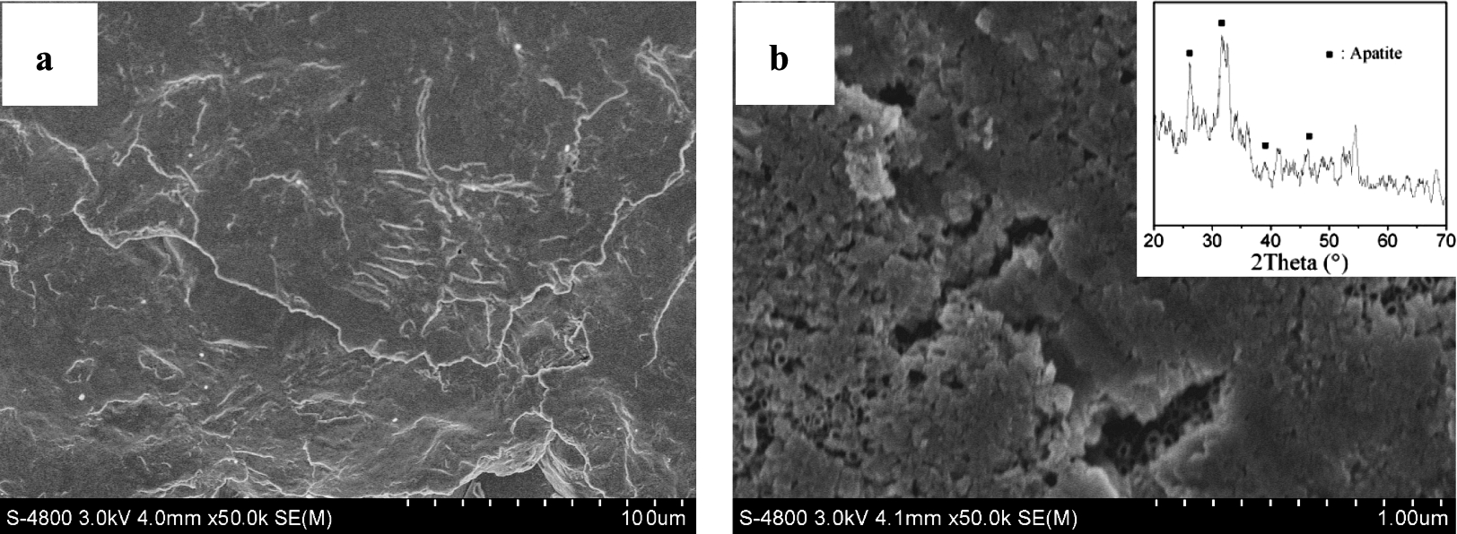
Surface morphologies of the as-polished (a) and anodized (b) Ti samples after immersion in SBF for four weeks.
After soaking the samples in SBF for four weeks, no new substance was formed on the as-polished Ti surface (Fig. 3a), showing similar morphology with the samples without immersion in SBF. While for the anodized Ti samples, a new layer of nanocrystals with a fine structure was observed on the surface (Fig. 3b). XRD pattern confirmed the formation of apatite on the anodized Ti surfaces by showing the characteristic peaks of apatite at around 26° and 32° (inset of Fig. 3b), indicating the good bioactivity of the anodized Ti surface. It is well known that the improved bioactivity can be attributed to the increased hydrophilicity. In our previous work, it has been reported that anodic oxidation can induce amount of hydroxyl groups on nanostructured surface, leading to the improved hydrophilicity [17]. The hydroxyl groups can combine with the Ca2+ ions in the SBF solution to form a positive charged surface, and then the Ca2+ ions, in turn, combine with the negative charged phosphate ions to form amorphous phosphate, which spontaneously transforms into the bone-like apatite. The apatite layer, the main inorganic composition of nanutal bone tissues, plays a vital role in forming the natural bone tissue and improving the bioactivity of the implant surface.
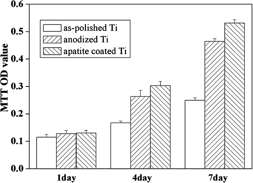
MTT assay representing the MC3T3-E1 cell proliferation on the as-polished, anodized and apatite-coated Ti surfaces.
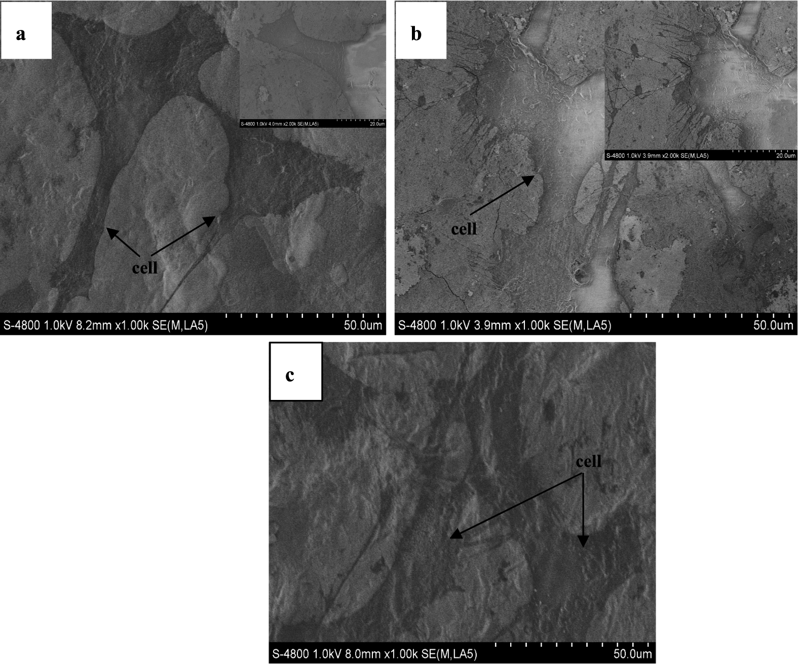
Cell morphologies on the as-polished (a), anodized (b) and apatite-coated (c) Ti samples.
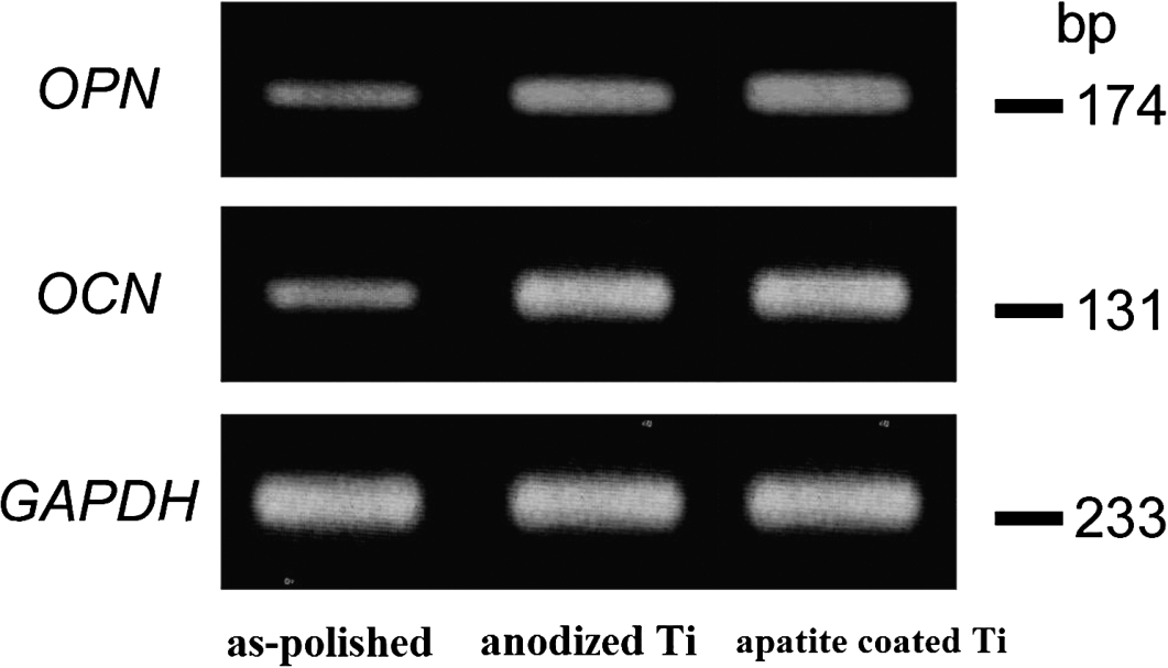
RT-PCR analysis of OPN and OCN mRNA levels in MC3T3-E1 cells cultured on the as-polished, anodized and apatite-coated Ti surfaces.
Co-cultured with the cells for 1 day, 4 days and 7 days, the cell proliferation on the as-polished and anodized Ti surfaces was shown in Fig. 4. For comparison, the cell proliferation on the apatite-coated Ti surface (the sample shown in Fig. 3b) was also investigated and shown in Fig. 4. It can be seen that, during the 7-day period, there was a progressive and significant increase in cell numbers. However, the cells proliferated much faster on anodized and apatite-coated Ti surfaces than those on the as-polished Ti surfaces (
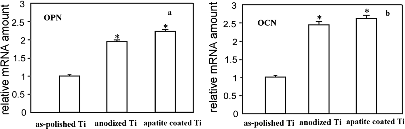
Real-time PCR analysis of OPN (a) and OCN (b) mRNA levels in MC3T3-E1 cells cultured on the as-polished, anodized and apatite-coated Ti surfaces.
Surface topography has already been proved to be responsible for changes of biocompatibility. The bone cells are accustomed to a nanoscale environment rather than to a microscale environment because the nanoscales mimic the dimensions of the constituents in natural bones [18]. For instance, osteoblast proliferation was observed to be significantly enhanced on nanostructured alumina, titania and hydroxyapatite in comparison to their conventional counterparts. Balasundaram et al. suggested that implants produced with conventional materials (or materials with constituent dimensions greater than 1 μm) could not invoke suitable cellular response to regenerate enough bone [19]. It was also proved that osteoblasts can form more calcium on nanosurfaces than on microsurfaces. In addition, the high surface area provided by nanostructures offers beneficial conditions for the interlocking with bone cells and the penetration of body fluid, which could effectively increase the bone fixation with implant. In our study, although better cell behavious were observed on the apatite-coated Ti surface, it also confirmed that the nanostructured surface obtained on Ti exhibited good biocompatibility.
The stiffness values and Young’s modulus of the anodized Ti surface were summarized in Table 2. For comparison, the corresponding mechanical properties of the as-polished Ti and cortical bone were also presented in Table 2. The stiffness values of the natural bone tissues, as-polished and anodized Ti surface are 0.2 GPa, 3.2 GPa and 0.7 GPa, respectively. Their corresponding Young’s modulus are 17–20 GPa, 67 GPa and 26 GPa. It can be concluded that, after anodic oxidation, the mechanical properties of the Ti surface is much closer to the natural bone tissues, indicating the beneficial effect of nanostructured surface on the mechanosensation of osteocytes. Although a number of studies investigating the influence of the cell-biomaterial interface on bone mechanotransduction appeared in recent years [6], there is no clear elucidation that how osteocytes sense mechanical loading and transduce it into cellular signal.
The stiffness values, Young’s modulus of the natural bone tissues, as-polished and anodized Ti surface
The sliding wear characteristics of the polished and anodized Ti surfaces were evaluated with an applied load of 5N and a speed of 0.3 m/s. The variation of friction coefficients was shown in Fig. 8. It can be seen that the friction coefficients of the as-polished and anodized Ti surfaces change gradually with time, and then tend to be stable. In addition, it is clear that the initial friction coefficient of the polished Ti is a little larger than that of the anodized Ti, however no obvious difference can be observed between them at the steady-stage. The steady-stage coefficients may result from the base material as the surface coating was abraded out.
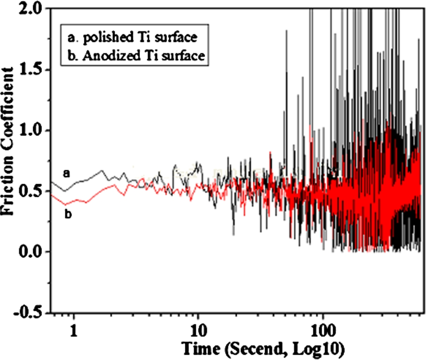
Friction coefficients of the as-polished (a) and anodized (b) Ti surfaces depending on time.
The anodic oxidation induced nanotubes can reduce the stiffness and Young’s modulus mismatches between implant and bone tissues. The friction coefficient of the anodized Ti was also inferior to that of the polished Ti surface, which may be explained by the following reason. Accompanying the formation of nanotubes during the anodization, structural defects like porosity and nanotube interfaces appear within the surface, which in turn increase the surface roughness. It has been well known that abrasion on these very fine scales is sensitive to extrinsic details such as surface roughness, or the presence and variable thickness of native oxide. Furthermore, grain morphology and texture are known to have a small effect on hardness, but a much more pronounced effect on wear properties, therefore, the decreased stiffness, Young’s modulus and friction coefficient were observed on the anodized Ti surface. The surface morphology of the porous implant promotes bone ingrowth into the pores and provides not only anchorage for biological fixation but also a system which enables stresses to be transferred from the implant to the bone, leading to long-term stability. To achieve better mechanical properties (similar values of stiffness, Young’s modulus and friction coefficient to cortical bone), the porous Ti surface can be optimized by controlling porosity, pore size and shape, as well as pore distribution to suit the natural bone, which will be investigated in our future work.
In this paper, anodic oxidation was applied to modify Ti surface to obtain a homogeneous and uniform array of nanotubes. The inner diameter of the nanotubes was about 70 nm, the wall thickness ranged around 20 nm, and the depth was about 200 nm. The formation of nanotubes led to the achievement of surface mechanical properties close to natural bone. And the nanotubed surface also resulted in excellent bioactivity and biocompatibility. Based on these results, it can be concluded that anodic oxidization endowed Ti surface with improved mechanical and biological properties, indicating that anodic oxidation might be a promising method to enhance the performance of Ti-based implants.
Footnotes
Acknowledgements
The authors gratefully acknowledge the support by the National Natural Science Foundation of China (No. 51201056, No. 81500886 and No. 51401146), college student innovation fund project (No. DC201510080068), Technology Foundation for returned overseas chinese scholars (No. C2015003038), and Tianjin Natural Science Foundation (No. 16JCYBJC28700).
Conflict of interest
The authors have no conflict of interest to report.