
Abstract
Mesenchymal stem cells (MSCs) are a common tool in regenerative medicine. The nanoscale extracellular vesicles (nEVs) secreted by these cells were recently brought up to light thanks to their therapeutic potential. In this study, we assessed the in vitro behaviour of human umbilical vein endothelial cells (HUVECs) exposed to nEVs derived from human umbilical cord mesenchymal stem cells (hUC-MSCs). Nanoscale extracellular vesicles were isolated and characterized by NanoSight® and flow cytometry. HUVECs were stimulated with various concentrations of nEVs. To assess nEV interactions with HUVECs, confocal microscopy and angiogenesis assay were performed. The use of nEVs derived from hUC-MSCs was able to produce positive outcomes on HUVECs by acting on their angiogenic potential.
Introduction
Mesenchymal stem cells (MSCs) are an attractive cell source for regenerative medicine thanks to their multipotency and immunomodulatory properties suitable for allogeneic therapies [1]. Despite these benefits, the therapeutic efficiency of infused MSCs is limited, because they are mainly trapped in the lungs rather than targeting the area in need of regeneration [2]. MSCs exert their regenerative action through the secretion of soluble factors and extracellular vesicles (EVs) [3]. Among EVs, those with the smallest size reach a nanoscale around 30 to 150 nm [4]. These nanoscale extracellular vesicles (nEVs) produce similar results to MSC infusion for immunomodulation and tissue regeneration [5].
The successful implantation of tissue-engineered construct is limited by the difficulty to mimic angiogenesis through the formation of endothelial tubular networks to generate new capillaries [6]. Human umbilical vein endothelial cells (HUVECs) are extensively used in research and have connections in situ with umbilical cord MSCs, which have superior capacities compared to adult MSCs due to their immaturity [1]. This cell-to-cell communication is particularly mediated by EVs [4] and could be a key element to promote angiogenesis.
In this work, the in vitro pro-angiogenic effect of nEVs derived from human umbilical cord mesenchymal stem cells (hUC-MSCs) was studied on HUVECs.
Material and methods
Cell culture and nanoscale extracellular vesicle preparation
HUVECs and hUC-MSCs were harvested and cultured as previously described [7,8]. After hUC-MSCs were put in contact with nanoscale particle-depleted culture medium for 48 h, nanoparticles were isolated from conditioned medium as reported before [9]. The size and concentration of nanoscale particles were assessed by nanoparticle tracking analysis (NTA, NanoSight® LM10). Then, the nanoparticles were characterized as nEVs by flow cytometry with specific antibodies for CD9, CD63 and CD81 (BD Pharmingen) as shown elsewhere [9].
Stimulation with nanoscale extracellular vesicles derived from hUC-MSCs
HUVECs were cultured in EBM™-2 (Lonza) supplemented with 10% FBS (Fetal Bovine Serum). This condition was used as control. HUVECs were also cultured with 10% nEV-depleted FBS and stimulated by nEVs (
Statistical analysis
Results are expressed by means ± SD (
Results
Characterization of nanoscale extracellular vesicles
NTA has shown that the particles contained in the medium are in the size range of nEVs (30–150 nm) with a concentration higher than
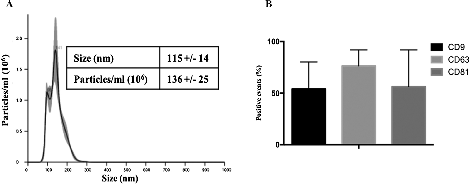
Physical (A) and biological (B) characterization of nanoscale extracellular vesicles (nEVs) secreted by human umbilical cord mesenchymal stem cells. (A) Representative NanoSight® plot and corresponding table for nanoparticle size and concentration. (B) Expression evaluated by flow cytometry of nEVs markers (CD9, CD63 and CD81) on the surface of nanoparticles. Results are means ± SD (

(A) Representative confocal microscopy picture of nanoscale extracellular vesicles (nEVs) uptake by human umbilical vein endothelial cells (HUVECs). HUVECs nuclei were labelled with To-Pro®-3 and nEVs with PKH67 (some of them are displayed here with white arrows). After 24 h of stimulation, nEVs were found inside HUVECs cytoplasm. (B) Effects on angiogenesis of nEVs stimulation (
HUVECs and nEVs labelling has permitted to observe that nEVs derived from hUC-MSCs have interactions with HUVECs and are found in their cytoplasm after 24 h of stimulation (Fig. 2(A)). After 4 h of culture on ECM gel solution, the formation of capillary-like tubes by HUVECs evaluated by node, junction, segment and branch formation, seems to be increased in the presence of nEVs derived from hUC-MSCs. The lower nEVs concentration seems to promote node formation (Fig. 2(B)).
Discussion
Stem cell grafts can induce some difficulties such as reject, acting on the wrong target or the development of cancers [10]. Nanoscale extracellular vesicles derived from MSCs have already shown their potential in tissue regeneration and their use could avoid the inconvenience associated with the presence of cells while maintaining the desired effects.
This work shows that the use of nEVs derived from hUC-MSCs is able to stimulate the angiogenesis potential of HUVECs compared to nEVs contained in FBS. This is also true for nEVs derived from other cell types such as endothelial progenitor and MSCs from bone marrow [11]. FBS cannot be used in human cell therapy. It will be interesting to explore the possibility of using platelet lysate completed with nEVs derived from MSCs for further experiments and thus suppressing animal products.
Conclusion
In this work, we showed that nEVs derived from hUC-MSCs could be used as cell culture additives without harmful effects. In addition, these nEVs have positive outcomes on HUVECs by acting on their angiogenic potential. The capacity of these nEVs may also be used to develop new concept in tissue engineering by functionalizing biomaterials.
Footnotes
Acknowledgements
G. Dostert and A.-S. Willemin are supported by a PhD scholarship granted by the French Ministry of National Education, Higher Education and Research (MENESR). É. Velot was supported by a grant from the Conseil Régional de Lorraine in association with the Pacte Lorraine (RNG14XNF-AOP14).
Conflict of interest
The authors have no conflict of interest to report.