
Abstract
Porous silk fibroin films can provide an optimal microenvironment for angiogenesis in vivo. Adhesion and migration of human umbilical vein endothelial cells (HUVEC) on porous silk films were observed by confocal laser scanning microscopy. The expression of fibronectin (FN), laminins (LN), intercellular adhesion molecule-l (ICAM-1) and vascular cell adhesion molecule-l (VCAM-1) after implanting Porous Silk Fibroin Films (PSFFs) as grafts for dermis regeneration in rat were studied. FN, LN, ICAM and VCAM may have direct relationships with angiogenesis. The result will help to design excellent PSFFs and study the process and mechanism of angiogenesis.
Keywords
Introduction
Immunochemistry plays an important role in observing angiogenesis in different fields, especially in biomaterials. Immunohistochemistry is widely used in biomaterials to observe wound healing and new tissue formation such as angiogenesis and skin and bone regeneration [1,2]. PSFFs as dermis substitutes were polypeptides consisting of heavy (H) and light (L) chains from Bombyx mori domestic silk and were applied in skin defects due to their high tensile strength, controllable biodegradability, haemostatic properties, non-cytotoxicity, low antigenicity and non-inflammatory characteristics [3,4]. During angiogenesis a dynamic interaction occurs among endothelial cells (EC), angiogenic cytokines such as vascular endothelial growth factor (VEGF), basic fibroblast growth factor (bFGF) and the extracellular matrix (ECM) environment. Endothelial cells make up the inner surface of the microvasculature or blood vessels which supply the cells in organs and tissues with oxygen and nutrients to survive and which remove waste products. VEGF increases permeability of EC monolayers in a dose-dependent fashion, and VEGF-induced permeability is mediated through PI-3 kinase-PKB and MAP-kinase signaling cascades. Especially endothelial cell ECM receptors are critical for these morphogenetic changes in blood vessels during wound repair. FN and LN mediate adhesion, spreading, and cell migration of endothelial cells (ECs) through integrin receptors, mainly through integrin and appear to be required for wound angiogenesis. The delivery of pro-angiogenic factors such as vascular endothelial growth factor (VEGF) or basic fibroblast growth factor (bFGF) encourage infiltration of host vasculature into the scaffolds and promote angiogenesis in situ. However, adhesion molecules may modulate endothelial cell migration and angiogenesis too. The increased expression of adhesion molecules such as ICAM-1 on host cells might promote the mobilization of endothelial progenitor cells to ischemic sites and contribute to neovascularization [5]. VCAM-1 can enhance angiogenesis by transmitting specific signals necessary for endothelial cell motility [6]. The relationship between FN, LN, VCAM and ICAM in PSFFs and angiogenesis is little known.
In this paper adhesion and migration of human umbilical vein endothelial cells (HUVEC) on porous silk films were studied and the expression of FN, LN, VCAM and ICAM in the skin of rat after implanting PSFFs was observed. Expression of VCAM and ICAM were used to evaluate the angiogenesis within the biomaterial in wounding healing. Biodegradable porous silk fibroin films may be engineered to provide an extracellular matrix mimicking environment for better cell adhesion and tissue in-growth in dermis regeneration.
Materials and methods
Culture of HUVEC
Human umbilical vein endothelial cells (HUVEC ) were cultured in PAA medium containing 2% FCS, 0.4% ECG supplement/heparin, 0.1 ng/ml epidermal growth factor (EGF), 1 ng/ml basic fibroblast growth factor (FGF), penicillin/streptomycin and 1 µg/ml hydroeortisone in a humidified atmosphere at 37°C and 5% CO2.
Preparation of PSFFs
Bombyx mori domestic silk was treated with Na2CO3 solution to remove sericin. PSFFs were obtained by freeze-drying and cut to pieces of 2 cm × 2 cm which were wrapped and irradiated to sterilize, and then stored at 4°C before using.
Preparation and culture of cells on fibroin nets
The porous silk fibroin films were prepared as previously described and stored in 4°C, and cut into 1 cm × 1 cm and put in distilled PBS for 4 days before using in 24 wells. Individual cell types were cultured in cell culture flasks, removed from the flasks with trypsin, and then 100 µl of medium containing 1 × 10⁶ cells or mixtures of HUVEC were added directly to a net for 4 hours, and then added to 1 ml medium for culture.
Cells on silk films for 12 days added VEGF were placed into medium or PBS containing 0.1 mM calcein-acetoxymethyl ester (calcein-AM), and incubated for 10–30 min at 37°C. Calcein-AM becomes fluorescent when taken up by viable cells and the fluorescence is spread throughout the cell. The stained net was examined by confocal laser scanning microscopy (CLSM).
SEM
To examine cell morphology, cells on porous silk fibroin films after 12 days were placed into glutaraldehyde solution, then Osmium tetroxide buffer, and dehydrated using graded ethanol for SEM. Specimens mounted onto aluminum stubs were sputter-coated with gold and next examined in a Leica Cambridge Stereoscan 360 microscope at an acceleration voltage of 3–7 kV.
In vivo experiment
All Sprague Dawley (SD) rats were SPF grade with a weight of 250–350 g, and raised in by the Experimental Animal Centre of Soochow University (License Number: SCXK (Su)2007-0035). Animal model was approved by the ethical committee in Hall of Jiangsu Province Science and Technology. All surgical procedures were performed under strict aseptic conditions. Animals were maintained under general anesthesia during surgery with intra-abdominal injection of pentobarbital sodium (30 mg/kg body weight). A 2 cm × 2 cm piece of dermis was removed from each rat, and a corresponding piece of PSFFs (Fig. 1). The split thickness skin was then sewed up.
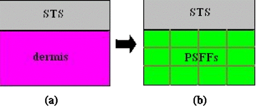
Animal model of implanting PSFFs or PVA. (a) Normal skin; (b) scheme after graft; STS – split thickness skin; PSFFs – porous silk fibroin films. (Colors are visible in the online version of the article;
Samples (2 cm × 2 cm) in vivo of the implanted area(s) were harvested at 1 day, 7 days, 15 days and 25 days after treatment respectively, washed with PBS buffer and fixed in 4% formalin, then to staple onto a plastic placard to keep the implant and skin from curling and embed in paraffin. 5 µm slides were cut (Leica BOND-MAX) and proceed with a SABC kit (Wuhan Boster Biological Technology, China) for immunohistochemistry. The sections were incubated overnight with (a) rabbit anti-rat CD34 antibody (Santa Cruz Biotechnology, Beijing Zhong Shan Jinqiao biotechnology agency, Beijing, diluted in PBS 1:200) for assessment of microvessels, (b) rabbit anti-rat FN, and LN antibody (Wuhan Boster Biological Technology, Wuhan, China, dilution 1:150) at 4°C, (c) rabbit anti-rat ICAM and VCAM antibody (Santa Cruz biotechnology, USA, diluted in PBS 1:200). The secondary antibody (Wuhan Boster Biological Technology, China, dilution in PBS 1:200) was incubated for 30 min, then with SABC (Wuhan Boster Biological Technology, Wuhan, China) reagent at 37°C for 20 min. The color was developed by 3-diaminobenzidine substrate (Wuhan Boster Biological Technology, Wuhan, China) at room temperature for 1–20 min under a microscope. Counterstaining of nuclei was done with hematoxylin for 5–10 min. Additional sections were incubated in diluents without primary antibody and served as negative controls. Brown particles in the cytoplasm and cell membrane were regarded as positive cells. Well-stained areas were chosen at high power (×100) in every visual field. The expression intensity of ICAM and VCAM were scored according to percentage of positive cells, Expression of FN and LN were scored according to intensity and divided into four levels: 4+ (95% visual fields in nigger-brown), 3+ (80% visual fields in brown), 2+ (60% visual fields in brown) and 1+ (40% visual fields in brown) [7]. One group chooses five slides but not less than four visual fields.
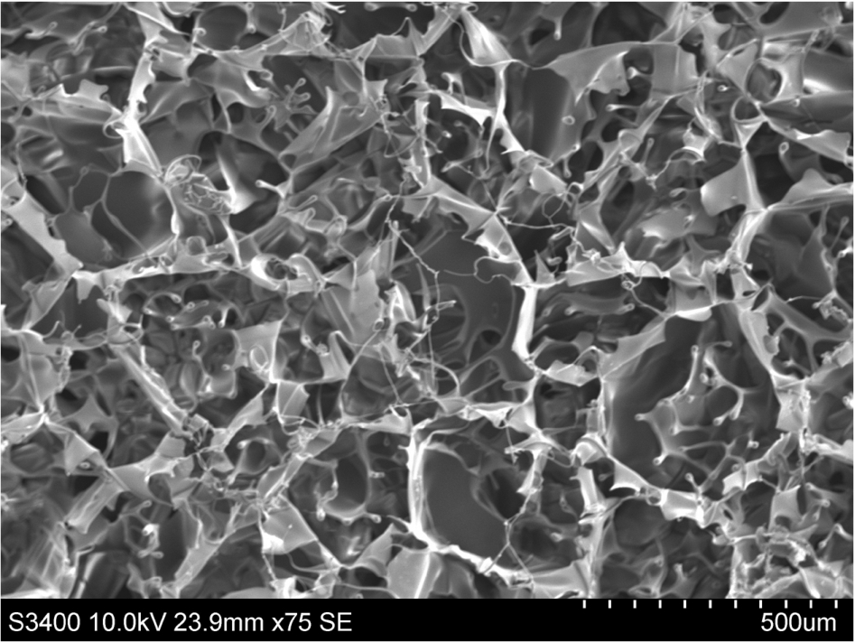
Structural and ultrastructural images of porous silk fibroin films without cell by SEM.
The PSFFs were soft, ivory white, 0.8–1.0 mm in thickness, and had symmetrical distribution of pores with average size of 50–70 µm, porosity of around 90% by SEM. In addition, micro pores could be found in the wall (Fig. 2).
By day 12 HUVECs stained with Calcein-AM method adhered to and spread along the porous structure and infiltrated into the materials. HUVECs began to move in line to form vessels and showed even distribution on silk fibroin films. The cells initially spread across individual fibers of the net and when the entire surface area of individual fibers was covered the cells spread across gaps in the net to form tissue-like structures (Fig. 3).
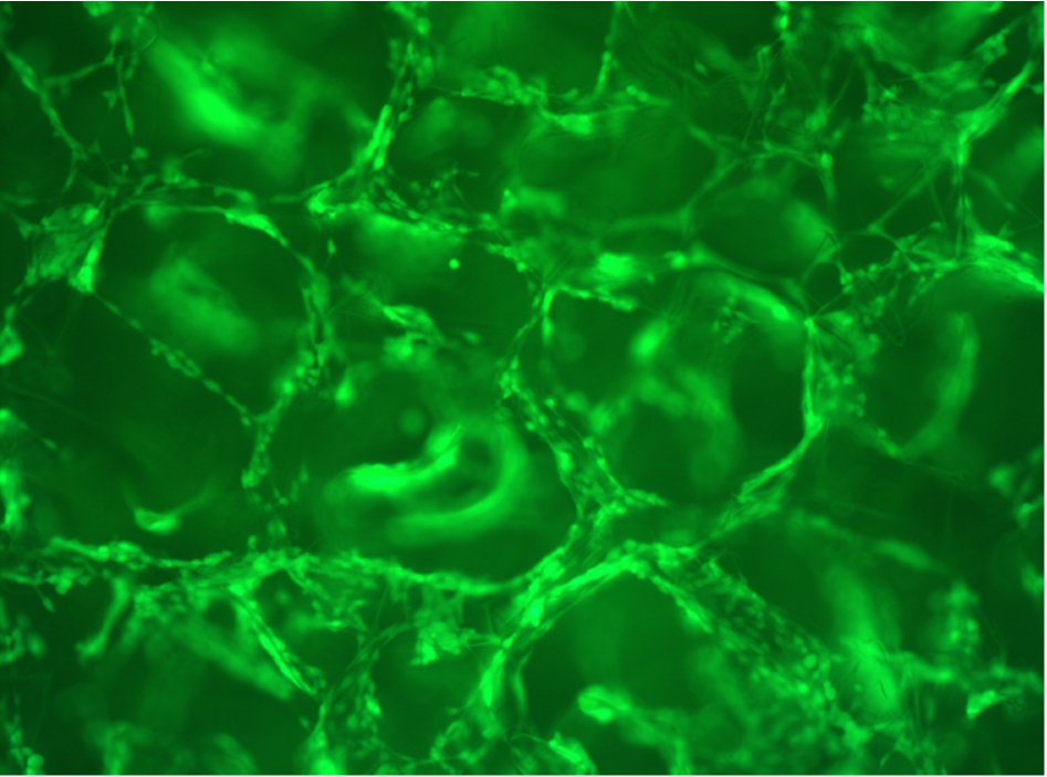
CLSM images of calcein-AM-stained adherent HUVEC after addition to porous silk fibroin films. (Colors are visible in the online version of the article;
Layers of cells on the round of degraded porous silk fibroin for 12 days can be seen. Some HUVECs were spread along with the surface of material, some HUVECs in the holes moved together, some crowded to migrate to the surface. Filopodias were attached strongly on the surface of silk films (Fig. 4).
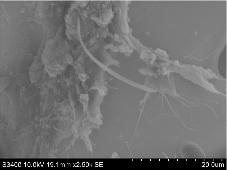
Endothelial cells were grown on fibroin films for 12 days were examined by SEM.
FN showed positive (1+) staining on almost all infiltrated inflammatory cells, especially in the vicinity of PSFFs. Amount of FN staining decreased from the margin of the tissues to the central portion of the PSFFs at day 1. All PSFFs were stained brown, which may absorb FN as a provisional matrix from serum and ECM near the existed tissues (Fig. 5(a)). On day 7, FN showed stronger positive (2+) staining than that of day 1, but more fibroblasts and less inflammatory cells stained with FN (Fig. 5(b)). By day 15, the strongest staining with FN (4+) was observed in fibroblasts which filled the further degraded PSFFs (Fig. 5(c)). On day 25 FN (3+) decreased which may represent the proper rebuilding of tissue in skin regeneration (Fig. 5(d)). LN expression was similar to FN (Fig. 6(a), (b), (c), (d)), but LN in basement membranes have strong staining by day 25.
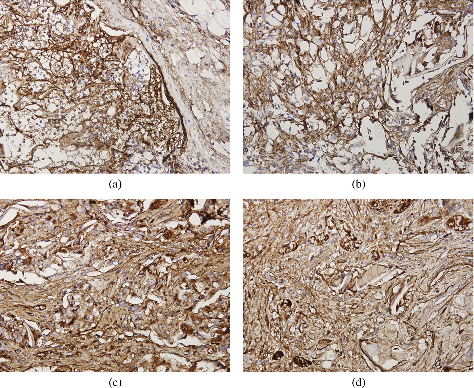
Expression of FN in cells at different times after implanting PSFFs. (a) 1 day (1+); (b) 7 days (2+); (c) 15 days (4+); (d) 25 days (3+); arrow = PSFFs. (Colors are visible in the online version of the article;

Expression of LN in cells at different times after implanting PSFFs. (a) 1 day (1+); (b) 7 days (2+); (c) 15 days (4+); (d) 25 days (3+); arrow = PSFFs. (Colors are visible in the online version of the article;
ICAM-1 showed strong positive staining on 78.6% of inflammatory cells and endothelial cells in the vicinity of or in the PSFFs by day 1. With the passage of inflammatory cells, the positive staining for ICAM-1 reduced significantly to 30.3% by day 7 and 10.7% by day 25.
VCAM-1 showed positive staining in 49.3% inflammatory cells by day 1. By day 7, day 15 and day 25, its expression was stable and did not increase significantly by day 25.
Silk fibroins were used in bone, skin, vascular tissue regeneration, neural tissue regeneration or drug delivery [8]. Morphological diversification of silk biomaterials for tissue regeneration can be obtained when degummed silk fiber is dissolved to obtain silk solution. Silk fibroins were prepared to films, 3D porous scaffolds, hydrogels, electro-spun and wet-spun fibers and particles for tissues engineering and medical application [1,9]. Different morphology of silk fibroin will affect the way of cell migration and attachment. The result showed HUVEC migrated and attached along with the pores of silk fibroin to form capillary-like structure. Pores of silk fibroin films look like vessel and can induce HUVEC to attach and migrate, proliferate without coating any protein or growth factor, it may be more excellent vascularization to form vessel if silk fibroin films coated with protein or growth factor. It is consistent with Professor Unger reported that cell will migrate according to the morphologies of silk fibroin coated with FN.
Angiogenesis is a vital component of normal wound healing and also requires a dynamic interaction among endothelial cells, inflammatory cell (leukocytes, monocytes), pro- and anti-angiogenic growth factors (VEGF) and the ECM environment (FN, LN). Otherwise, it can be induced by the activation of the innate immune response. Gu proposed that biomaterials can induce tissue and vessel regeneration [8]. Neovascularization of biomaterial consists of the interaction between biomaterial and cell, intercellular, ECM and angiogenic growth factors. bFGF may set the stage for angiogenesis during the first 3 days wound repair, whereas VEGF has potent angiogenesis as well as permeability and may be critical for angiogenesis during granulation tissue formation from day 4 [9]. Most of wound healing involves both inflammation and angiogenesis, but the mechanisms of cross talk between inflammatory and angiogenic pathways is developing in research and understanding. Inflammatory cell adhesion molecules may modulate endothelial cell migration and angiogenesis. Intercellular adhesion molecule-l (ICAM-1; CD54) is an adhesive protein belonging to the immunoglobulin superfamily, localized in the endothelial cell membrane, and regulates the adhesion process of leukocytes to these cells. Transplantation of human adipose-derived stromal cells (hADSC) spheroids into ischemic tissue resulted in the increased expression of the adhesion molecules ICAM-1, VCAM-1, and this possibly contributes to the increased recruitment of circulating angiogenic progenitor cells into the ischemic site as well as subsequent blood vessel formation [1]. ICAM-1 plays a novel role in modulating VEGF-A-induced angiogenesis and endothelial nitric oxide synthase (eNOS) activity through regulation of phosphatase and tensin homolog deleted on chromosome ten (PTEN) expressions via modulation of intracellular GSH status [10]. VCAM-1 (the immunoglobulin superfamily as well) can enhance angiogenesis by transmitting specific signals necessary for endothelial cell motility. VCAM-1 participates in the adhesion of circulating leukocytes to endothelial cells at the early stage of wound healing. The rapid new tissue development observed in wound healing depends not only on the cells and cytokines present but also on the ECM microenvironment. FN not only mediates endothelial cell migration but also controls the availability of growth factors TGF [11]. VEGF-A dependent migration of retinal vessels requires PI3K activity and astrocytic FN possibly functions to retain VEGF protein on the astrocytic matrix [12]. Laminins are large trimeric ECM glycoproteins in forming the basement membrane sheet through polymerization and plays a role in cell migration, differentiation and proliferation.
Now the PSFFs, FN and LN complex will provide ECM to adhere to inflammatory cells and endothelial cells to a new environment and induce fibroblast and endothelial cell migration and differentiation by day 1. At the same time VCAM and ICAM has a strong effect on cells to induce new vessel growth and invade the provisional ECM to form capillaries. The process of endothelial cells distinguishing provisional ECM is the subject of further study. VEGF, Hypoxia-Inducible Factor (HIF) also takes part in angiogenesis at the early stage of wound healing. VCAM and ICAM had returned to normal levels in PSFFs. By day 15, more and more capillaries, FN, and LN were formed in PSFFs with the degradation of PSFFs. New ECM may play an important role in angiogenesis. By day 25, the diameter of the lumen increased and formed a vessel net in PSFFs. FN and LN expression was reduced and began to rebuild ECM. But these studies do not know whether the inflammatory factors ICAM and VCAM up regulated important VEGF or VEGF regulated ICAM in angiogenesis. The mechanism of angiogenesis in PSFFs need further study. The experiments describe the process of angiogenesis in PSFFs by immunohistochemistry as follows:
Provisional ECM (FN, LN PSFFs) as a microenvironment filled with ICAM, VCAM and VEGF. Pre-existing capillaries invade the provisional ECM to the central portion of degraded PSFFs. FN and LN from fibroblasts filled the degraded PSFFs. Vessel net formed and ECM is rebuilt.
The PSFFs, FN and LN complex functioned as provisional ECM to attach to inflammatory cells and endothelial cells in vivo. PSFF induced endothelial cell migration and differentiation along with the pores and then to the centre of PSFFs in vitro. PSFFs will be used to dermal regeneration as the excellent vascularization biomaterials.
Footnotes
Acknowledgements
This work was supported by a grant pre-research of national funds and open funds of National Engineering Laboratory for Modern Silk of China. Thanks to Di-Silvio Lucy in King College London and Jie Huang in University College London.