
Abstract
In tissue engineering approaches, the quality of substitutes is a key element to determine its ability to treat cartilage defects. However, in clinical practice, the evaluation of tissue-engineered cartilage substitute quality is not possible due to the invasiveness of the standard procedure, which is to date histology. The aim of this work was to validate a new innovative system performed from two-photon excitation laser adapted to an optical macroscope to evaluate at macroscopic scale the collagen network in cartilage tissue-engineered substitutes in confrontation with gold standard histologic techniques or immunohistochemistry to visualize type II collagen. This system permitted to differentiate the quality of collagen network between ITS and TGF-β1 treatments. Multiscale large field imaging combined to multimodality approaches (SHG-TCSPC) at macroscopical scale represent an innovative and non-invasive technique to monitor the quality of collagen network in cartilage tissue-engineered substitutes before in vivo implantation.
Introduction
Tissue-engineering (TE) is an emerging field, whose goal is to replace damaged tissues with artificial tissues constructs grown in vitro to mimic the composition and properties of the cartilage in a controlled laboratory environment by delivering the appropriate cells, biomaterials and signaling factors to diseased or damaged areas. The quality of in vitro tissue-engineered substitute is a key element to determine the success of the cartilaginous graft in vivo [1]. The differences in remodeling outcome using implanted engineered cartilage in an animal osteochondral defect model depend on the degree of cartilage maturity in vitro [2]. The collagen network inside TE-substitute is one crucial criteria of quality control. However, current evaluation methods are destructive and reduce the amount of graft material finally available. Conventional optical techniques, like histology (Sirius red) and fluorescence (specific antibody), remained a reference for structural characterization.
Unfortunately, their invasive character leads to development and validation of non-invasive techniques to detect collagen molecules based on near-infrared illumination as two-photon excitation (TPE) and Second Harmonic Generation (SHG). Combined with multiphoton fluorescence excitation, SHG microscopy has been demonstrated to be effective in numerous label-free tissue imaging applications without any exogenous dye, and without damaging the collagen proteins to observe non-centrosymmetric molecular assemblies, such as collagen fibers discrimination [3] and collagen architecture in cartilage [4].
Recently, this approach using SHG imaging constitutes a diagnostic application for detecting the initial symptom of OA, including degeneration of the cartilage matrix and pathological changes in other connective tissues [5]. Moreover, as SHG is based on a light scattering and not on fluorescence, photo-bleaching and toxicity are significantly reduced. This multimodality technique could be considered as a precise tool for biodiagnosis to detect in safety conditions collagen synthesized by cells seeded in scaffolds in presence of chondrogenic growth factors [6].
In this work, we showed for the first time that multiscale large field imaging (macroscopy) combined to multimodality approaches (SHG-TCSPC) at macroscopical scale could be an innovative and non-invasive technique to monitor the quality of collagen network in cartilage TE substitutes before in vivo implantation. TE substitutes were compared to empty scaffolds and MSC-seeded sponges without growth factor (TGF-β1).
Materials and methods
Preparation of cartilaginous TE substitute
Bone marrows were obtained in informed patients who underwent total hip replacement surgery in collaboration with the “Service de Chirurgie Orthopédique at Traumatologique” and “Unité de Thérapie Cellulaire et Tissus” of Nancy Universitary Hospital. Mesenchymal stem cells (MSCs) from bone marrow were plated at 37°C at 5% CO2 with Dulbecco’s modified Eagle Medium (DMEM-LG, 31885, Gibco) supplemented with 10% of fetal bovine serum (FBS, Sigma), 1 ng/ml bFGF (R&D), glutamine and penicillin-streptomycin. During the last passage before seeding in biomaterials (passage 3), MSCs were cultured with DMEM 4.5 g/ml supplemented with sodium pyruvate (110 μg/ml), bFGF (1 ng/ml) penicillin-streptomycin, Proline (P), L-Ascorbic acid-2-Phosphate (A) and Dexamethasone (D). MSCs were seeded in collagen sponges composed of type I and III collagens from bovine dermis (Symatese) at 5 × 10⁵ cells/sponge cultured in two different medium supplements: ITS 1% (non-chondrogenic medium) or ITS 1% + TGF-β1 (10 ng/ml) (chondrogenic medium).
Multiscale and multimodality imaging
Immunofluorescence
Collagen II produced by MSCs embedded in scaffold was marked by indirect immunostaining. The primary antibodies were a type II anti-collagen with the suitable secondary antibody, rabbit polyclonal antibody associated to Alexa488™. After 15 min washing with PBS, scaffolds were fixed with PFA 4% during 20 min and then deposed on glass slides 2 hours for observation at room temperature.
Two-photon excitation in macroscopy (TPE-Ma)
For Two Photon Excitation system, a femtosecond oscillator (Mira 900F, Coherent), pumped with a solid laser (Verdi 8 W, Coherent) was used to generate some ultra-short infrared pulses (120 fs, 76 MHz) at
TCSPC-SHG imaging and F-SHG ratio
Decay times were measured in the backward direction by a time-correlated single photon counting method (SPC-730 TCSPC Imaging module, Becker & Hickl, Berlin) coupled to scan-head of CLSM TCS SP2-AOBS (Leica Microsystems). A signal coming from the scan controller of the microscope enabled to trace decay curves for each pixel of an image (128 × 128). The signal has to be deconvoluted with calculated instrumental response function and analyzed for fitting with SPC Image software (Becker & Hickl, Berlin). The time slop of the decay is usually a sum of different exponential decays and an index (F-SHG ratio) was described as the ratio between the long lifetime (extrinsic fluorescence relative to Alexa488™ labeling) and very fast lifetime (SHG due to collagen) which is obtained by two exponential components fitting. Images were coded with a color LUT according to F/SHG index values.
Second Harmonic Generation in macroscopy
For the two photon excitation system, a femtosecond oscillator (Mira 900F from 600 to 1100 nm, Coherent), pumped with a solid laser (Verdi 8 W, Coherent) was used to generate some ultra-short infrared pulses (120 fs, 76 MHz) at
Results
Two-photon excitation in macroscopy
On Fig. 1 Part 1, three different scaffolds were observed by Multiphoton Excitation Macroscopy (TPE-Ma) in fluorescence at 488 nm excitation after Alexa488™ fluorescence indirect labeling for type II collagen. Native sponge without cells ((A): SWC) presented just the basal autofluorescence of native collagen fibers of sponge. With no chondrogenic medium ((B): ITS), the labeling inside sponge corresponded to autofluorescence of cells and scaffold. In contrary, a strong signal of type II collagen was observed in ECM synthesized by MSCs inside the scaffold with chondrogenic medium ((C): TGF-β1) after 28 days of culture.
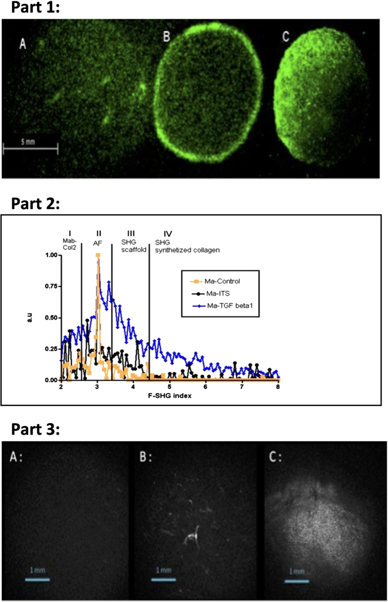
Study of 3 different scaffolds after 28 days of culture: sponge without cell (A), MSC-seeded sponge cultured with ITS (B), MSC-seeded sponge cultured with TGF-β1 (C). Part 1. Direct observation of scaffolds by Multiphoton Excitation Macroscopy (TPE-Ma) in fluorescence at 488 nm excitation after Alexa488TM fluorescence indirect labeling for type II collagen. Part 2. Samples were analyzed in TCSPC-SHG macroscopy to extract F-SHG index, after immunolabeled with Mab directed against type II collagen. Distribution of F-SHG index is defined in four defined areas (segments I, II, III and IV) respectively in close relation with detection of type II collagen only (Mab-Col2), global autofluorescence (AF), SHG scaffold and SHG synthesized collagen. Part 3. Direct observation of MSC-seeded scaffolds by SHG-Macroscopy (original figure: http://link.springer.com/article/10.1186/s40634-015-0026-0#copyrightInformation, © Cucchiarini et al.; licensee Springer. 2015, no changes were made).
For MSCs seeded in 3D scaffolds, challenge was to distinguish changes in collagen providing from fluorescence (F) signal and from Second Harmonic Generation (SHG) signal. The change in F-SHG index was monitored according to medium culture (supplemented or not) at 28 days and confirmed identity of type II neo-synthesized fiber collagen in scaffold and enhanced expression in the presence of TGF-β1. The results revealed a precise range of F-SHG index (from 2 to 8), distributed among 4 defined areas (I, II, III, IV) in relation with cellular behavior and metabolism (Fig. 1 Part 2). The range detection of Alexa488™ fluorophore coupled to Mab against type II collagen (Mab col II) was restricted in part I (from 2 to 2.3).
Then, part II (from 2.3 to 3.3) concerned more specifically global autofluorescence (AF) coming from scaffold (type I collagen) and cells. SHG signal from the scaffold (with cells seeded or not) was strictly confined in area III with a range from 3.3 to 4.4. Finally, SHG signal relative to synthesized collagen by MSCs was significatively well distributed in the last range IV (from 4.4 to 8). For Fig. 1, for MSCs cultured in the presence of TGF-β1 or not (ITS), type II collagen has been specifically detected by immunolabeling in part (A). However, coll II-SHG signal appeared only in the presence of TGF-β1 in part (D), showing the abundant extracellular collagen synthesis and as an indication on organized and structured collagen fibers. This result indicated that a change in the collagen conformation and its ability to be detected as a harmonophore by TCSPC-SHG imaging. The part (B) was also informative as an indication on the cellular density in scaffold and proliferation and its effect on global autofluorescence.
SHG-imaging in macroscopy
Direct observation of three different scaffolds by TCSPC-SHG macroscopy for the detection of collagen as a harmonophore is presented on Fig. 1 Part 3. In our three different culture conditions (SWC, ITS and TGF-β1), the SHG signal is detected only in sponges treated with TGF-β1 in comparison with sponges treated with ITS characterized by absence of type II collagen synthesis.
Discussion
In cartilage bioengineering, early detection according to degree of organization of collagen network could be a crucial step in the differentiation process of autologous cells seeded in a biomaterial. The amount of produced matrix and especially the synthesis of type II collagen and otherwise standardization of preclinical step of setting-up of one biomaterial is a crucial step.
In this work, we showed that SHG signal specific to collagen network is a non-invasive technology to monitor the quality of collagen network of TE substitutes before in vivo graft, in comparison with classical techniques as immunofluorescence using a specific anti-collagen type II antibody to localize specifically this synthesized protein. The major disadvantage of use antibodies was their cost, their limited penetration in tissue and the introduction of an exogenous dye. So, they are almost exclusively used on fixed sections and absolutely not on a precious biological sample reserved for graft in patients. The interest and the difficulty of this approach were to work on a biomaterial with collagen. The collagen sponges, already used in hospital in other applications, shows to be a biomaterial of choice for the tissue repair of the cartilage being given the very good preliminary results already obtained in vitro and in vivo (in comparison with a biomaterial with alginate for example, easier to study).
It will thus be necessary to consider for quite used techniques the obtained signal corresponding to the sponge it even and the signal corresponding to the collagen synthesized by cells. In conclusion, we have demonstrated with regard to the conventional techniques that SHG at the macroscopically scale is an innovative technique to evaluate noninvasively collagen network of cartilage TE substitute. This technique could be used in a clinical step to screen easily and rapidly a large number of TE substitutes in a same time without exogenous probe and without alteration of substitute to select the better for the graft.
Footnotes
Acknowledgements
This work was supported by grants from the Région Lorraine, the Communauté Urbaine du Grand Nancy and the Conseil Général de Meurthe at Moselle. This work was supported by grants from Agence Nationale de la Recherche – Programme Blanc 2009. Project: « Cartilage Engineering and MRI: Assessment of Biointegration and Biofunctionality in the Rat » (CEMABIR) and by GIS IBISA.
Conflict of interest
The authors have no conflict of interest to report.